Introduction
Pancreatic neuroendocrine tumors (PNETs) are rare
neoplasms, with an incidence of 0.48 cases/100,000 individuals each
year between 2000 and 2012 in the United States of America
(1–2).
A trend towards increasing incidence and prevalence rates has been
documented by previous studies (3,4). Although
functional PNETs may be present in a variety of hormone syndromes
and detected at an earlier stage, numerous non-functional PNETs are
diagnosed late in the disease course, with symptoms associated with
local mass effects or metastatic disease (5). A biomarker with a high sensitivity and
specificity is mandatory for the early and accurate diagnosis of
PNETs, particularly for those with vague symptoms.
Chromogranin A (CgA) is an acidic glycoprotein
stored in the dense granules of the NETs and co-released with
peptide hormones (6). CgA has been
suggested to be a reliable biomarker for NETs in terms of
diagnostic value, prognosis prediction and treatment response
evaluation (7,8). In western countries, a low diagnostic
accuracy but good prognostic value of CgA in patients with
resectable non-functional PNETs has been demonstrated (9,10).
Concerning differences in biomarker performance across racial
groups, there are a small number of studies examining the
diagnostic value of CgA in Asian populations. Plasma CgA has been
suggested to be a useful biomarker for PNETs in Asian populations
(11–14), but few studies have evaluated the
diagnostic role of plasma CgA in early-stage PNETs. The present
study aimed to evaluate the clinical utility of plasma CgA in the
diagnostic confirmation and evaluation of treatment response in
Asian patients with PNETs, particularly those with early-stage
tumors.
Materials and methods
Study design and population
The study protocol was approved by the Institutional
Review Board of National Taiwan University Hospital (Taipei,
Taiwan). Written informed consent for participation in the study
was obtained from all participants. From August 2010 to April 2014,
35 patients with PNETs according to tissue-based diagnosis were
prospectively enrolled consecutively. The age, gender, clinical
presentation and plasma CgA level were recorded prior to
tissue-based diagnosis Patients presenting with symptoms suggestive
of excessive hormone production were considered to have functional
PNETs, and the other patients were considered to have
non-functional tumors. Endoscopic ultrasound-guided fine-needle
aspiration (EUS-FNA) cell blocks or surgical specimens were used
for a definite diagnosis. The tissue coagulum clot method was
adapted for cell block preparation (15). The steps included fixation by
transferring tissue coagulum clot strips into the 10% formalin
solution at room temperature for between 10 and 24 h, subsequent
centrifugation, and final transfer of the pellet for paraffin
embedding as a cell block. Each cell clock was examined via
hematoxylin and eosin staining and immunohistochemical staining
with antibodies against CgA and synaptophysin, as previously
described (16). Histopathological
characteristics, including tumor location, size, grade,
Tumor-Node-Metastasis (TNM) status and tissue CgA immunoreactivity
were also recorded. Tumor grade was classified according to the
recommendations of the World Health Organization (WHO) (17). TNM status was based on the 7th edition
of the American Joint Commission on Cancer Staging System (18). Inclusion criteria included patients
aged ≥20 years old with tissue-based diagnosis of PNETs
consecutively within the study period. Participants were recruited
on a rolling basis as and when they were identified using
tissue-based diagnosis. Exclusion criteria included end-stage renal
disease, liver failure and the presence of any other malignancies.
Serial CgA measurement and imaging studies were used to evaluate
treatment response. Response Evaluation Criteria In Solid
Tumors (RECIST) (19) was used to
evaluate the treatment response of the image studies.
Measurement of plasma CgA
Blood samples were obtained following overnight
fasting and collected prior to definite tissue-based diagnosis
either by EUS-FNA or by surgical resection. The plasma CgA level
was measured with a commercial kit (Chromoa R assay; CIS Bio
International S.A., Saclay, France; cat. no. CGA-ELISA), according
to the manufacturer's protocol. The recommended cutoff value was
set at 94 U/l, according to a previous study (9).
Statistical analysis
Comparison of values from independent groups was
performed using a Mann-Whitney test. Pearson's χ2 test
was used to measure the strength of the association between pairs
of variables without specifying dependencies. To determine whether
CgA level was an independent predictor of metastasis, hazard ratios
were calculated using the Cox proportional hazards model. The
sensitivity and specificity of CgA level to discriminate metastatic
from localized PNETs were analyzed using the receiver operating
characteristic (ROC) curve, and the optimal cutoff value was
determined. Kendall's τ correlation test was applied to estimate
the correlation between change of CgA level and treatment response
by RECIST. Data were analyzed with SPSS version 17.0 (SPSS Inc.,
Chicago, IL, USA). P<0.05 was considered to indicate a
statistically significant difference.
Results
Clinical characteristics of enrolled
patients
Among the 35 patients with PNET, there were 18 males
and 17 females. The mean age of these patients was 53.0 (range,
31–80) years. A total of 29 patients received surgical resection
only, and 1 patient received surgical resection and additional
targeted therapy. A total of 4 patients received systemic antitumor
therapies, including target therapy and cytotoxic chemotherapy, and
1 patient received best supportive care. The CgA levels among
patients with PNET with different clinicopathological
characteristics were compared (Table
I). There was no association between sex and CgA value. The
patients usually presented asymptomatically, and those PNETs were
identified incidentally by health examination (21/35, 60%). The
majority of these tumors were well differentiated (G1; 22/35, 63%)
and localized tumors (27/35, 77%). Among patients with the
localized disease, the majority of the tumors were >2 cm in
diameter (16/27, 59%). The majority of the tumors were located in
single areas of the pancreatic body or tail where they were more
easily detected by routine abdominal sonography, with the exception
of 3 cases of multiple endocrine neoplasia type 1 (MEN-1), which
presented with multiple tumors from the pancreatic head to tail.
The majority of the patients with PNETs were immunoreactive to
tissue CgA.
 | Table I.Plasma CgA levels in patients with
PNETs. |
Table I.
Plasma CgA levels in patients with
PNETs.
| Variables | n | Median CgA level
(range), U/l | P-value |
|---|
| Sex |
|
| 0.22 |
|
Female | 17 | 84.4
(12.1–16465.7) |
|
|
|
Male | 18 | 66.5
(33.6–3117.5) |
|
|
| Age, years |
|
| 0.26 |
|
<50 | 17 | 67.9
(12.1–16465.7) |
|
|
|
≥50 | 18 | 107.5
(33.6–3117.5) |
|
|
| Clinical
symptoms |
|
| 0.71 |
|
Absent | 21 | 53.5
(12.1–3117.5) |
|
|
|
Present | 14 | 70.5
(99.5–16465.7) |
|
|
| Proton pump
inhibitor |
|
| 0.39 |
|
Absent | 33 | 84.4
(12.1–3117.5) |
|
|
|
Present | 2 | 8264.4
(63.0–16465.7) |
|
|
| MEN-1 |
|
| 0.68 |
|
Absent | 32 | 85.0
(12.1–3117.5) |
|
|
|
Present | 3 | 67.9
(59.9–16465.7) |
|
|
| Pancreatic
locationa |
|
| 0.62 |
| Body +
tail | 21 | 72.7
(33.6–3117.5) |
|
|
| Head +
neck | 11 | 86.5
(12.1–2637.5) |
|
|
| Size, cm |
|
| 0.03 |
| ≤2 | 16 | 64.0
(12.1–1506.5) |
|
|
|
>2 | 19 | 115.4
(33.6–16465.7) |
|
|
| Metastasis |
|
| 0.02 |
|
Absent | 27 | 67.9
(12.1–16465.7) |
|
|
|
Present | 8 | 308.7
(35.9–2637.5) |
|
|
| Pathological
stage |
|
| 0.03 |
| I | 24 | 66.5
(12.1–16465.7) |
|
|
|
II+III+IV | 11 | 221.0
(35.9–2637.5) |
|
|
| WHO grade |
|
| 0.59 |
| G1 | 22 | 72.7
(42.5–16465.7) |
|
|
|
G2+G3 | 13 | 132.0
(12.1–3117.5) |
|
|
| Tissue CgA |
|
| 0.20 |
|
Negative | 2 | 51.4
(35.9–67.9) |
|
|
|
Positive | 33 | 85.5
(12.1–16465.7) |
|
|
Univariate analysis of CgA levels based on the
clinicopathological variables demonstrated that plasma CgA levels
were significantly higher in patients with larger tumor size (size
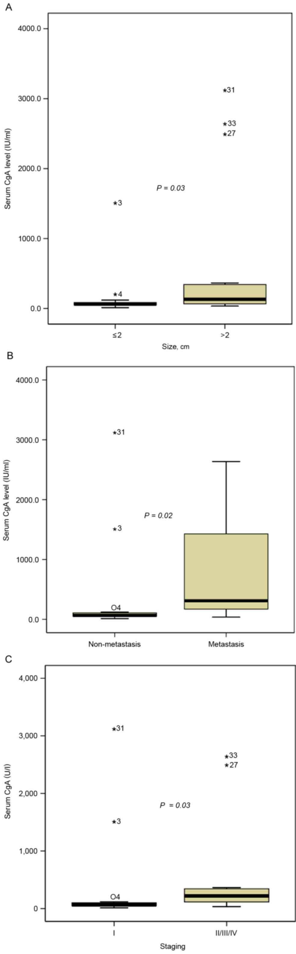
>2 cm vs. size ≤2 cm: 115.4 vs. 64.0 U/l, P=0.03; Fig. 1A), distant metastasis (metastasis vs.
non-metastasis: 308.7 vs. 67.9 U/l, P=0.02; Fig. 1B) and advanced pathological stage
(stage II/III/IV vs. stage I: 221.0 vs. 66.5 U/l, P=0.03; Fig. 1C). CgA level did not differ in terms
of sex, age, clinical symptoms, proton pump inhibitor use, MEN-1
presence, tumor location, tumor grade or tissue CgA
immunoreactivity.
Diagnostic value of CgA
Table II summarizes
the sensitivities of plasma CgA levels of PNETs under the different
cutoff values suggested by previous studies (8,12,16). When the cutoff value was 94 U/l, the
overall sensitivity of plasma CgA was only 42.9%. Subgroup analysis
indicated that the sensitivity of plasma CgA for patients with
metastatic tumors was 87.5%, while the sensitivity for patients
with localized tumors was 29.6%. On evaluation under different
cutoff values, all the sensitivities for the patient with localized
PNETs were poor (74 U/l, 40.7%; 65.7 U/l, 51.9%).
| Table II.Sensitivity of CgA under different
cutoff values. |
Table II.
Sensitivity of CgA under different
cutoff values.
|
| Tumor location |
|
|---|
|
|
|
|
|---|
| CgA cutoff level,
U/l |
Localizeda |
Metastasisa | Overall
sensitivitya |
|---|
| 94 | 29.6 (8/27) | 87.5 (7/8) | 42.9 (15/35) |
| 74 | 40.7 (11/27) | 87.5 (7/8) | 51.4 (18/35) |
| 64.3 | 51.9 (14/27) | 87.5 (7/8) | 60.0 (21/35) |
CgA as a predictor of metastasis
Table III summarizes
the univariate and multivariate analyses of predictors of
metastasis. Larger tumor size (P=0.019), WHO grade 2 or 3 (P=0.006)
and higher plasma CgA level (≥94 U/l; P=0.014) were associated with
a higher risk of metastasis according to the univariate analysis.
Multivariate analysis of significant factors indicated that only
higher plasma CgA level was significantly associated with a higher
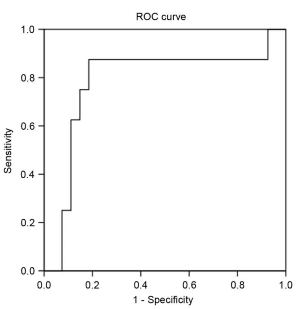
risk of metastasis (P=0.045). To distinguish between patients with
localized disease or metastasis, a ROC curve analysis was applied
(Fig. 2). When the cutoff value was
94 U/l, the sensitivity and specificity were 87.5 and 70.4%,
respectively. Using 126 U/l as the optimal cutoff value, the
sensitivity and specificity were 87.5 and 81.5%, respectively.
| Table III.Univariate and multivariate analyses
of predictors of metastasis. |
Table III.
Univariate and multivariate analyses
of predictors of metastasis.
| Variables | HR (95% CI) | P-value | HR (95% CI) | P-value |
|---|
| Sex |
|
|
|
|
| Male vs.
female | 0.93
(0.19–4.50) | 0.927 |
|
|
| Age, years |
|
|
|
|
| ≥50 vs. <50 | 0.93
(0.19–4.50) | 0.927 |
|
|
| Clinical
symptoms |
|
|
|
|
| Present vs.
absent | 3.33
(0.65–17.18) | 0.150 |
|
|
| Location |
|
|
|
|
| Head/neck vs.
body/tail | 0.53
(0.09–3.18) | 0.490 |
|
|
| Size, cm |
|
|
|
|
| >2 vs. ≤2 | 2.05
(1.13–3.74) | 0.019 | 1.72
(0.66–4.50) | 0.266 |
| WHO grade |
|
|
|
|
| G1 vs. G2/3 | 0.04
(0.004–0.40) | 0.006 | 0.09
(0.57–202.8) | 0.112 |
| Plasma CgA,
U/l |
|
|
|
|
| ≥94 vs. <94 | 16.63
(1.75–158.09) | 0.014 | 18.31
(1.073–312.56) | 0.045 |
Correlation between plasma CgA change
and treatment response
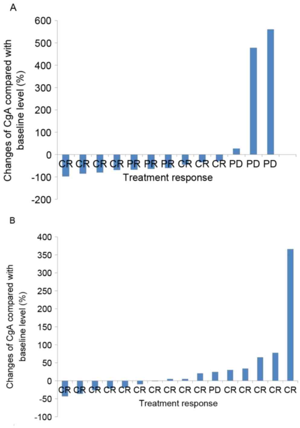
A total of 29 out of 35 patients underwent serial
CgA measurement and imaging studies prior and subsequent to
treatment (6 patients did not receive serial CgA measurement as
they were lost to follow-up). A total of 13 patients exhibited high
baseline plasma CgA levels (≥94 U/l) and 16 patients exhibited low
baseline levels (<94 U/l). Among the patients with high baseline
CgA levels, 7 patients achieved a complete response and 3 patients
demonstrated a partial response. A total of 3 patients exhibited
progressive disease. Serial plasma CgA change was correlated with
treatment response. A decrease of ≥30% in CgA levels was observed
in patients with complete and partial responses (P=0.025; Fig. 3A). Among the patients with low
baseline CgA levels, 15 patients achieved complete response and 1
patient exhibited progressive disease. However, serial CgA level
was not correlated with treatment response in this patient group
(P=0.587; Fig. 3B).
Discussion
The present study demonstrated that plasma CgA
exhibited limited diagnostic value in early PNETs. Smaller and
localized PNETs tended to exhibit lower plasma CgA levels. Although
plasma CgA change was correlated with treatment response, this
result was limited to the patients with high baseline CgA levels
(≥94 U/l).
CgA is an acidic glycoprotein stored in the dense
granules of NETs and co-released with peptide hormones (6). CgA is considered the most accurate tumor
marker in the diagnosis of gastro-entero-pancreatic NETs
(GEP-NETs), in comparison with other tumor markers, including
urinary 5-hydroxyindoleacetic acid, neuron-specific enolase and
carcinoembryonic antigen (7). The
sensitivity and specificity of CgA differs and depends on numerous
factors, such as the type of assay used, the cutoff value, tumor
burden and organs involved (20). A
small number of studies concerning the diagnostic value of plasma
CgA in Asian populations are summarized in Table IV (11–14). The
sensitivities and specificities of plasma CgA were 53.6–86.0 and
78.6–91.9% respectively. However, these studies only enrolled a
small number of early-stage PNETs. In the present study, the
overall sensitivity was only 42.9%, and the sensitivity of
localized PNETs was even lower (29.6%). The low sensitivity of
plasma CgA identified in the patients was probably due to the small
size of the tumors and the early stage. Among the localized tumors,
the majority were >2 cm (59%). A previous study indicated that
small tumors may be associated with normal CgA levels (8). In concordance with previous studies, the
present study demonstrated that plasma CgA levels were
significantly associated with tumor size. Smaller tumors tended to
exhibit lower plasma CgA levels. Conversely, the sensitivity of
plasma CgA for patients with metastatic cancer was 87.5%, which was
comparable with a previous study conducted in Asia (11). CgA levels and their associated
sensitivities depended on metastasis (21). Previous studies suggested that plasma
CgA level was correlated with tumor mass and disease extent. CgA
levels were higher in patients with extensive liver metastases
compared with localized disease (22,23). The
present study also indicated that those patients with metastatic
tumors tended to exhibit higher plasma CgA levels. The sensitivity
of plasma CgA for patients with metastatic PNETs was relatively
good in comparison to patients with localized PNETs.
| Table IV.Summary of previous studies on the
diagnostic value of CgA in Asian populations. |
Table IV.
Summary of previous studies on the
diagnostic value of CgA in Asian populations.
| First author | Study design | Assay type | Cutoff value,
U/l | Sensitivity, % | Specificity, % | (Ref.) |
|---|
| Han et
al | Non-functional PNET
(n=51) | Chromoa assay,
ELISA | 64.3 | 63.2 | 91.2 | (12) |
| Qiao et
al | Non-insulinoma PNET
(n=32) | Chromoa assay,
ELISA | 74.0 | 65.6 | 91.9 | (14) |
| Chou et
al | GEP-NET (n=44) | Chromoa assay,
ELISA | 94.0 | 86.0 | 88.0 | (11) |
| Hijioka et
al | PNET (n=69) | Chromoa assay,
ELISA | 78.7 | 53.6 | 78.6 | (13) |
Distant metastasis predicts a higher risk of
mortality in PNETs (24,25). Identification of reliable predictors
of metastasis has been the aim of numerous studies. Tumor size was
the most commonly documented clinical predictor of metastasis
(26). The risk of metastasis
increased significantly if the tumor size exceeded 15 mm. A small
number of studies evaluated the association between the plasma CgA
level and the risk of metastasis. Paik et al demonstrated
that a high level of CgA (>156.5 U/l) predicted distant
metastasis in those patients with PNETs (27). In concordance with this study, the
present study also demonstrated that plasma CgA was a reliable
indicator of metastasis. When the cutoff value was 94 U/l, the
sensitivity and specificity were 87.5 and 70.4%, respectively.
Using 126 U/l as the best cutoff value, the sensitivity and
specificity were 87.5 and 81.5%, respectively. Identification of
any metastatic tumors should thus be performed carefully once the
PNET patient has demonstrated high CgA levels.
Korse et al (28), demonstrated that a higher sensitivity
of plasma CgA was noted in patients with well-differentiated
tumors. The sensitivity was ~68% for G1 NETs and 74% for G2 NETs. A
much lower sensitivity (37%) was noted in the patients with poorly
differentiated NETs, which was probably due to the relative lack of
large dense-core granules. The present study did not indicate a
significant association between grade and plasma CgA level. This
may be as only 2 cases of G3 NETs were enrolled in the series. The
single patient with metastatic PNET and lower plasma CgA level
exhibited grade G3 NET. Although plasma CgA was revealed to be
useful in the detection of metastatic NETs (11), it may not diagnose those patients with
poorly differentiated NETs.
Plasma CgA has been shown to be valuable in
evaluating the treatment response of different therapies (2,29). For
patients with hepatic metastases from functional carcinoid tumors,
Jensen et al (29),
demonstrated that a reduction in CgA of ≥80% following
cytoreductive surgery was predictive of the stabilization of
disease. For patients with advanced PNETs, Yao et al
(2) revealed that those patients with
an early CgA response following treatment of everolimus experienced
longer progression-free survival. Similar to previous studies, the
present study also indicated that changes in plasma CgA were
correlated with treatment response in those patients with high
baseline CgA levels. However, this was not true for those patients
with low baseline CgA levels, which implied that the role of CgA in
evaluating treatment efficacy may be limited to patients with high
baseline CgA levels.
Functional PNETs may present with various hormone
syndromes, so they are usually detected at an earlier stage. By
contrast, non-functional PNETs usually present with symptoms
following local mass effects or metastatic disease at more advanced
stages (5). In the present study, the
majority of PNETs were non-functional and detected incidentally
(60%), with a localized (77%) status. This is likely due to the
widespread use of abdominal ultrasound in Taiwan. Routine
ultrasound screening for patients with chronic hepatitis or
abnormal liver function tests is extremely popular. PNETs localized
in the body or tail may be easily detected by screening ultrasounds
at an earlier stage.
A previous study indicated that high CgA levels
(150-fold higher compared with the normal upper limit) in GEP-NETs
were associated with MEN-1 (6).
However, low diagnostic accuracy of plasma CgA in the detection of
PNETs in patients with MEN-1 has been demonstrated by two other
studies (30,31). The plasma CgA test cannot replace the
other established diagnostic tools in screening for early PNETs
among patients with MEN-1. The results of the present study also
indicated a limited use of plasma CgA in patients with MEN-1,
although only 3 patients with MEN-1 were included and only 1 of
these exhibited a higher CgA level.
There are a number of non-neoplastic causes of CgA
elevation, including renal insufficiency, chronic hepatitis and
drug use. Certain adenocarcinomas may also account for CgA
elevation (6,7). In the present study, the patients with
liver failure, renal failure and other types of cancer were
excluded. Although proton pump inhibitor use has been suggested to
be a common cause for CgA elevation (32), only 2 cases in the present study were
administered proton pump inhibitors and they exhibited different
CgA responses (63.0 and 16,465.7 U/l). Additional interpretation
with subgroup analysis was not feasible due to a relatively small
number of patients with PNET in the present study. As PNET is an
uncommon disease, future studies with multi-center cooperation may
provide a more comprehensive view.
To conclude, the present study demonstrated that
plasma CgA levels were associated with tumor size, metastasis
status and tumor stage in diagnosing patients with PNET. Changes in
plasma CgA levels were correlated with treatment response only in
those patients with high baseline CgA levels. For early-stage
PNETs, CgA exhibited a limited role in the diagnosis and evaluation
of treatment response.
Acknowledgements
Not applicable.
Funding
Pinancial support was provided by Novartis (Taiwan)
Co., Ltd., (Taipei, Taiwan) for the CgA test.
Availability of data and materials
All the datasets generated and analyzed in the
present study are included in this published article.
Authors' contributions
CMT designed the study, reviewed the article
(Tables. 2 and 4) and drafted the manuscript. TYC drafted
and revised the manuscript, collected data (FNA and tissue
staining) and performed statistic analysis (Tables. 1 and 3). T-BC performed statistical analysis and
revised the figures and tables (Tables.
1 and 3). YWT contributed to
data collection (surgical tissue) and provided technical support.
C-CC contributed to data collection (plasma chromogranin A) and was
involved in the interpretation of data. JTL revised the study
protocol and revised the manuscript. H-PW designed the study,
contributed to data collection (patient's background) and performed
critical revisions of the manuscript.
Ethics approval and consent to
participate
The study protocol was approved by the Institutional
Review Board of National Taiwan University Hospital (Taipei,
Taiwan). Written informed consent for participation in the study
was obtained from all participants.
Consent for publication
All study participants provided consent for the data
to be published.
Competing interests
The authors declare that they have no competing
interests.
Glossary
Abbreviations
Abbreviations:
|
CgA
|
chromogranin A
|
|
EUS-FNA
|
endoscopic ultrasound-guided
fine-needle aspiration
|
|
NET
|
neuroendocrine tumor
|
|
GEP-NET
|
gastro-entero-pancreatic NET
|
|
MEN-1
|
multiple endocrine neoplasia type
1
|
|
PNET
|
pancreatic NET
|
|
RECIST
|
Response Evaluation Criteria In Solid
Tumors
|
|
ROC
|
receiver operating characteristic
|
|
WHO
|
World Health Organization
|
References
|
1
|
Dasari A, Shen C, Halperin D, Zhao B, Zhou
S, Xu Y, Shih T and Yao JC: Trends in the incidence, prevalence,
and survival outcomes in patients with neuroendocrine tumors in the
United States. JAMA Oncol. 3:1335–1342. 2017. View Article : Google Scholar : PubMed/NCBI
|
|
2
|
Yao JC, Pavel M, Phan AT, Kulke MH, Hoosen
S, St Peter J, Cherfi A and Öberg KE: Chromogranin A and
neuron-specific enolase as prognostic markers in patients with
advanced pNET treated with everolimus. J Clin Endocrinol Metab.
96:3741–3749. 2011. View Article : Google Scholar : PubMed/NCBI
|
|
3
|
Milan SA and Yeo CJ: Neuroendocrine tumors
of the pancreas. Curr Opin Oncol. 24:46–55. 2012. View Article : Google Scholar : PubMed/NCBI
|
|
4
|
Tsai HJ, Wu CC, Tsai CR, Lin SF, Chen LT
and Chang JS: The epidemiology of neuroendocrine tumors in taiwan:
A nation-wide cancer registry-based study. PLoS One. 8:e624872013.
View Article : Google Scholar : PubMed/NCBI
|
|
5
|
Burns WR and Edil BH: Neuroendocrine
pancreatic tumors: Guidelines for management and update. Curr Treat
Options Oncol. 13:24–34. 2012. View Article : Google Scholar : PubMed/NCBI
|
|
6
|
Modlin IM, Gustafsson BI, Moss SF, Pavel
M, Tsolakis AV and Kidd M: Chromogranin A-biological function and
clinical utility in neuro endocrine tumor disease. Ann Surg Oncol.
17:2427–2443. 2010. View Article : Google Scholar : PubMed/NCBI
|
|
7
|
Lawrence B, Gustafsson BI, Kidd M, Pavel
M, Svejda B and Modlin IM: The clinical relevance of chromogranin A
as a biomarker for gastroenteropancreatic neuroendocrine tumors.
Endocrinol Metab Clin North Am. 40(111–134): viii2011.
|
|
8
|
Vinik AI, Woltering EA, Warner RR, Caplin
M, O'Dorisio TM, Wiseman GA, Coppola D and Go VL: North American
Neuroendocrine Tumor Society (NANETS): NANETS consensus guidelines
for the diagnosis of neuroendocrine tumor. Pancreas. 39:713–734.
2010. View Article : Google Scholar : PubMed/NCBI
|
|
9
|
Jilesen AP, Busch OR, van Gulik TM, Gouma
DJ and van Dijkum Nieveen EJ: Standard pre- and postoperative
determination of chromogranin a in resectable non-functioning
pancreatic neuroendocrine tumors-diagnostic accuracy: NF-pNET and
low tumor burden. Dig Surg. 31:407–414. 2014. View Article : Google Scholar : PubMed/NCBI
|
|
10
|
Shanahan MA, Salem A, Fisher A, Cho CS,
Leverson G, Winslow ER and Weber SM: Chromogranin A predicts
survival for resected pancreatic neuroendocrine tumors. J Surg Res.
201:38–43. 2016. View Article : Google Scholar : PubMed/NCBI
|
|
11
|
Chou WC, Hung YS, Hsu JT, Chen JS, Lu CH,
Hwang TL, Rau KM, Yeh KY, Chen TC and Sun CF: Chromogranin A is a
reliable biomarker for gastroenteropancreatic neuroendocrine tumors
in an Asian population of patients. Neuroendocrinology. 95:344–350.
2012. View Article : Google Scholar : PubMed/NCBI
|
|
12
|
Han X, Zhang C, Tang M, Xu X, Liu L, Ji Y,
Pan B and Lou W: The value of serum chromogranin A as a predictor
of tumor burden, therapeutic response, and nomogram-based survival
in well-moderate nonfunctional pancreatic neuroendocrine tumors
with liver metastases. Eur J Gastroenterol Hepatol. 27:527–535.
2015. View Article : Google Scholar : PubMed/NCBI
|
|
13
|
Hijioka M, Ito T, Igarashi H, Fujimori N,
Lee L, Nakamura T, Jensen RT and Takayanagi R: Serum chromogranin A
is a useful marker for Japanese patients with pancreatic
neuroendocrine tumors. Cancer Sci. 105:1464–1471. 2014. View Article : Google Scholar : PubMed/NCBI
|
|
14
|
Qiao XW, Qiu L, Chen YJ, Meng CT, Sun Z,
Bai CM, Zhao DC, Zhang TP, Zhao YP, Song YL, et al: Chromogranin A
is a reliable serum diagnostic biomarker for pancreatic
neuroendocrine tumors but not for insulinomas. BMC Endocr Disord.
14:642014. View Article : Google Scholar : PubMed/NCBI
|
|
15
|
Yung RC, Otell S, Illei P, Clark DP,
Feller-Kopman D, Yarmus L, Askin F, Gabrielson E and Li QK:
Improvement of cellularity on cell block preparations using the
so-called tissue coagulum clot method during endobronchial
ultrasound-guided transbronchial fine-needle aspiration. Cancer
Cytopathol. 120:185–195. 2012. View Article : Google Scholar : PubMed/NCBI
|
|
16
|
Chatzipantelis P, Salla C, Konstantinou P,
Karoumpalis I, Sakellariou S and Doumani I: Endoscopic
ultrasound-guided fine-needle aspiration cytology of pancreatic
neuroendocrine tumors: A study of 48 cases. Cancer. 114:255–262.
2008. View Article : Google Scholar : PubMed/NCBI
|
|
17
|
Bosman FT, Carneiro F, Hruban RH and
Theise ND: WHO classification of tumours of the digestive system:
World Health Organization. 2010.
|
|
18
|
Edge SB, Byrd DR, Compton CC, Fritz AG,
Greene F and Trotti A: AJCC Cancer Staging Manual. New York, NY:
Springer; 2010
|
|
19
|
Eisenhauer EA, Therasse P, Bogaerts J,
Schwartz LH, Sargent D, Ford R, Dancey J, Arbuck S, Gwyther S,
Mooney M, et al: New response evaluation criteria in solid tumours:
Revised RECIST guideline (version 1.1). Eur J Cancer. 45:228–247.
2009. View Article : Google Scholar : PubMed/NCBI
|
|
20
|
Vinik AI, Silva MP, Woltering EA, Go VL,
Warner R and Caplin M: Biochemical testing for neuroendocrine
tumors. Pancreas. 38:876–889. 2009. View Article : Google Scholar : PubMed/NCBI
|
|
21
|
Nölting S, Kuttner A, Lauseker M, Vogeser
M, Haug A, Herrmann KA, Hoffmann JN, Spitzweg C, Göke B and
Auernhammer CJ: Chromogranin a as serum marker for
gastroenteropancreatic neuroendocrine tumors: A single center
experience and literature review. Cancers (Basel). 4:141–155. 2012.
View Article : Google Scholar : PubMed/NCBI
|
|
22
|
Campana D, Nori F, Piscitelli L,
Morselli-Labate AM, Pezzilli R, Corinaldesi R and Tomassetti P:
Chromogranin A: Is it a useful marker of neuroendocrine tumors? J
Clin Oncol. 25:1967–1973. 2007. View Article : Google Scholar : PubMed/NCBI
|
|
23
|
Walter T, Chardon L, Chopin-laly X,
Raverot V, Caffin AG, Chayvialle JA, Scoazec JY and Lombard-Bohas
C: Is the combination of chromogranin A and pancreatic polypeptide
serum determinations of interest in the diagnosis and follow-up of
gastro-entero-pancreatic neuroendocrine tumours? Eur J Cancer.
48:1766–1773. 2012. View Article : Google Scholar : PubMed/NCBI
|
|
24
|
Cherenfant J, Stocker SJ, Gage MK, Du H,
Thurow TA, Odeleye M, Schimpke SW, Kaul KL, Hall CR, Lamzabi I, et
al: Predicting aggressive behavior in nonfunctioning pancreatic
neuroendocrine tumors. Surgery. 154:785–793. 2013. View Article : Google Scholar : PubMed/NCBI
|
|
25
|
Metz DC and Jensen RT: Gastrointestinal
neuroendocrine tumors: Pancreatic endocrine tumors.
Gastroenterology. 135:1469–1492. 2008. View Article : Google Scholar : PubMed/NCBI
|
|
26
|
Kishi Y, Shimada K, Nara S, Esaki M,
Hiraoka N and Kosuge T: Basing treatment strategy for
non-functional pancreatic neuroendocrine tumors on tumor size. Ann
Surg Oncol. 21:2882–2888. 2014. View Article : Google Scholar : PubMed/NCBI
|
|
27
|
Paik WH, Ryu JK, Song BJ, Kim J, Park JK,
Kim YT and Yoon YB: Clinical usefulness of plasma chromogranin a in
pancreatic neuroendocrine neoplasm. J Korean Med Sci. 28:750–754.
2013. View Article : Google Scholar : PubMed/NCBI
|
|
28
|
Korse CM, Taal BG, Vincent A, van
Velthuysen ML, Baas P, Buning-Kager JC, Linders TC and Bonfrer JM:
Choice of tumour markers in patients with neuroendocrine tumours is
dependent on the histological grade. A marker study of Chromogranin
A, Neuron specific enolase, Progastrin-releasing peptide and
cytokeratin fragments. Eur J Cancer. 48:662–671. 2012. View Article : Google Scholar : PubMed/NCBI
|
|
29
|
Jensen EH, Kvols L, McLoughlin JM, Lewis
JM, Alvarado MD, Yeatman T, Malafa M and Shibata D: Biomarkers
predict outcomes following cytoreductive surgery for hepatic
metastases from functional carcinoid tumors. Ann Surg Oncol.
14:780–785. 2007. View Article : Google Scholar : PubMed/NCBI
|
|
30
|
de Laat JM, Pieterman CR, Weijmans M,
Hermus AR, Dekkers OM, de Herder WW, van der Horst-Schrivers AN,
Drent ML, Bisschop PH, Havekes B, et al: Low accuracy of tumor
markers for diagnosing pancreatic neuroendocrine tumors in multiple
endocrine neoplasia type 1 patients. J Clin Endocrinol Metab.
98:4143–4151. 2013. View Article : Google Scholar : PubMed/NCBI
|
|
31
|
Granberg D, Stridsberg M, Seensalu R,
Eriksson B, Lundqvist G, Oberg K and Skogseid B: Plasma
chromogranin A in patients with multiple endocrine neoplasia type
1. J Clin Endocrinol Metab. 84:2712–2717. 1999. View Article : Google Scholar : PubMed/NCBI
|
|
32
|
Giusti M, Sidoti M, Augeri C, Rabitti C
and Minuto F: Effect of short-term treatment with low dosages of
the proton-pump inhibitor omeprazole on serum chromogranin A levels
in man. Eur J Endocrinol. 150:299–303. 2004. View Article : Google Scholar : PubMed/NCBI
|