Introduction
At present, uterine cancer is the most common tumor
in the female reproductive system, and is also one of the most
common female malignant tumors (1).
Changes in living conditions and the increased incidence of obesity
have led to an increase in morbidity of uterine cancer, and this
disease also tends to affect younger patients (2). According to the report of the World
Health Organization, uterine cancer currently ranks fourth among
all cancers in females (3). Thus,
studies on the treatment of uterine cancer have attracted
increasing attention. At present, serum molecular markers are not
available in the diagnosis of uterine cancer. Therefore, this study
aimed to identify new indicators for the diagnosis of uterine
cancer, thereby improving early diagnosis and treatment.
Micro-ribonucleic acid (miRNA) is a class of long
non-coding RNA with a length of about 18–22 bp. miRNA can regulate
gene expression at the transcriptional level to regulate cell
proliferation, differentiation and apoptosis. Findings have shown
that miRNAs are closely-related to the occurrence, invasion and
metastasis of tumors (4).
Additionally, that miRNA-93 is abnormally expressed in breast
(5), gastric (6), lung (7)
and other malignant tumors, but its relationship with uterine
cancer has yet to be reported. This study aimed to examine the
differential expression of miRNA-93 in serum of patients with
uterine cancer, and to analyze the correlation between the
expression of miRNA-93 and the clinical features of this
disease.
Materials and methods
General materials
A total of 176 patients who received uterine cancer
surgery from May, 2009 to January, 2011 in Hubei Cancer Hospital
were selected. At the same time, 100 healthy individuals were
selected from the Physical Examination Center of Hubei Cancer
Hospital (Hubei, China) to serve as the control group. The mean age
of the patients was 55±11 years, and the median age was 55 years,
and the mean age of the control group was 53±9 years, and the
median age was 53 years. The difference in age between the two
groups was not statistically significant (P=0.08). Inclusion
criteria were: i) Patients with uterine cancer confirmed by
histopathological examination; ii) patients received preoperative
radiotherapy, chemotherapy and drug therapy; and iii) operations
were conducted in accordance with the 8th edition of 2017 American
Joint Committee on Cancer Clinical Staging.
Sample collection
Blood (5 ml) was extracted from each patient. All
blood samples were processed within 2 h after collection to prepare
serum samples through centrifugation for 5 min at 3,800 × g at 4°C.
Serum samples were stored at an ultra-low temperature refrigerator
(−80°C) before use. This study was approved by the Ethics Committee
of Hubei Cancer Hospital, and all participants signed informed
consent.
Instruments and reagents
The mirVana™ PARIS™ kit was purchased from Ambion,
Inc.; Thermo Fisher Scientific, Inc. (Waltham, MA, USA). Reverse
transcription kit and Maxima SYBR-Green quantitative polymerase
chain reaction (qPCR) kit were purchased from Thermo Fisher
Scientific, Inc. Primers and internal references were produced by
Guangzhou Shangeng Biotechnology Co., Ltd. (Guangzhou, China). The
spectrophotometer (SMA5000) was purchased from Merinton Instrument,
Ltd. (Beijing, China) and qPCR was purchased from Applied
Biosystems; Thermo Fisher Scientific, Inc. Other conventional
materials and instruments were provided by our hospital (Table I).
 | Table I.Reverse transcription of miRNA-93 and
primers of reverse transcription-polymerase chain reaction. |
Table I.
Reverse transcription of miRNA-93 and
primers of reverse transcription-polymerase chain reaction.
| Items | Primer sequence |
|---|
| miRNA-93 |
|
| Reverse |
5′-GTCGTATCCAGTGCAGGGTCCGAG |
|
|
GTATTGGCACTGGATACGACCTACC TGC-3 |
| Upstream |
5′-CGTAGTTCGCCAAAGTGCTGTTC-3′ |
| Downstream U6 |
5′-ACGATGTAGGGTCCGAGGTATTC-3′ |
| Reverse |
5′-AACGCTTCACGAATTTGCGT-3′ |
| Upstream |
5′-CTCGCTTCGGCAGCACA-3′ |
| Downstream |
5′-AACGCTTCACGAATTTGCGT-3′ |
RNA extraction
Total RNA was extracted from serum according to
instructions of the mirVana™ PARIS™ kit, and the purity and
concentration of extracted miRNAs in serum were determined using
the spectrophotometer. Only RNA samples with an A260/A280 ratio
between 1.9 and 2.1 were used in reverse transcription to
synthesize cDNA.
RT-PCR
Reverse transcription kit was used to synthesize
cDNA. RTase M-MLV (RNase H-), dNTP Mixture, 5×M-MLV Buffer was
used. Reaction volume was 20 µl. Reaction conditions were: 37°C for
60 min and 95°C for 5 min. cDNA samples were stored at −20°C before
use.
qPCR
Maxima SYBR-Green qPCR kit was used to prepare
reaction system according to the instructions. cDNA, (2 µl) 10 µl
Maxima SYBR-Green qPCR Master Mix (2X), 0.5 µl upstream primer, 0.5
µl downstream primer and 7 µl RNase double-distilled water were
mixed to make a final volume of 20 µl. PCR reaction conditions
were: initial denaturation 95°C for 3 min, followed by 40 cycles of
annealing 95°C for 15 sec and elongation of 60°C for 45 sec. Each
reaction was repeated 3 times and the mean value was calculated. Cq
values were processed using 2−ΔΔCq method, and the
relative expression of miRNA-93 was normalized to endogenous
control U6.
Follow-up
All patients were followed up once every 2 months
within 1 year after surgery. Then patients were followed up once
every 3 months until the third year. Then patients were followed up
once every six months until the fifth year. After that, patients
were followed up once per year until December 31, 2016.
Statistical analysis
All data were analyzed by SPSS statistical software
(SPSS, Inc., Chicago, IL, USA) and processed by the non-parametric
rank-sum test and independent-samples t-test. Data are expressed as
mean ± standard deviation. Correlation between the expression level
of miRNA-93 and clinical factors was analyzed using the Chi-square
test. The survival analysis was conducted using the Kaplan-Meier
survival analysis. P<0.05 indicated that the difference was
statistically significant.
Results
Expression of miRNA-93 in patients
with uterine cancer and healthy controls
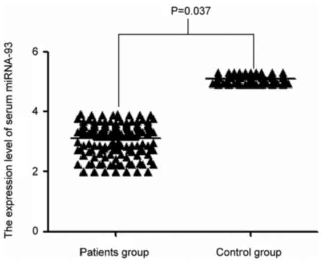
As shown in Fig. 1,
compared with the control group, the expression level of miRNA-93
in serum of patients with uterine cancer was significantly
decreased (P=0.037).
Relationship between the expression of
miRNA-93 and clinical factors
Expression level of miRNA-93 was significantly
correlated with pathological staging and lymph node metastasis
(P<0.05). Lower expression level of serum miRNA-93 was detected
in patients at higher pathological stage (P=0.010; P=0.026). Other
clinicopathological factors such as age, tumor size,
tumor-node-metastasis (TNM) staging, distant metastasis and smoking
status were not significantly correlated with the expression level
of miRNA-93 (P>0.05) (Table
II).
| Table II.Clinicopathological characteristics of
patients with uterine cancer. |
Table II.
Clinicopathological characteristics of
patients with uterine cancer.
| Characteristics | No. | Expression level of
miRNA-93 | P-value |
|---|
| Age (years) |
|
| 0.573 |
|
<55 | 94 | 3.72±1.87 |
|
| ≥55 | 82 | 3.64±1.55 |
|
| Pathological
staging |
|
| 0.010 |
| Stage
I | 76 | 3.54±0.941 |
|
| Stage
II | 61 | 3.19±0.862 |
|
| Stage
III | 31 | 2.84±0.764 |
|
| Stage
IV | 9 | 2.53±0.841 |
|
| Tumor size |
|
| 0.121 |
| ≤30
mm | 124 | 2.87±1.21 |
|
| >30
mm | 53 | 3.02±1.47 |
|
| TNM staging |
|
| 0.061 |
| I/II | 131 | 2.94±1.47 |
|
|
III/IV | 45 | 2.76±1.51 |
|
| Lymph node
metastasis |
|
| 0.009 |
|
Positive | 43 | 2.61±1.41 |
|
|
Negative | 133 | 3.24±1.71 |
|
| Distant
metastasis |
|
| 0.057 |
|
Positive | 50 | 3.04±0.901 |
|
|
Negative | 216 | 3.41±0.857 |
|
| Smoking
condition |
|
| 0.173 |
| Yes | 20 | 3.25±1.24 |
|
| No | 156 | 3.17±1.19 |
|
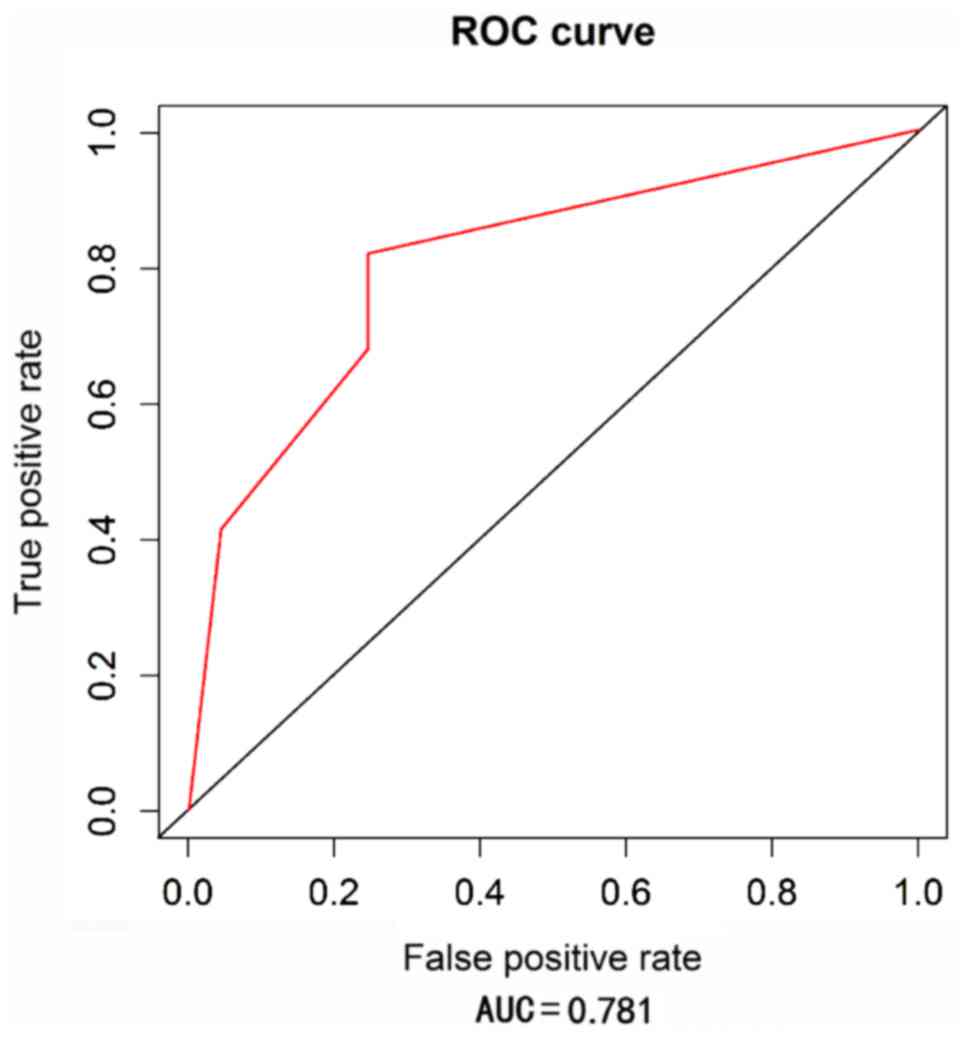
Diagnostic value of miRNA-93 for
uterine cancer
Receiver operating characteristic (ROC) curve
analysis was performed to analyze the diagnostic value of miRNA-93
for uterine cancer. As shown in Fig.
2, area under curve (AUC) was 0.781, and the 95% confidence
interval was 0.724–0.842, indicating that miRNA-93 can be used to
accurately predict uterine cancer.
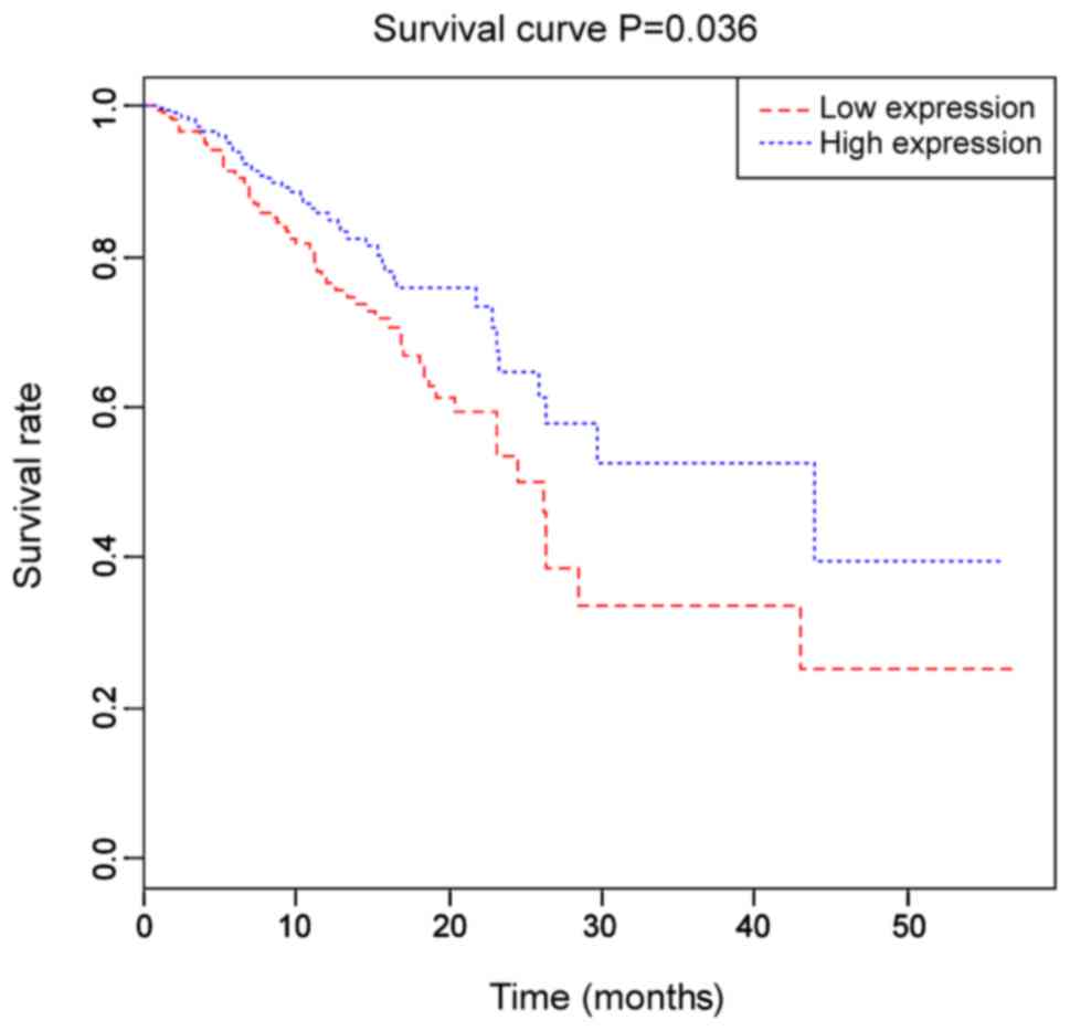
Prognostic values of miRNA-93 for
uterine cancer
Patients were divided into two groups based on the
median expression level of miRNA-93. Survival rate of the high
miRNA-93 expression group was significantly higher than that of the
low miRNA-93 expression group (P=0.036). As shown in Fig. 3, the Kaplan-Meier survival curve
showed that miRNA-93 expression was correlated with the prognosis
of patients with uterine cancer.
Discussion
Uterine cancer is currently one of the most common
malignant tumors in the female reproductive system and poses a
serious threat to women's health and life (8–11). Early
diagnosis is still the key for the treatment of this disease.
Radiotherapy combined with chemotherapy is the first choice of
treatment of uterine cancer after surgery (12). However, the early diagnosis of uterine
cancer is performed through colposcopy and visual observation,
which mainly depends on clinician's experience. Consequently, the
rate of misdiagnosis is high. This study aimed to identify novel
molecular markers for the diagnosis of uterine cancer to improve
the diagnosis and treatment of this disease. miRNA expression is
closely-related to the occurrence and development of tumors
(13). Findings have shown that
miRNAs can be stably expressed in urine, serum and other body
fluids (14–16). Expression level of miRNAs in cancer
tissues is basically the same as that in plasma, suggesting that
circulating miRNAs can reflect miRNA expression level in tumor
tissues (17). Serum is the most
convenient and relatively non-invasive biological sample. Test with
serum samples can be performed in vitro and avoid the side
effects caused by surgeries.
miRNA-93 is located on human chromosome 7q.22.1 and
is a miRNA produced by the transcription of miRNA-106b-25. miRNA-93
can participate in many inflammatory and immune reactions through
the interactions with downstream target proteins MMP-2, integrin-β8
and E2F1 (18). To the best of our
knowledge, the expression of miRNA-93 in uterine cancer has yet to
be reported. Therefore, we detected the differential expression of
miRNA-93 in patients with uterine cancer to identify a new
biomarker for this disease.
At present, correlation between miRNA-93 expression
and clinicopathological characteristics of uterine cancer has not
been reported. To demonstrate the potential relationship between
miRNA-93 and uterine cancer, RT-qPCR was conducted to detect the
expression of miRNA-93 in serum of each participant. miRNA-93 was
significantly downregulated in serum of patients with uterine
cancer compared with the control group. Expression level of
miRNA-93 in pathological stage III/IV was significantly lower than
that in stage I/II. Expression level of miRNA-93 in patients with
lymph node metastasis was also downregulated compared with the
healthy controls. The above results suggested that the low
expression of miRNA-93 is closely related to the occurrence and
development of uterine cancer. It has been reported that miRNA-93
can mediate the downregulation of transforming growth factor β
receptor 2, thus participating in nasopharyngeal carcinoma
aggressiveness (18). Singh et
al (19) reported that miRNA-93
reduced apoptosis of mammary epithelial cells and increased colony
formation, mammary ball formation and cell migration. Silencing of
miRNA-93 in these cells inhibited the development of cancer. Li
et al (20) showed that
miRNA-93 could promote angiogenesis by increasing endothelial cell
proliferation and migration, and inhibition of miRNA-93 expression
inhibited the secretion of vascular endothelial growth factor. The
downregulation of c-Myc expression by TSA (acetylase inhibitor) can
directly regulate the expression of miRNA-93 host gene MCM7 to
induce cell cycle arrest and apoptosis (21). This finding can be explained by the
changed miRNA-93 gene locus, which can affect the function of
miRNA-93 in the tumor.
In this experiment, analysis of the Kaplan-Meier
survival prognosis and ROC curve analysis were conducted. The
Kaplan-Meier survival analysis showed that the survival rate of the
miRNA-93 high expression group was higher than that in the low
expression group (P=0.036). AUC (0.781) of ROC curve indicated that
miRNA-93 could be used as a clinical indicator for uterine
cancer.
This study is still limited by the small sample
size, and in addition, this study only detected the expression
level of miRNA-93 in serum. The mechanism of the function of
miRNA-93 in uterine cancer was not investigated. Thus, further
studies are needed.
In summary, expression level of miRNA-93 is
significantly higher in the serum of patients with uterine cancer
than in the healthy controls, and the expression level of miRNA-93
is significantly correlated with clinical stage and other
pathological characteristics of patients with uterine cancer.
miRNA-93 can be used as a potential molecular marker for the
diagnosis of uterine cancer. However, the clinical applications of
miRNA-93 needs to be further studied and confirmed.
Acknowledgements
Not applicable.
Funding
No funding was received.
Availability of data and materials
The datasets used and/or analyzed during the current
study are available from the corresponding author on reasonable
request.
Authors' contributions
SF wrote the manuscript, treated patients and
collected blood sample. MG helped with RNA extraction. SX and QC
performed PCR and qPCR. HZ recorded and analyzed follow-up. All
authors have read and approved the final manuscript.
Ethics approval and consent to
participate
This study was approved by the Ethics Committee of
Hubei Cancer Hospital (Hubei, China), and all participants signed
informed consent.
Consent for publication
Not applicable.
Competing interests
The authors declare that they have no competing
interests.
References
|
1
|
Church DN, Stelloo E, Nout RA, Valtcheva
N, Depreeuw J, ter Haar N, Noske A, Amant F, Tomlinson IP, Wild PJ,
et al: Prognostic significance of POLE proofreading mutations in
endometrial cancer. J Natl Cancer Inst. 107:4022014.PubMed/NCBI
|
|
2
|
Gunderson CC, Java J, Moore KN and Walker
JL: The impact of obesity on surgical staging, complications, and
survival with uterine cancer: A Gynecologic Oncology Group LAP2
ancillary data study. Gynecol Oncol. 133:23–27. 2014. View Article : Google Scholar : PubMed/NCBI
|
|
3
|
Torre LA, Bray F, Siegel RL, Ferlay J,
Lortet-Tieulent J and Jemal A: Global cancer statistics, 2012. CA
Cancer J Clin. 65:87–108. 2015. View Article : Google Scholar : PubMed/NCBI
|
|
4
|
Medina PP and Slack FJ: microRNAs and
cancer: An overview. Cell Cycle. 7:2485–2492. 2008. View Article : Google Scholar : PubMed/NCBI
|
|
5
|
Singh B, Ronghe AM, Chatterjee A, Bhat NK
and Bhat HK: MicroRNA-93 regulates NRF2 expression and is
associated with breast carcinogenesis. Carcinogenesis.
34:1165–1172. 2013. View Article : Google Scholar : PubMed/NCBI
|
|
6
|
Li F, Liu J and Li S: MicroRNA 106b
approximately 25 cluster and gastric cancer. Surg Oncol. 22:7–10.
2013. View Article : Google Scholar
|
|
7
|
Savita U and Karunagaran D:
MicroRNA-106b-25 cluster targets β-TRCP2, increases the expression
of Snail and enhances cell migration and invasion in H1299 (non
small cell lung cancer) cells. Biochem Biophys Res Commun.
434:841–847. 2013. View Article : Google Scholar : PubMed/NCBI
|
|
8
|
Previs RA and Bodurka DC: Diagnosis and
management of stage II endometrial cancer. Springer; New Delhi: pp.
293–305. 2015
|
|
9
|
Hampton T: Critics of fibroid removal
procedure question risks it may pose for women with undetected
uterine cancer. JAMA. 311:891–893. 2014. View Article : Google Scholar : PubMed/NCBI
|
|
10
|
Hammer SM, Brown JC, Segal S, Chu CS and
Schmitz KH: Cancer-related impairments influence physical activity
in uterine cancer survivors. Med Sci Sports Exerc. 46:2195–2201.
2014. View Article : Google Scholar : PubMed/NCBI
|
|
11
|
Elit LM, O'Leary EM, Pond GR and Seow HY:
Impact of wait times on survival for women with uterine cancer. J
Clin Oncol. 32:27–33. 2014. View Article : Google Scholar : PubMed/NCBI
|
|
12
|
Colombo N, Preti E, Landoni F, Carinelli
S, Colombo A, Marini C and Sessa C: ESMO Guidelines working group:
endometrial cancer: ESMO Clinical practice guidelines for
diagnosis, treatment and follow-up. Ann Oncol. 24:33–38. 2013.
View Article : Google Scholar
|
|
13
|
Turchinovich A, Tonevitsky AG, Cho WC and
Burwinkel B: Check and mate to exosomal extracellular miRNA: New
lesson from a new approach. Front Mol Biosci. 2:112015. View Article : Google Scholar : PubMed/NCBI
|
|
14
|
Mitchell PS, Parkin RK, Kroh EM, Fritz BR,
Wyman SK, Pogosova-Agadjanyan EL, Peterson A, Noteboom J, O'Briant
KC, Allen A, et al: Circulating microRNAs as stable blood-based
markers for cancer detection. Proc Natl Acad Sci USA.
105:10513–10518. 2008. View Article : Google Scholar : PubMed/NCBI
|
|
15
|
Gilad S, Meiri E, Yogev Y, Benjamin S,
Lebanony D, Yerushalmi N, Benjamin H, Kushnir M, Cholakh H, Melamed
N, et al: Serum microRNAs are promising novel biomarkers. PLoS One.
3:e31482008. View Article : Google Scholar : PubMed/NCBI
|
|
16
|
Calin GA and Croce CM: MicroRNA signatures
in human cancers. Nat Rev Cancer. 6:857–866. 2006. View Article : Google Scholar : PubMed/NCBI
|
|
17
|
Tsujiura M, Ichikawa D, Komatsu S,
Shiozaki A, Takeshita H, Kosuga T, Konishi H, Morimura R, Deguchi
K, Fujiwara H, et al: Circulating microRNAs in plasma of patients
with gastric cancers. Br J Cancer. 102:1174–1179. 2010. View Article : Google Scholar : PubMed/NCBI
|
|
18
|
Lyu X, Fang W, Cai L, Zheng H, Ye Y, Zhang
L, Li J, Peng H, Cho WC, Wang E, et al: TGFβR2 is a major target of
miR-93 in nasopharyngeal carcinoma aggressiveness. Mol Cancer.
13:512014. View Article : Google Scholar : PubMed/NCBI
|
|
19
|
Singh B, Ronghe AM, Chatterjee A, Bhat NK
and Bhat HK: MicroRNA-93 regulates NRF2 expression and is
associated with breast carcinogenesis. Carcinogenesis.
34:1165–1172. 2013. View Article : Google Scholar : PubMed/NCBI
|
|
20
|
Li F, Liang X, Chen Y, Li S and Liu J:
Role of microRNA-93 in regulation of angiogenesis. Tumour Biol.
35:10609–10613. 2014. View Article : Google Scholar : PubMed/NCBI
|
|
21
|
Zhao ZN, Bai JX, Zhou Q, Yan B, Qin WW,
Jia LT, Meng YL, Jin BQ, Yao LB, Wang T, et al: TSA suppresses
miR-106b-93-25 cluster expression through downregulation of MYC and
inhibits proliferation and induces apoptosis in human EMC. PLoS
One. 7:e451332012. View Article : Google Scholar : PubMed/NCBI
|