Introduction
Oral cavity cancer is the most common head and neck
cancer (1). It was estimated that
300,400 new cases of oral cavity cancer and 145,400 cases of oral
cavity cancer-induced mortality have occurred in 2012 globally
(2). Oral leukoplakia (OL) is one of
the most common potentially malignant disorders of the oral cavity
(3), with a malignant transformation
rate of 17–35% (4). The prognosis and
overall survival rate of patients with oral cavity cancer depend on
the early detection of any lesion that may identify a patient with
increased risk or with early infiltration prior to metastatic
disease (3).
DNA doublestrand repair (DDR) is associated with
cancer occurrence and progression (5,6). DDR
activation occurs almost universally in the earliest stages of
carcinogenesis (7,8). Three DDR proteins, ATM serine/threonine
kinase (ATM), checkpoint kinase 2 (CHEK2) and γH2AFX have been
observed in numerous premalignant lesions and are associated with
the DNA damage response (7,9–11).
ATM activates checkpoint signaling at doublestrand
breaks (DSBs), following apoptosis and in response to genotoxic
stresses, and thereby functions as a DNA damage sensor. ATM
responds to DSBs by phosphorylating numerous substrates and may
initiate DSB signaling (12).
CHEK2 is a crucial downstream target of ATM
(13). Following DNA damage, ATM
preferentially activates CHEK2 (14).
Subsequently, activated CHEK2 modulates the activity of cell
division cycle 25C, which either facilitates DNA repair or directs
the cell to the apoptotic pathway (15,16). The
expression of CHEK2 is aberrant in numerous human premalignant and
malignant lesions (8,17,18).
H2A histone family member X (H2AFX) is a key DDR
component. Within minutes of DNA damage, H2AFX is phosphorylated at
its carboxyl terminus to form γH2AFX at DSB sites (19). The formation of numerous DDR proteins
requires H2AFX, indicating that H2AFX serves a key function in the
early stages of DDR. H2AFX protein is phosphorylated by ATM, and
the level of γH2AFX is positively associated with the degree of DNA
damage (20).
Accordingly, the present study hypothesized that
alterations to ATM, CHEK2 and γH2AFX may influence the
carcinogenesis of OL. As the epithelium of OL is a useful model for
monitoring abnormalities and exploring oral carcinogenesis
(9), the present study evaluated the
protein expression of ATM, CHEK2 and γH2AFX in OL and OSCC tissues
using immunohistochemistry. In addition, the present study assessed
the association between the clinicopathological data and expression
of these proteins, and their usefulness as biomarkers for
predicting the oral carcinogenesis.
Patients and methods
Patients and collection of clinical
specimens
In the present study, all patients with a clinical
and pathological diagnosis of OL or OSCC at the Department of Oral
Mucosal Diseases at Shanghai Ninth People's Hospital, Shanghai Jiao
Tong University School of Medicine (Shanghai, China) were enrolled.
The patients underwent biopsy or surgery between January 2005 and
December 2014. Normal oral mucosa tissues were obtained during
teeth extraction, gingivectomy or other minor surgical procedures.
All the study specimens were 10% formalin-fixed for 24 h at room
temperature and paraffin-embedded. Age, sex, lesion site, dietary
habit, smoking history and alcohol use were also collated.
Study design
The present study was approved by the Institutional
Review Board of Shanghai Ninth People's Hospital, Shanghai Jiao
Tong University School of Medicine. The patients enrolled in the
present study were divided into two cohorts. Cohort 1 consisted of
61 OL patients, 33 OSCC patients and 15 healthy individuals. The OL
patients were classified into the low risk dysplasia group (OL low
risk, n=41) and the high-risk dysplasia group (OL high risk, n=20)
according to the degree of epithelial dysplasia:
No⁄questionable⁄mild dysplasia (low risk) and moderate or severe
dysplasia (high risk) (21). All
examinations of tissues were determined by light microscope in 4
random fields (magnification, ×400). A total of 33 OSCC specimens
were confirmed as grade I without lymph node metastasis. The
exclusion criteria for patients with OL and OSCC were as follows:
(I) Any patient without an initial histopathological examination of
OL and OSCC, (II) any patient treated with radiotherapy or
chemotherapy prior to sampling and (III) any patient diagnosed with
OL and concomitant OSCC at the first visit. The clinical
characteristics of cohort 1 were summarized in Table I. Cohort 2 was based on a case-control
study and included 99 patients clinically and pathologically
diagnosed with OL with low risk dysplasia confirmed by the first
biopsy. The inclusion criteria were as follows: (I) Patients were
treated with Vita A (7.5 mg once a day for 3 months; Shanghai
Donghai Pharmaceuticals Co. Ltd., Shanghai, China) and mouth
rinsing (primary ingredient is gallnut containing gallic acid, 5 ml
three times a day when necessary; Xinjiang Qikang Habowei
Pharmaceutical Co. Ltd.) during the disease course, (II) all
patients underwent two biopsies and the interval between biopsies
was ≥3 years and (III) the lesion sites of each biopsy should
remain the same. The exclusion criteria were the same as described
for cohort 1. According to the results of the second biopsy, the 99
patients were classified either into the untransformed (UT) group
or the malignant-transformed (MT) group.
 | Table I.Clinical characteristics of cohort
1. |
Table I.
Clinical characteristics of cohort
1.
| Characteristic | Normal (n=15) | OL low risk
(n=41) | OL high risk
(n=20) | OSCC (n=33) |
|---|
| Age |
|
|
|
|
| Mean ±
SD | 44.80±15.48 | 56.10±11.86 | 57.10±10.92 | 56.36±13.74 |
|
Range | 26–70 | 35–79 | 31–82 | 26–81 |
| Sex |
|
|
|
|
|
Male | 4 | 21 | 13 | 11 |
|
Female | 11 | 20 | 7 | 12 |
| Lesion site |
|
|
|
|
|
Tongue | 5 | 18 | 15 | 26 |
|
Buccal | 5 | 18 | 5 | 6 |
|
Gingiva | 3 | 2 | 0 | 1 |
|
Palate | 1 | 1 | 0 | 0 |
| Mouth
floor | 0 | 0 | 0 | 0 |
|
Lip | 1 | 2 | 0 | 0 |
| Smoking
history |
|
|
|
|
|
Never | 11 | 22 | 12 | 24 |
| Past
and present | 4 | 19 | 8 | 9 |
| Alcohol intake |
|
|
|
|
|
Never | 8 | 16 | 8 | 14 |
| Past
and present | 7 | 25 | 12 | 19 |
| Dietary habits |
|
|
|
|
|
Bland | 10 | 21 | 14 | 15 |
|
Spicy | 5 | 20 | 6 | 18 |
Immunohistochemical analysis of the
expression of ATM, CHEK2 and γH2AFX
Serial tissue sections (3 µm) from the paraffin
blocks of normal oral tissues, OL and OSCC were placed in xylene
for deparaffinization and in graded alcohol dilutions (ethanol
concentration was 80, 95 and 100%, respectively) for hydration.
Antigen retrieval was performed with 1 mM Tris-EDTA (pH 8.0) in a
100°C water bath for 20 min and endogenous peroxidase activity was
blocked with 3% hydrogen peroxide for 10 min at room temperature.
Immunohistochemical analysis was performed for the sections.
Primary, monoclonal antibodies against ATM (cat. no. ab78;
1:1,000), CHEK2 (cat. no. ab109413; 1:100) and γH2AFX (cat. no.
ab22551; 1:200; all Abcam, Cambridge, UK) (0.01 mol/l; pH 7.4;
Wuhan Boster Biological Engineering Co., Ltd., Wuhan, China) were
used for 1 h at room temperature. Following rinses with PBS three
times for 10 min, a Peroxidase/DAB, K5007 EnVision™
Detection System kit (ready-to-use, Dako; Agilent Technologies,
Inc., Santa Clara, CA, USA) was used to detect the primary
antibodies for 1 h at room temperature, according to the
manufacturer's instructions. The sections were subsequently
counterstained with 4.8 mg/ml Harris Hematoxylin for 2 min at room
temperature. Overall, at least three sections were stained to
confirm reproducibility. The staining intensity of the cells was
observed under a light microscope (Axio Scope A1; Carl Zeiss AG,
Oberkochen, Germany). The mean percentage of positive cells was
determined in 4 random fields (magnification, ×400). To ensure
pathological diagnoses were standardized, the cellular
localization, intensity, and the percentage of cells with positive
ATM, CHEK2 and γH2AFX staining were assessed by two oral
pathologists (Department of Oral Pathology, Shanghai Ninth People's
Hospital, Shanghai Jiao Tong University School of Medicine) in a
doubleblind manner and a consensus was reached in cases of
discrepancy. Cell nuclear and/or cytoplasmic immunoreactivity in
the epithelium was considered to indicate positive expression of
ATM. Cell nuclear immunoreactivity in the epithelium was considered
to indicate positive expression of CHEK2 and γH2AFX. Positive
controls for the antibodies were used according to the
manufacturer's instructions. Negative control slides omitted
primary antibodies. Positive staining intensity was defined as 0,
1, 2, and 3 for no staining, light yellow, yellow brown, and brown,
respectively. The scoring method used for ATM and γH2AFX was
referred to by Hu et al (22).
The positive cell percentages of 0, 1–25, 26–50, 51–75, and >75
were defined as 0, 1, 2, 3, and 4, respectively. The scoring method
used for CHEK2 was a modified version of that used by Alkema et
al (23). The positive cell
percentages of 0–5, 6–25, 26–50, 51–75, and >75 were defined as
0, 1, 2, 3, and 4, respectively. The semiquantitative expression
level was evaluated by multiplying the distribution and intensity
score. A final score of <5 was defined as low expression of ATM
and γH2AFX, of ≥5 as high expression of ATM and γH2AFX, of <7 as
low expression of CHEK2, and of ≥7 as high expression of CHEK2.
Statistical analysis
The SPSS 19.0 software package (IBM Corp., Armonk,
NY, USA) was used for all statistical analysis. ATM, CHEK2 and
γH2AFX expression levels in the normal tissue, OL low risk, OL high
risk, and OSCC groups were compared using the Kruskal-Wallis test
followed by Dunn's Test. The association between the expression of
the proteins and clinicopathological features were assessed using a
χ2 test. Pearson correlation analysis was used to
evaluate the association between protein expressions for patients
in cohort 1. The Fisher's exact test was used to assess the
statistical difference between the expression levels of certain
proteins in cohort 2. A logistic regression model was used to
evaluate the relative risk of OL malignant transformation. Ranked
data were presented as percentage. All tests were two-sided.
P<0.05 was considered to indicate a statistically significant
difference.
Results
ATM, CHEK2 and γH2AFX expression
levels in cohort 1
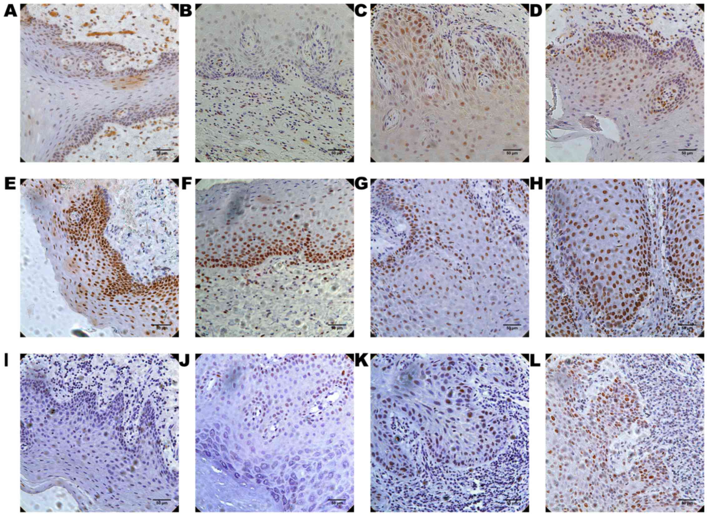
ATM was primarily expressed in the nucleus and
cytoplasm in the epithelium, while CHEK2 and γH2AFX were primarily
expressed in the nucleus. Cells positive for ATM, CHEK2 and γH2AFX
expression were detected not only in the stratum basale and stratum
spinosum, but also in the stratum granulosum, and in the stratum
corneum of certain patients (Fig. 1).
ATM expression tended to increase gradually in the normal tissue,
OL low risk, OL high risk and OSCC groups during carcinogenesis
(P=0.005; Table II). ATM expression
was significantly increased in 29/33 of the samples of the OSCC
group, compared with the normal tissue group (P=0.008). In
addition, ATM expression was significantly increased in 23/41 of
the samples of the OL low risk group compared with the OSCC group
(P=0.027). In the OL high-risk group, ATM expression in 13/20 of
the samples exhibited no statistically significant difference
compared with that in the other three groups, respectively. γH2AFX
expression increased in the groups as carcinogenesis progressed
(P=0.001; Table II). A comparison of
the groups revealed that there was a significant difference between
the OL low risk and the OSCC groups (P=0.014), and between the
normal tissue and the OSCC groups in γH2AFX expression (P= 0.001).
There was no significant difference in CHEK2 expression among the
four groups (P=0.074; Table II). The
expression of ATM and γH2AFX increased in the groups as
carcinogenesis progressed. To assess this association, Pearson
correlation analysis was performed. The result was statistically
significant [(P=0.045; Pearson correlation coefficient (r)=0.192)]
among the groups. correlation demonstrated in the OSCC group
exhibited [r=0.383 (P=0.028)]
| Table II.ATM, CHEK2 and γH2AFX expression in
cohort 1 (n=109). |
Table II.
ATM, CHEK2 and γH2AFX expression in
cohort 1 (n=109).
| Expression | Total (n) | Normal control
(n) | OL low risk
(n) | OL high risk
(n) | OSCC (n) | P-value |
|---|
| ATM |
|
|
|
|
| 0.005 |
|
Low | 38 | 9 | 18 | 7 | 4 |
|
|
High | 71 | 6 | 23 | 13 | 29 |
|
| CHEK2 |
|
|
|
|
| 0.074 |
|
Low | 20 | 1 | 12 | 1 | 6 |
|
|
High | 89 | 14 | 29 | 19 | 27 |
|
| γH2AFX |
|
|
|
|
| 0.001 |
|
Low | 79 | 15 | 33 | 15 | 16 |
|
|
High | 30 | 0 | 8 | 5 | 17 |
|
Correlation and clinical significance
of ATM and γH2AFX expression in normal oral mucosa, OL and OSCC
tissues in cohort 1
The association between ATM and γH2AFX expression
and multiple clinical characteristics of OL in cohort 1 (n=109) was
assessed using the χ2 test. Multiple degrees of
epithelial dysplasia revealed different expression levels of ATM
(P=0.004; Table III). OSCC and
normal tissues demonstrated the highest and lowest percentage,
respectively, of ATM expression among the tissues. No correlation
was demonstrated between ATM expression and the other clinical
characteristics: Age, sex, lesion site, dietary habits, smoking
history and alcohol use. γH2AFX expression was also associated with
the degree of epithelial dysplasia (P=0.001; Table IV). OSCC and normal tissues revealed
the highest and lowest percentage, respectively, of γH2AFX
expression among the tissues. No clinical characteristics revealed
association with γH2AFX expression.
| Table III.Association between ATM expression
and clinicopathological features in cohort 1 (n=109). |
Table III.
Association between ATM expression
and clinicopathological features in cohort 1 (n=109).
| Clinicopathological
feature | Total (n) | Low ATM expression
(n, %) | High ATM expression
(n, %) | P-value |
|---|
| Age, years |
|
|
|
|
| 0.866 |
|
≤60 | 70 | 24 | 34.3 | 46 | 65.7 |
|
|
>60 | 39 | 14 | 35.9 | 25 | 64.1 |
|
| Sex |
|
|
|
|
| 0.711 |
|
Male | 49 | 18 | 36.7 | 31 | 63.3 |
|
|
Female | 60 | 20 | 33.3 | 40 | 66.7 |
|
| Lesion site |
|
|
|
|
| 0.345 |
|
Tongue | 64 | 20 | 31.3 | 44 | 68.8 |
|
|
Non-tongue | 45 | 18 | 40.0 | 27 | 60.0 |
|
| Dietary habits |
|
|
|
|
| 0.333 |
|
Bland | 62 | 24 | 38.7 | 38 | 61.3 |
|
|
Spicy | 47 | 14 | 29.8 | 33 | 70.2 |
|
| Smoking
history |
|
|
|
|
| 0.261 |
|
Never | 68 | 21 | 30.9 | 47 | 69.1 |
|
| Past
and present | 41 | 17 | 41.5 | 24 | 58.5 |
|
| Alcohol intake |
|
|
|
|
| 0.345 |
|
Never | 45 | 18 | 40.0 | 27 | 60.0 |
|
| Past
and present | 64 | 20 | 31.3 | 44 | 68.8 |
|
| Epithelial
dysplasia |
|
|
|
|
| 0.004 |
|
Normal | 15 | 9 | 60.0 | 6 | 40.0 |
|
| OL low
risk | 41 | 18 | 43.9 | 23 | 56.1 |
|
| OL high
risk | 20 | 7 | 35.0 | 13 | 65.0 |
|
|
OSCC | 33 | 4 | 12.1 | 29 | 87.9 |
|
| Table IV.Association between γH2AFX expression
and clinicopathological features in cohort 1 (n=109). |
Table IV.
Association between γH2AFX expression
and clinicopathological features in cohort 1 (n=109).
| Clinicopathological
feature | Total (n) | Low γH2AFX
expression (n, %) | High γH2AFX
expression (n, %) | P-value |
|---|
| Age, years |
|
|
|
|
| 0.905 |
|
≤60 | 70 | 51 | 72.9 | 19 | 27.1 |
|
|
>60 | 39 | 28 | 71.8 | 11 | 28.2 |
|
| Sex |
|
|
|
|
| 0.834 |
|
Male | 49 | 36 | 73.5 | 13 | 26.5 |
|
|
Female | 60 | 43 | 71.7 | 17 | 28.3 |
|
| Lesion site |
|
|
|
|
| 0.056 |
|
Tongue | 64 | 42 | 65.6 | 22 | 34.4 |
|
|
Non-tongue | 45 | 37 | 82.2 | 8 | 17.8 |
|
| Dietary habits |
|
|
|
|
| 0.402 |
|
Bland | 62 | 43 | 69.4 | 19 | 30.6 |
|
|
Spicy | 47 | 36 | 76.6 | 11 | 23.4 |
|
| Smoking
history |
|
|
|
|
| 0.9 |
|
Never | 68 | 49 | 72.1 | 19 | 27.9 |
|
| Past
and present | 41 | 30 | 73.2 | 11 | 26.8 |
|
| Alcohol intake |
|
|
|
|
| 0.14 |
|
Never | 45 | 36 | 80.0 | 9 | 20.0 |
|
| Past
and present | 64 | 43 | 67.2 | 21 | 32.8 |
|
| Epithelial
dysplasia |
|
|
|
|
| 0.001 |
|
Normal | 15 | 15 | 100.0 | 0 |
0.0 |
|
| OL low
risk | 41 | 33 | 80.5 | 8 | 19.5 |
|
| OL high
risk | 20 | 15 | 75.0 | 5 | 25.0 |
|
|
OSCC | 33 | 16 | 48.5 | 17 | 51.5 |
|
Identifying candidate DDR proteins in
cohort 1
The selection criteria for candidate DDR proteins
were as follows: The expression of candidate DDR proteins increased
or decreased successively with respect to increasing grades of
carcinogenesis, with significance set at P<0.05 and assessed
using the Kruskal-Wallis test. According to the results of the
present study, not only did none of the three proteins assessed
demonstrate decreased expression as carcinogenesis progressed but
ATM and γH2AFX expression increased as carcinogenesis progressed.
ATM and γH2AFX expression levels were also significantly different
between the normal tissue and the OSCC groups, and between the OL
low risk and the OSCC groups. Therefore, ATM and γH2AFX expression
was assessed further and validated in the case-control study.
ATM and γH2AFX expression in cohort
2
Cohort 2 consisted of 99 patients with OL low risk.
On the basis of the second biopsy, these patients were assigned to
the UT group (n=81) or the MT group (n=18). Table V summarizes the characteristics of
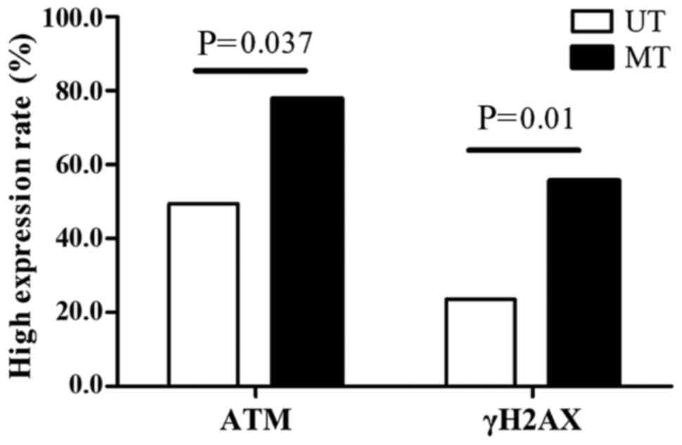
these patients. High expression of ATM was demonstrated in 54 of
the 99 patients (54.5%). Increased expression of ATM was detected
in 40 of the 81 (49.4%) patients in the UT group and in 14 of the
18 (77.8%) patients in the MT group (P=0.037; Fig. 2). In addition, 29 of the 99 patients
(29.3%) demonstrated high expression of γH2AFX. Increased
expression of γH2AFX was detected in 19 of the 81 (23.5%) patients
in the UT group and in 10 of the 18 (55.6%) patients in the MT
group (P=0.01; Fig. 2).
| Table V.Clinical characteristics of cohort
2. |
Table V.
Clinical characteristics of cohort
2.
| Characteristic | UT (n=81) | MT (n=18) |
|---|
| Age, years |
|
|
| Mean ±
SD | 56.53±11.27 | 54.83±11.63 |
|
Range | 31–82 | 35–71 |
| Sex |
|
|
|
Male | 44 | 8 |
|
Female | 37 | 10 |
| Lesion site |
|
|
|
Tongue | 44 | 13 |
|
Buccal | 32 | 3 |
|
Gingiva | 2 | 1 |
|
Palate | 1 | 0 |
| Mouth
floor | 0 | 0 |
|
Lip | 2 | 1 |
| Dietary habits |
|
|
|
Bland | 50 | 11 |
|
Spicy | 31 | 7 |
| Smoking
history |
|
|
|
Never | 50 | 11 |
| Past
and present | 31 | 7 |
| Alcohol intake |
|
|
|
Never | 33 | 6 |
| Past
and present | 48 | 12 |
| ATM expression |
|
|
|
Low | 41 | 4 |
|
High | 40 | 14 |
| γH2AFX
expression |
|
|
|
Low | 62 | 8 |
|
High | 19 | 10 |
High expression of ATM as an
independent factor for OL malignant transformation in cohort 2
To evaluate the risk of OL malignant transformation,
clinicopathological parameters, and ATM and γH2AFX expression were
assessed using logistic regression (Table VI). In the univariate analysis, age,
sex, lesion site, smoking history, and alcohol use were not
significant risk factors for transformation in cohort 2, and high
expression of ATM and γH2AFX was associated with a 3.59-fold [(95%
confidence interval (CI), 1.09–11.83; P=0.036)] and a 4.08-fold
(95% CI, 1.41–11.80; P=0.009), increase in the risk of malignant
transformation, respectively. In the multivariate analysis, high
expression of ATM and γH2AFX was also significantly associated with
an increased risk of malignant transformation. The adjusted odds
ratio for malignant transformation was 4.29 for high ATM expression
(95% CI, 1.22–15.07; P=0.023) and 4.79 for high γH2AFX expression
(95% CI, 1.56–14.73; P=0.006).
| Table VI.Logistic regression analysis of the
potential risk of oral cancer. |
Table VI.
Logistic regression analysis of the
potential risk of oral cancer.
| Characteristic | OR (95% CI) | P-value |
|---|
| Univariate
analysis |
|
|
|
Age | 1.36
(0.48–3.82) | 0.560 |
|
Sex | 0.67
(0.24–1.88) | 0.449 |
| Lesion
site | 2.19
(0.71–6.70) | 0.171 |
| Dietary
habits | 1.03
(0.36–2.93) | 0.961 |
| Smoking
history | 1.03
(0.36–2.93) | 0.961 |
| Alcohol
intake | 1.38
(0.47–4.03) | 0.562 |
| High
ATM expression | 3.59
(1.09–11.83) | 0.036 |
| High
γH2AFX expression | 4.08
(1.41–11.80) | 0.009 |
| Multivariate
analysis |
|
|
| High
ATM expression | 4.29
(1.22–15.07) | 0.023 |
| High
γH2AFX expression | 4.79
(1.56–14.73) | 0.006 |
Discussion
To the best of our knowledge, the present study is
the first to evaluate ATM, CHEK2 and γH2AFX expression in patients
with OL with multiple degrees of epithelial dysplasia and to assess
the functions of these proteins in predicting the risk of OSCC in
two independent cohorts using immunohistochemical analysis. ATM
serves a key function in the DNA DSB-induced signaling cascade.
Tumorigenic events that occur early in the progression of major
types of human cancer activate ATM-regulated cell cycle checkpoints
and thereby an inducible barrier that inhibits tumor progression
and genetic instability (7,24). A previous study has demonstrated that
ATM expression was increased in certain types of cancer tissue
compared with that in benign tumorous lesions and normal tissues
(22). Other previous studies have
suggested that ATM potentially represents a promising indicator for
hyperplasia and cancer, and may serve as a useful marker for
identifying patients with poor prognosis (25,26).
Raynaud et al (10)
demonstrated that the difference in ATM activation between normal
and precancerous tissues was not significant, though ATM expression
differed significantly between precancerous and cancerous tissues.
The results of the aforementioned studies support those of the
present study.
He et al (9)
indicated that ATM protein expression was higher in OL compared
with that in normal oral tissue, but demonstrated no significant
difference between OL and OSCC tissues in ATM protein expression.
The present study dynamically observed the activation of the DNA
damage signaling pathway in normal oral mucosa, OL (low risk
dysplasia and high risk dysplasia) and OSCC tissues in a large
population. In contrast to the results demonstrated by He et
al (9) those demonstrated in the
present study revealed that ATM expression gradually increased as
OL progressed to OSCC. In addition, the present study revealed a
significant difference in ATM expression between OL with low risk
dysplasia and OSCC. In the present study, ATM expression correlated
with the degree of epithelial dysplasia during carcinogenesis and
age, sex, lesion site, dietary habits, smoking history, and alcohol
use were not significant factors in the expression of ATM.
Therefore, the results of the present study indicated that ATM was
activated in oral precancerous lesions and served a function in the
early stages of oral carcinogenesis.
One of the proteins phosphorylated following DNA
damage, a process initiated by ATM, is H2AFX, which, in
phosphorylated form (γH2AFX), functions as a specific indicator for
the presence of DSBs (27). Increased
γH2AFX expression may result in increased radiosensitivity
(28). Multiple studies have revealed
that γH2AFX expression is increased in certain types of cancer and
their premalignant lesions (8,22,29,30), which
supports the results of the present study. Overexpression of γH2AFX
may represent an independent prognostic indicator of a poor overall
patient survival rate (31,32). In contrast to the results of the
present study, Chou et al (33) reported that γH2AFX expression was
increased in dysplastic epithelium and significantly decreased in
OSCC tissue. The results of the present study revealed that γH2AFX
expression increased in OSCC tissue with increasing disease
severity; this discrepancy between the aforementioned and present
study may be due to the difference in the OSCC tumor
differentiation grade selected. In the present study, similar to
ATM expression, γH2AFX expression correlated with the degree of
epithelial dysplasia, according to the results of the associations
between clinicopathological features and γH2AFX expression.
Detecting γH2AFX expression may help to evaluate precancerous oral
cavity lesions and monitor cancer progression.
CHEK2 serves a key function in inhibiting cell cycle
progression in response to the DNA damage pathway (34). In multiple types of solid tumor, CHEK2
expression was decreased compared with that in normal tissues
(35–37). CHEK2 expression in oral precancerous
lesions is rarely assessed. In the present study, the expression of
CHEK2 protein altered during oral carcinogenesis. There were no
significant differences between any two groups of the four during
carcinogenesis. Based on these conflicting results, the present
study suggested that: (I) Aberrant CHEK2 protein may be
functionally defective and regulated by other, unknown upstream
proteins during DDR; (II) CHEK2 protein expression is regulated
differently depending on the type of carcinoma; (III) the ATM-CHEK2
pathway may not be associated with oral carcinoma or precancerous
lesions or (IV) more complex signaling pathways may participate in
the DNA damage response.
Pearson correlation analysis of cohort 1
demonstrated that γH2AFX and ATM expression was correlated
(P=0.045; r=0.192), particularly in OSCC tissue (P=0.028; r=0.383).
Therefore, the present study suggested that the ATM-γH2AFX pathway
contributes to the DNA damage response.
For cohort 2, the present study assessed the
prognostic value of ATM and γH2AFX expression and evaluate whether
the prognostic value was independent of clinicopathological
factors. Univariate and multivariate analysis revealed that
increased ATM and γH2AFX expression was significantly associated
with an increased risk of transformation (P<0.05). The results
of the present study indicated that increased ATM and γH2AFX
expression served as an independent predictor of carcinogenesis.
However, age, sex, lesion site, dietary habits, smoking history,
and alcohol use were not revealed to be significant risk factors
for OL malignant transformation in the cohort 2, a result that
reflects that of multiple previous studies (38–40).
To conclude, the results of the present study
suggested that ATM and γH2AFX expression in OL tissue was
associated with oral cancer progression. Immunohistochemical
staining of ATM and γH2AFX may represent a promising technique for
the early identification and risk evaluation of OSCC in patients
with precancerous oral lesions. Further studies are required to
assess the function of ATM and γH2AFX in oral carcinogenesis,
including for grade I, II and III OSCC. Further study of the
mechanisms underlying DNA damage and response in OL tissue is also
required.
Acknowledgements
The abstract was presented at the 35th Annual
Meeting of the International Association for Dental Research Korean
Division on June 25th 2016 in Seoul, Republic of Korea. The authors
would like to thank Professor Jiang Li and Dr Lizhen Wang
(Department of Oral Pathology, Shanghai Ninth People's Hospital,
Shanghai Jiao Tong University School of Medicine) for their
technical assistance.
Funding
The present study was supported by the National
Natural Science Foundation of China (grant no. 81400513) and the
Science and Technology Commission of Shanghai (grant no.
14401931600).
Availability of data and materials
The datasets used and/or analyzed during the current
study are available from the corresponding author on reasonable
request.
Authors' contributions
LW, ZZ and WL conceived and designed the study. MZ,
LS, XX and WW performed the experiments. MZ analyzed the data and
wrote the manuscript. LW and ZZ reviewed and edited the manuscript.
All authors read and approved the final manuscript.
Ethics approval and consent to
participate
The present study was approved by the Institutional
Review Board of Shanghai Ninth People's Hospital, Shanghai Jiao
Tong University School of Medicine.
Consent for publication
The subjects or parent/guardian that participated in
the present study provided written informed consents for
publication.
Competing interests
The authors declare that they have no competing
interests.
Authors' information
LW, Department of Oral Mucosal Diseases, Shanghai
Ninth People's Hospital, Shanghai Jiao Tong University School of
Medicine, committee member of the Oral Mucosal Disease Professional
Committee of Shanghai Stomatological Association; committee member
of Chinese and Western medicine professional committee of Chinese
Stomatological Association.
ZZ, Department of Oral Mucosal Diseases, Shanghai
Ninth People's Hospital, Shanghai Jiao Tong University School of
Medicine, former chairman of the oral mucosal disease professional
committee of Chinese Stomatological Association; former chairman of
the oral mucosal disease professional committee of Shanghai
Stomatological Association.
References
|
1
|
Chi AC, Day TA and Neville BW: Oral cavity
and oropharyngeal squamous cell carcinoma-an update. CA Cancer J
Clin. 65:401–421. 2015. View Article : Google Scholar : PubMed/NCBI
|
|
2
|
Torre LA, Bray F, Siegel RL, Ferlay J,
Lortet-Tieulent J and Jemal A: Global cancer statistics, 2012. CA
Cancer J Clin. 65:87–108. 2015. View Article : Google Scholar : PubMed/NCBI
|
|
3
|
Boy SC: Leukoplakia and erythroplakia of
the oral mucosa-a brief overview. SADJ. 67:558–560. 2012.PubMed/NCBI
|
|
4
|
van der Waal I, Schepman KP, van der Meij
EH and Smeele LE: Oral leukoplakia: A clinicopathological review.
Oral Oncol. 33:291–301. 1997. View Article : Google Scholar : PubMed/NCBI
|
|
5
|
Khanna KK and Jackson SP: DNA doublestrand
breaks: Signaling, repair and the cancer connection. Nat Genet.
27:247–254. 2001. View
Article : Google Scholar : PubMed/NCBI
|
|
6
|
Peng A and Maller JL: Serine/threonine
phosphatases in the DNA damage response and cancer. Oncogene.
29:5977–5988. 2010. View Article : Google Scholar : PubMed/NCBI
|
|
7
|
Bartkova J, Horejsi Z, Koed K, Krämer A,
Tort F, Zieger K, Guldberg P, Sehested M, Nesland JM, Lukas C, et
al: DNA damage response as a candidate anti-cancer barrier in early
human tumorigenesis. Nature. 434:864–870. 2005. View Article : Google Scholar : PubMed/NCBI
|
|
8
|
Gorgoulis VG, Vassiliou LV, Karakaidos P,
Zacharatos P, Kotsinas A, Liloglou T, Venere M, Ditullio RA Jr,
Kastrinakis NG, Levy B, et al: Activation of the DNA damage
checkpoint and genomic instability in human precancerous lesions.
Nature. 434:907–913. 2005. View Article : Google Scholar : PubMed/NCBI
|
|
9
|
He Y, Chen Q and Li B: ATM in oral
carcinogenesis: Association with clinicopathological features. J
Cancer Res Clin Oncol. 134:1013–1020. 2008. View Article : Google Scholar : PubMed/NCBI
|
|
10
|
Raynaud CM, Hernandez J, Llorca FP,
Nuciforo P, Mathieu MC, Commo F, Delaloge S, Sabatier L, André F
and Soria JC: DNA damage repair and telomere length in normal
breast, preneoplastic lesions, and invasive cancer. Am J Clin
Oncol. 33:341–345. 2010. View Article : Google Scholar : PubMed/NCBI
|
|
11
|
Yuan J, Adamski R and Chen J: Focus on
histone variant H2AFX: To be or not to be. FEBS Lett.
584:3717–3724. 2010. View Article : Google Scholar : PubMed/NCBI
|
|
12
|
Shiloh Y: ATM and related protein kinases:
Safeguarding genome integrity. Nat Rev Cancer. 3:155–168. 2003.
View Article : Google Scholar : PubMed/NCBI
|
|
13
|
Bartek J and Lukas J: Chk1 and CHEK2
kinases in checkpoint control and cancer. Cancer Cell. 3:421–429.
2003. View Article : Google Scholar : PubMed/NCBI
|
|
14
|
Zhou BB and Elledge SJ: The DNA damage
response: Putting checkpoints in perspective. Nature. 408:433–439.
2000. View
Article : Google Scholar : PubMed/NCBI
|
|
15
|
Peng CY, Graves PR, Thoma RS, Wu Z, Shaw
AS and Piwnica-Worms H: Mitotic and G2 checkpoint control:
Regulation of 14-3-3 protein binding by phosphorylation of Cdc25C
on serine-216. Science. 277:1501–1505. 1997. View Article : Google Scholar : PubMed/NCBI
|
|
16
|
Chang CC, Hung CM, Yang YR, Lee MJ and Hsu
YC: Sulforaphane induced cell cycle arrest in the G2/M phase via
the blockade of cyclin B1/CDC2 in human ovarian cancer cells. J
Ovarian Res. 6:412013. View Article : Google Scholar : PubMed/NCBI
|
|
17
|
Matthews TP, Jones AM and Collins I:
Structure-based design, discovery and development of checkpoint
kinase inhibitors as potential anticancer therapies. Expert Opin
Drug Discov. 8:621–640. 2013. View Article : Google Scholar : PubMed/NCBI
|
|
18
|
Lantuejoul S, Raynaud C, Salameire D,
Gazzeri S, Moro-Sibilot D, Soria JC, Brambilla C and Brambilla E:
Telomere maintenance and DNA damage responses during lung
carcinogenesis. Clin Cancer Res. 16:2979–2988. 2010. View Article : Google Scholar : PubMed/NCBI
|
|
19
|
Bonner WM, Redon CE, Dickey JS, Nakamura
AJ, Sedelnikova OA, Solier S and Pommier Y: GammaH2AFX and cancer.
Nat Rev Cancer. 8:957–967. 2008. View
Article : Google Scholar : PubMed/NCBI
|
|
20
|
Rogakou EP, Boon C, Redon C and Bonner WM:
Megabase chromatin domains involved in DNA doublestrand breaks in
vivo. J Cell Biol. 146:905–916. 1999. View Article : Google Scholar : PubMed/NCBI
|
|
21
|
Warnakulasuriya S, Reibel J, Bouquot J and
Dabelsteen E: Oral epithelial dysplasia classification systems:
Predictive value, utility, weaknesses and scope for improvement. J
Oral Pathol Med. 37:127–133. 2008. View Article : Google Scholar : PubMed/NCBI
|
|
22
|
Hu JL, Hu SS, Hou XX, Zhu X, Cao J, Jiang
LH and Ge MH: Abnormal expression of DNA Double-Strand breaks
related genes, ATM and GammaH2AFX, in thyroid carcinoma. Int J
Endocrinol. 2015:1368102015. View Article : Google Scholar : PubMed/NCBI
|
|
23
|
Alkema NG, Tomar T, van der Zee AG, Everts
M, Meersma GJ, Hollema H, de Jong S, van Vugt MA and Wisman GB:
Checkpoint kinase 2 (Chek2) supports sensitivity to platinum-based
treatment in high grade serous ovarian cancer. Gynecol Oncol.
133:591–598. 2014. View Article : Google Scholar : PubMed/NCBI
|
|
24
|
Bartkova J, Rezaei N, Liontos M,
Karakaidos P, Kletsas D, Issaeva N, Vassiliou LV, Kolettas E,
Niforou K, Zoumpourlis VC, et al: Oncogene-induced senescence is
part of the tumorigenesis barrier imposed by DNA damage
checkpoints. Nature. 444:633–637. 2006. View Article : Google Scholar : PubMed/NCBI
|
|
25
|
Wang YH, Li F, Luo B, Wang XH, Sun HC, Liu
S, Cui YQ and Xu XX: A side population of cells from a human
pancreatic carcinoma cell line harbors cancer stem cell
characteristics. Neoplasma. 56:371–378. 2009. View Article : Google Scholar : PubMed/NCBI
|
|
26
|
Dou J, Wen P, Hu W, Li Y, Wu Y, Liu C,
Zhao F, Hu K, Wang J, Jiang C, et al: Identifying tumor stem-like
cells in mouse melanoma cell lines by analyzing the characteristics
of side population cells. Cell Biol Int. 33:807–815. 2009.
View Article : Google Scholar : PubMed/NCBI
|
|
27
|
Takahashi A and Ohnishi T: Does gammaH2AFX
foci formation depend on the presence of DNA double strand breaks?
Cancer Lett. 229:171–179. 2005. View Article : Google Scholar : PubMed/NCBI
|
|
28
|
Qiang L, Yang Y, Ma YJ, Chen FH, Zhang LB,
Liu W, Qi Q, Lu N, Tao L, Wang XT, et al: Isolation and
characterization of cancer stem like cells in human glioblastoma
cell lines. Cancer Lett. 279:13–21. 2009. View Article : Google Scholar : PubMed/NCBI
|
|
29
|
Wasco MJ and Pu RT: Utility of
antiphosphorylated H2AFX antibody (gamma-H2AFX) in diagnosing
metastatic renal cell carcinoma. Appl Immunohistochem Mol Morphol.
16:349–356. 2008. View Article : Google Scholar : PubMed/NCBI
|
|
30
|
Mah LJ, El-Osta A and Karagiannis TC:
GammaH2AFX as a molecular marker of aging and disease. Epigenetics.
5:129–136. 2010. View Article : Google Scholar : PubMed/NCBI
|
|
31
|
Matthaios D, Foukas PG, Kefala M, Hountis
P, Trypsianis G, Panayiotides IG, Chatzaki E, Pantelidaki E, Bouros
D, Karakitsos P and Kakolyris S: γ-H2AFX expression detected by
immunohistochemistry correlates with prognosis in early operable
non-small cell lung cancer. Onco Targets Ther. 5:309–314. 2012.
View Article : Google Scholar : PubMed/NCBI
|
|
32
|
Oliveira-Costa JP, Oliveira LR, Zanetti R,
Zanetti JS, da Silveira GG, Buim Chavichiolli ME, Zucoloto S,
Ribeiro-Silva A and Soares FA: BRCA1 and γH2AFX as independent
prognostic markers in oral squamous cell carcinoma. Oncoscience.
1:383–391. 2014. View Article : Google Scholar : PubMed/NCBI
|
|
33
|
Chou SJ and Alawi F: Expression of DNA
damage response biomarkers during oral carcinogenesis. Oral Surg
Oral Med Oral Pathol Oral Radiol Endod. 111:346–353. 2011.
View Article : Google Scholar : PubMed/NCBI
|
|
34
|
Inoue K, Fry EA and Taneja P: Recent
progress in mouse models for tumor suppressor genes and its
implications in human cancer. Clin Med Insights Oncol. 7:103–122.
2013. View Article : Google Scholar : PubMed/NCBI
|
|
35
|
Sullivan A, Yuille M, Repellin C, Reddy A,
Reelfs O, Bell A, Dunne B, Gusterson BA, Osin P, Farrell PJ, et al:
Concomitant inactivation of p53 and Chek2 in breast cancer.
Oncogene. 21:1316–1324. 2002. View Article : Google Scholar : PubMed/NCBI
|
|
36
|
Zhang P, Wang J, Gao W, Yuan BZ, Rogers J
and Reed E: CHEK2 kinase expression is down-regulated due to
promoter methylation in non-small cell lung cancer. Mol Cancer.
3:142004. View Article : Google Scholar : PubMed/NCBI
|
|
37
|
Kilpivaara O, Bartkova J, Eerola H,
Syrjäkoski K, Vahteristo P, Lukas J, Blomqvist C, Holli K, Heikkilä
P, Sauter G, et al: Correlation of CHEK2 protein expression and
c.1100delC mutation status with tumor characteristics among
unselected breast cancer patients. Int J Cancer. 113:575–580. 2005.
View Article : Google Scholar : PubMed/NCBI
|
|
38
|
Liu W, Wu L, Shen XM, Shi LJ, Zhang CP, Xu
LQ and Zhou ZT: Expression patterns of cancer stem cell markers
ALDH1 and CD133 correlate with a high risk of malignant
transformation of oral leukoplakia. Int J Cancer. 132:868–874.
2013. View Article : Google Scholar : PubMed/NCBI
|
|
39
|
Liu W, Bao ZX, Shi LJ, Tang GY and Zhou
ZT: Malignant transformation of oral epithelial dysplasia:
Clinicopathological risk factors and outcome analysis in a
retrospective cohort of 138 cases. Histopathology. 59:733–740.
2011. View Article : Google Scholar : PubMed/NCBI
|
|
40
|
Liu W, Wang YF, Zhou HW, Shi P, Zhou ZT
and Tang GY: Malignant transformation of oral leukoplakia: A
retrospective cohort study of 218 Chinese patients. BMC Cancer.
10:6852010. View Article : Google Scholar : PubMed/NCBI
|