Introduction
Breast cancer derived from mammary gland epithelial
tissue has had an increasing morbidity rate since the 1980s
(1). Breast cancer is also the most
common malignant tumor in China, and accounts for 12.2% of the
novel diagnosed cases and 9.6% of mortalities (2).
A microRNA (miRNA) is a small non-coding single
strand RNA containing ~22 nucleotides, with a function of combining
the 3′untranslated region (UTR) of target genes. With the
combination of intracellular proteins to form a RNA interference
silencing ribonucleoprotein complex, the target mRNA is bound to
and degraded via complete complementary sequence specific binding;
however, it can inhibit translation by incomplete complementary
combination, to regulate the expression of gene at the post
transcriptional level (3,4). At present, nearly 1,000 miRNA sequences
have been cloned in vivo, including those involved in: The
development of the organism (5); cell
differentiation and proliferation (6); cell apoptosis (7); tumorigenesis (8); inflammation (9); and numerous other physiological and
pathological processes (3). It is
speculated that 1/3 of gene expression may be regulated by miRNAs
(10).
Numerous studies have indicated that miRNAs serve an
important part in genesis and progression of breast cancer
(11–15). In the previous experimental study
(16), it was determined that the
expression of miR-146a-5p in breast cancer tissue was significantly
higher, compared with paraneoplastic tissue.
The present study further investigated the
expression of miR-146a-5p in breast cancer and its association with
the proliferation of breast cancer cells to search for the target
gene and understand the molecular mechanism underlying miR-146a-5p,
which regulates the proliferation of breast cancer cells (17), providing a novel strategy for the
prevention and treatment of breast cancer.
Materials and methods
Breast cancer tissue and
paraneoplastic tissue
The surgical specimens of carcinoma tissue and
paraneoplastic tissue (<3 cm from the edge of tumor specimens)
were collected from 32 clinical diagnosed female patients with
breast cancer from January 2014 to June 2015 in The Department of
Oncology, Changhai Hospital (Shanghai, China). The median age of
the patients was 49 years, and the age ranged from 27–69 years. Any
patient underwent chemotherapy, immunotherapy or radiotherapy were
excluded. All specimens were frozen in liquid nitrogen rapidly and
stored at −80°C.
Cell culture
Human breast cancer cell line MCF-7, normal breast
epithelial cells MCF 10A and human embryonic kidney cell line,
293T, were purchased from Shanghai Bioleaf Biotech Co., Ltd.
(Shanghai, China). Cells were maintained in RPMI-1640 medium
(Thermo Fisher Scientific, Inc., Waltham, MA, USA) with 10% FBS in
a 5% CO2 incubator at 37°C. The medium was replaced
every 2–3 days. When the cells were at 70–80% confluency, the
transfection experiment was carried out.
Reverse transcription-quantitative
polymerase chain reaction (RT-qPCR)
Total RNA was extracted from breast cancer tissue,
paraneoplastic tissue, human breast cancer cell line MCF-7 and
normal breast epithelial cells MCF 10A. RT-qPCR was used to detect
the expression of miR-146a-5p. Primers of miR-146a-5p and β-actin
U6 were designed and synthesized by Sangon Biotech Co., Ltd.
(Shanghai, China). RT primers were of an miRNA specific stem loop
structure. OligodT20 were used as RT primers for reverse
transcription. The relative expression of miRNA and U6 were
detected by using in SYBR Premix Ex Taq enzyme (Takara Biomedical
Technology (Beijing) Co., Ltd., Beijing, China) and specific qPCR
primer. The primer sequences were as follows: miR-146a-5p forward,
5′-cgagtccagttttcccagga-3′, and reverse, 5′-gtcgtatccagtgcaggg-3′;
U6 forward, 5′-ctcgcttcggcagcaca-3′, and reverse,
5′-aacgcttcacgaatttgcgt-3′.
RT-qPCR reaction system: 7.5 µl 2X SYBR master Mix,
0.3 µl forward Primer, 0.3 µl reverse Primer, 1.0 µl cDNA, 5.9 µl
ddH2O and a total volume of 15 µl.
RT-qPCR reaction conditions: 95°C for 10 min, then
95°C 15 sec, annealing temperature (55°C) depending on the primer
for 20 sec and 72°C for 30 sec, for 40 cycles, this was following
with 72°C for 10 min. The 2−∆∆Cq method was used for
quantification (18).
Transfection and experimental
groups
The logarithmic growth phase cells were divided into
blank control group, mock group, over expression group and
suppression group. The blank control group had no intervention,
whilst the mock, over expression and suppression group were
transfected with mock (empty vector pSuper plasmid gifted from
Shanghai Institutes for Biological Sciences), 100 nmol/l mimic
miR-146a-5p and 100 nmol/l inhibitor miR-146a-5p (Shanghai
GenePharma Co., Ltd, China), respectively, according to the
Lipofectamine® 2000 (Invitrogen; Thermo Fisher
Scientific, Inc. CA, USA) specification. Mimic miR-146a-5p and
inhibitor miR-146a-5p were synthesized by Shanghai GenePharma Co.,
Ltd. (Shanghai, China). The effects of transfections were evaluated
through RT-qPCR by detecting the miR-146a-5p expression level of
each group.
MTT
MTT was used to detect the proliferation rate at 1
day (D1), 2 days (D2) and 3 days (D3) following transfection. The
cells in the logarithmic growth phase were seeded into a 96 well
plate (5×10³ cells/well). A total of 20 µl 5 µg/µl MTT
solution was added into each well at D1, D2 and D3 following
transfection, then 150 µl dimethyl sulfoxide (Henan Tianfu Chemical
Co., Ltd., Henan, China) was also added into each well following a
4-h incubation at room temperature, value the optical density of
each group at detection wavelength of 490 nm. The proliferation
ratio of the other groups were calculated based on comparison with
the blank control group value and the experiment was repeated three
times.
Bioinformatics prediction
Using TargetScan (http://www.targetscan.org/vert_71/), PicTar
(http://www.pictar.org), MiRBase (http://www.mirbase.org) and BibiServ (https://bibiserv.cebitec.uni-bielefeld.de/index.html)
software to predict the potential target gene of miR-146a-5p. The
results were screened according to the matching condition between
miRNA seed sequence and target sequence, RNA double chain free
energy and miRNA target sequence conservation among different
species.
Dual fluorescence reporter gene
assay
The 3′UTR region of BRCA1 gene sequence that can
interact with 3′UTR region of miR-146a-5p was cloned into the same
region in reporter gene Luciferase of plasmid pGl3. If miR-146a-5p
can act on the target gene sequence, then the translation process
of report gene luciferase will be inhibited, the amount of
luciferase protein with activity will be reduced, thus the
catalytic substrate luminescence signal will be reduced; therefore,
the inhibitory effect of miR-146a-5p on the target gene sequence
can be reflected indirectly by the luminescence signal strength.
Pre-miR-146a-5p was amplified with the 293T cell genome as a
template, subcloned into pSuper plasmid H1 RNA promoter into
multiple downstream cloning sites, and the recombinant pSuper
plasmid expressing pre-miR-146a-5p was constructed. Cell lysis
following transfection into 293T cells for 24 h, then the
luciferase detection kit (Promega Corporation, Madison, WI, USA)
was used to detect the relative expression of luciferase.
Statistical analysis
SPSS 13.0 (SPSS, Inc. Chicago, IL, USA) statistical
software was applied to determine to statistical significance. Data
are presented as mean ± standard deviation. Two groups were
compared by Student's t-test and the comparison between multiple
groups was conducted by one-way analysis of variance followed by
Student-Neuman-Keuls post hoc comparisons.
Results
Expression of miR-146a-5p in breast
cancer and breast cancer cell lines
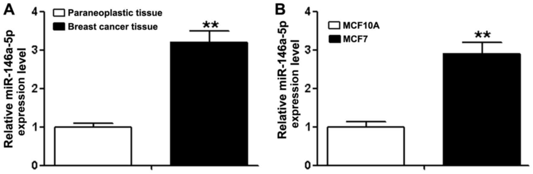
The results of RT-qPCR demonstrated that the
expression of miR-146a-5p in breast cancer tissue was 3.2±0.3 times
more, compared with paraneoplastic tissue (Fig. 1A, P<0.01), the expression in MCF-7
cell lines was 2.9±0.3 times more, compared with MCF 10A cells
(Fig. 1B, P<0.01).
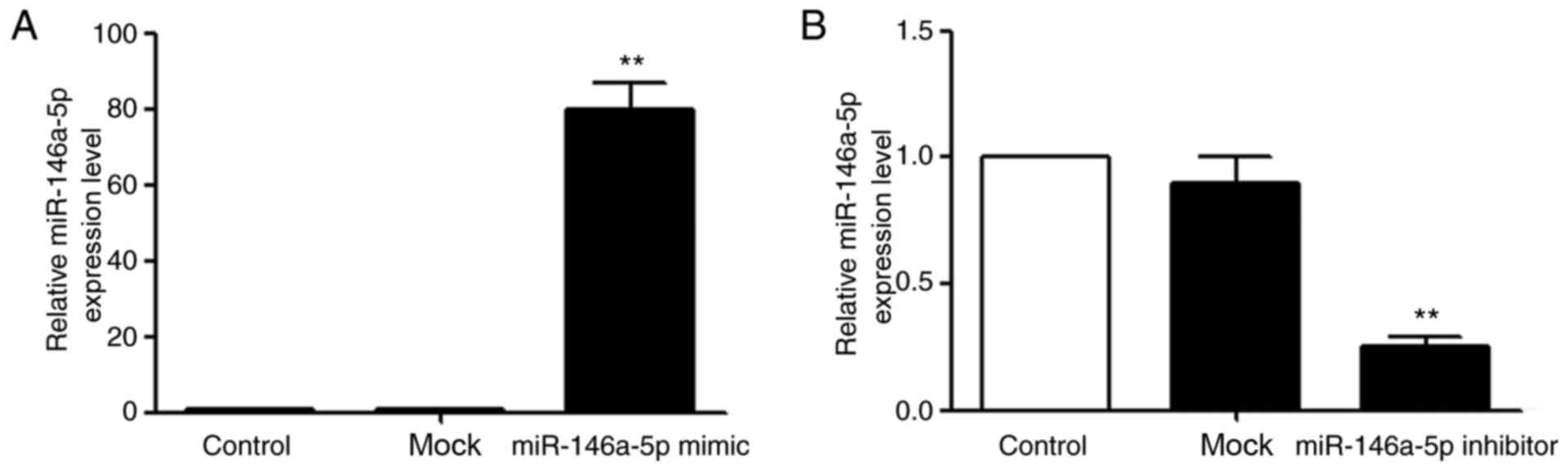
miR-146a-5p mimic and inhibitor
transfection into MCF-7 cells
According to the miR mimic and inhibitor
transfection introduction, the transfection concentration was 50
nM. The results indicated that the expression of miR-146a-5p in
MCF-7 cells transfected with miR-146a-5p mimic was 80±7 times more,
compared with original expression (Fig.
2A, P<0.01), whilst cells transfected with miR-146a-5p
inhibitor decreased to about 0.25±0.04 times compared with original
expression (Fig. 2B, P<0.01).
These results indicated that the transfections were successful.
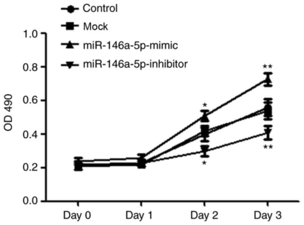
Effect of miR-146a-5p on the
proliferation of breast cancer cell line MCF-7
The proliferation (OD 490) of MCF-7 cells increased
gradually following D3. The proliferation of the overexpression
group significantly increased following transfection of the
miR-146a-5p mimic at D2 (0.51±0.03, P<0.05) and D3 (0.73±0.04,
P<0.01), whilst the mock group cells at D2 and D3 (D2,
0.42±0.03; D3, 0.54±0.05) had no significant difference (Fig. 3) with the blank control group (D2,
0.40±0.04; D3, 0.56±0.05). By contrast, the proliferation of
suppression group following the transfection of miR-146a-5p
inhibitor was significantly decreased, compared with the blank
control group at D2 (0.30±0.03, P<0.05) and D3 (0.41±0.04,
P<0.01). These results indicated that high expression of
miR-146a-5p in MCF-7 cells promoted proliferation, and low
expression of miR-146a-5p inhibited proliferation significantly
(Fig. 3).
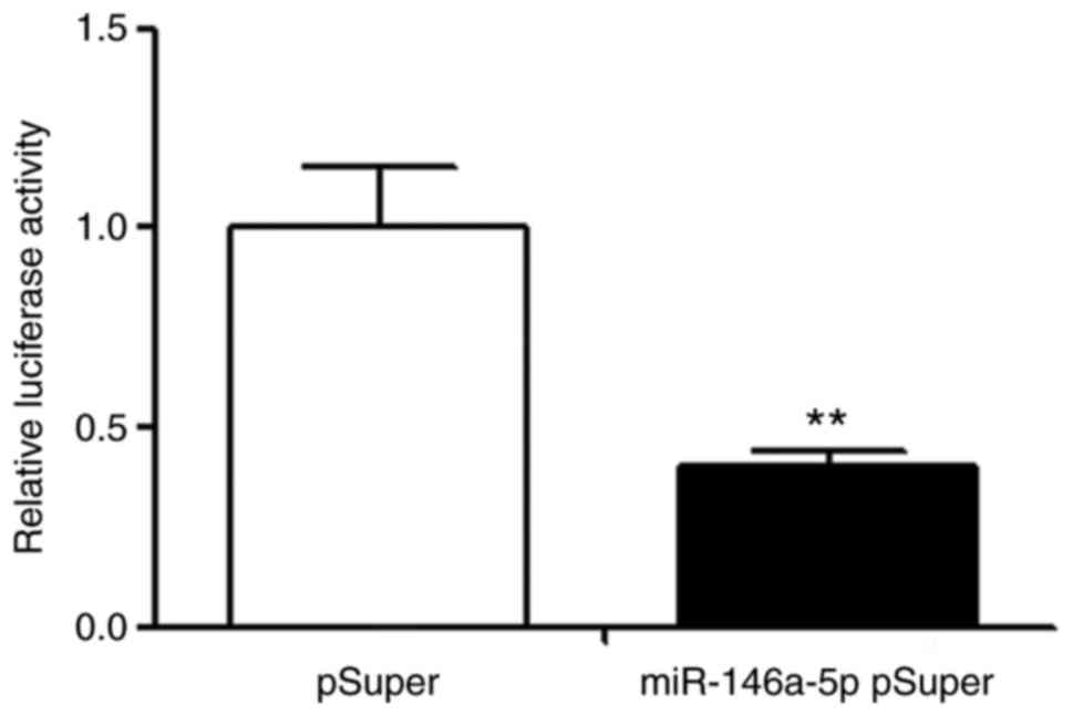
Detection of miR-146a-5p target
genes
Through further analysis of bioinformatic methods,
it was determined that the 3′UTR of tumor suppressor gene BRCA1
existed at the acting site of miR-146a-5p (Fig. 4). Furthermore, the interacting
sequence was highly conserved between species. All examples
indicated that BRCA1 may be the target gene of miR-146a-5p. The
pGL3 plasmid was recombined with luciferase expression of BRCA1
3′UTR. The luciferase results demonstrated that the luciferase
activity (0.40±0.04) of transfected miR-146a-5p group was
significantly lower, compared with the control group transfected
with mock (Fig. 5, P<0.01),
indicating that BRCA1 was the target gene of miR-146a-5p.
Discussion
miRNA is a type of non-coding small single stranded
RNA containing 20–24 nucleotides involved in gene transcription and
expression (19). miRNA is not
encoding protein, but has the ability to direct degrade the target
mRNA or inhibit its translation by complete or incomplete
complementary combination with the target mRNA (20). Numerous studies have demonstrated that
miRNA is involved in the process of tumor growth, metastasis and
angiogenesis by regulating the expression of oncogenesis, migration
and other associated genes (21–23).
In 2005, Iorio et al (24) first reported the change of miRNAs
expression profile in human breast cancer and determined that there
were 29 kinds of miRNAs with an expression disorder, in which the
expression of miRNA-21 and miRNA-155 were significantly
upregulated, whilst the expression of miRNA-10b, miRNA-125b and
miRNA-145 were significantly reduced. Additionally, a number were
associated with the clinical and pathological features of breast
cancer, including estrogen and progesterone receptor expression and
vascular invasion. In 2007, Blenkiron et al (25) determined that there were 133 kinds of
miRNA expression in normal breast tissue and/or cancer tissues, a
number of which were associated with molecular subtype of breast
cancer. Following this, the role of miRNAs in breast cancer gained
more attention.
In the previous work, it was determined that the
expression of miR-146a-5p in breast cancer tissue was significantly
higher, compared with paraneoplastic tissue (16). It was confirmed through RT-qPCR in the
present study and also discovered that the expression of
miR-146a-5p in breast cancer cell line MCF-7 was also significantly
higher, compared with control cells. High expression of miR-146a-5p
in MCF-7 could significantly promote the proliferation, and low
expression of miR-146a-5p could significantly inhibit it. BRCA1 was
preliminarily confirmed as the target gene of miR-146a-5p by
bioinformatics prediction and fluorescence reporter gene
detection.
BRCA1 is a tumor suppressor gene, which frequently
mutated in hereditary breast cancer (26). BRCA1 serves an important role in DNA
repair, cell cycle control, transcriptional activation, ubiquitin
and other processes (27,28). In the present study, it was determined
that the proliferation of MCF-7 cells was enhanced following high
expression of miR-146a-5p, whilst the proliferation of MCF-7 cells
decreased following low expression of miR-146a-5p. It was
hypothesized that the decreased expression of BRCA1, in consequence
of the high expression of miR-146a-5p, weakened the anti-tumor
ability and caused the increase of tumor cell proliferation.
Following miR-146a-5p low expression, the expression of BRCA1 was
increased, which caused the inhibition of tumor and the decrease of
the proliferation ability.
In conclusion, high expression of miR-146a-5p in
breast cancer and breast cancer cell lines may regulate the
proliferation of MCF-7 by regulating the expression of BRCA1.
Acknowledgements
Not applicable.
Funding
The present study was supported by the natural
science foundation of Science and Technology Commission of Shanghai
Municipality (grant no. 14ZR1408800).
Availability of data and materials
The datasets used and analyzed during the current
study are available from the corresponding author on reasonable
request.
Authors' contributions
GW, HJ and YY designed this study. JZ, DJ, HZ, DY
and LQ performed the experiments and interpreted the data. GW and
HJ analyzed and interpreted the data. GW and HJ performed the
histological examination of the breast and were major contributors
towards the writing of the manuscript. All authors read and
approved the final manuscript.
Ethics approval and consent to
participate
The Changhai Hospital Scholastic Ethics Committee
approved the research. All methods were performed in accordance
with the relevant guidelines and regulations.
Consent for publication
All subjects signed informed consent for
publication.
Competing interests
The authors declare that they have no competing
interests.
References
|
1
|
Rodriguez-Barrueco R, Nekritz EA, Bertucci
F, Yu J, Sanchez-Garcia F, Zeleke TZ, Gorbatenko A, Birnbaum D,
Ezhkova E, Cordon-Cardo C, et al: miR-424(322)/503 is a breast
cancer tumor suppressor whose loss promotes resistance to
chemotherapy. Genes Dev. 31:553–566. 2017. View Article : Google Scholar : PubMed/NCBI
|
|
2
|
Fan L, Strasser-Weippl K, Li JJ, St Louis
J, Finkelstein DM, Yu KD, Chen WQ, Shao ZM and Goss PE: Breast
cancer in China. Lancet Oncol. 15:e279–e289. 2014. View Article : Google Scholar : PubMed/NCBI
|
|
3
|
Cheng Y, Dong L, Zhang J, Zhao Y and Li Z:
Recent advances in microRNA detection. Analyst. 2018. View Article : Google Scholar
|
|
4
|
Rapado-González Ó, Majem B, Muinelo-Romay
L, Álvarez-Castro A, Santamaría A, Gil-Moreno A, López-López R and
Suárez-Cunqueiro MM: Human salivary microRNAs in Cancer. J Cancer.
9:638–649. 2018. View Article : Google Scholar : PubMed/NCBI
|
|
5
|
Horak M, Novak J and Bienertova-Vasku J:
Muscle-specific microRNAs in skeletal muscle development. Dev Biol.
410:1–13. 2016. View Article : Google Scholar : PubMed/NCBI
|
|
6
|
Harding RL and Velleman SG: MicroRNA
regulation of myogenic satellite cell proliferation and
differentiation. Mol Cell Biochem. 412:181–195. 2016. View Article : Google Scholar : PubMed/NCBI
|
|
7
|
Nakagawa R, Leyland R, Meyer-Hermann M, Lu
D, Turner M, Arbore G, Phan TG, Brink R and Vigorito E:
MicroRNA-155 controls affinity-based selection by protecting c-MYC+
B cells from apoptosis. J Clin Invest. 126:377–388. 2016.
View Article : Google Scholar : PubMed/NCBI
|
|
8
|
Saito Y, Nakaoka T and Saito H:
microRNA-34a as a therapeutic agent against human cancer. J Clin
Med. 4:1951–1959. 2015. View Article : Google Scholar : PubMed/NCBI
|
|
9
|
Singh RP, Massachi I, Manickavel S, Singh
S, Rao NP, Hasan S, Mc Curdy DK, Sharma S, Wong D, Hahn BH and
Rehimi H: The role of miRNA in inflammation and autoimmunity.
Autoimmun Rev. 12:1160–1165. 2013. View Article : Google Scholar : PubMed/NCBI
|
|
10
|
Lewis BP, Burge CB and Bartel DP:
Conserved seed pairing, often flanked by adenosines, indicates that
thousands of human genes are microRNA targets. Cell. 120:15–20.
2005. View Article : Google Scholar : PubMed/NCBI
|
|
11
|
Bertoli G, Cava C and Castiglioni I:
MicroRNAs: New biomarkers for diagnosis, prognosis, therapy
prediction and therapeutic tools for breast cancer. Theranostics.
5:1122–1143. 2015. View Article : Google Scholar : PubMed/NCBI
|
|
12
|
Hou LK, Yu Y, Xie YG, Wang J, Mao JF,
Zhang B, Wang X and Cao XC: miR-340 and ZEB1 negative feedback loop
regulates TGF-β-mediated breast cancer progression. Oncotarget.
7:26016–26026. 2016. View Article : Google Scholar : PubMed/NCBI
|
|
13
|
Shimono Y, Mukohyama J, Nakamura S and
Minami H: MicroRNA regulation of human breast cancer stem cells. J
Clin Med. 5:2015. View Article : Google Scholar : PubMed/NCBI
|
|
14
|
Takahashi RU, Miyazaki H and Ochiya T: The
roles of micrornas in breast cancer. Cancers(Basel). 7:598–616.
2015.PubMed/NCBI
|
|
15
|
Wang B, Teng Y and Liu Q: MicroRNA-153
regulates NRF2 expression and is associated with breast
carcinogenesis. Clin Lab. 62:39–47. 2016. View Article : Google Scholar : PubMed/NCBI
|
|
16
|
Meshkat M, Tanha HM, Naeini MM, Ghaedi K,
Sanati MH, Meshkat M and Bagheri F: Functional SNP in stem of
mir-146a affects Her2 status and breast cancer survival. Cancer
Biomark. 17:213–222. 2016. View Article : Google Scholar : PubMed/NCBI
|
|
17
|
Majer A, Blanchard AA, Medina S, Booth SA
and Myal Y: Claudin 1 expression levels affect miRNA dynamics in
human basal-like breast cancer cells. DNA Cell Biol. 35:328–339.
2016. View Article : Google Scholar : PubMed/NCBI
|
|
18
|
Livak KJ and Schmittgen TD: Analysis of
relative gene expression data using real-time quantitative PCR and
the 2(-Delta Delta C(T)) Method. Methods. 25:402–408. 2001.
View Article : Google Scholar : PubMed/NCBI
|
|
19
|
Schanza LM, Seles M, Stotz M, Fosselteder
J, Hutterer GC, Pichler M and Stiegelbauer V: MicroRNAs associated
with von hippel-lindau pathway in renal cell carcinoma: A
comprehensive review. Int J Mol Sci. 18:2017. View Article : Google Scholar : PubMed/NCBI
|
|
20
|
Kagiya T: MicroRNAs: Potential biomarkers
and therapeutic targets for alveolar bone loss in periodontal
disease. Int J Mol Sci. 17:E13172016. View Article : Google Scholar : PubMed/NCBI
|
|
21
|
Jansson MD and Lund AH: MicroRNA and
cancer. Mol Oncol. 6:590–610. 2012. View Article : Google Scholar : PubMed/NCBI
|
|
22
|
Lin S and Gregory RI: MicroRNA biogenesis
pathways in cancer. Nat Rev Cancer. 15:321–333. 2015. View Article : Google Scholar : PubMed/NCBI
|
|
23
|
Ohtsuka M, Ling H, Doki Y, Mori M and
Calin GA: MicroRNA processing and human cancer. J Clin Med.
4:1651–1667. 2015. View Article : Google Scholar : PubMed/NCBI
|
|
24
|
Iorio MV, Ferracin M, Liu CG, Veronese A,
Spizzo R, Sabbioni S, Magri E, Pedriali M, Fabbri M, Campiglio M,
et al: MicroRNA gene expression deregulation in human breast
cancer. Cancer Res. 65:7065–7070. 2005. View Article : Google Scholar : PubMed/NCBI
|
|
25
|
Blenkiron C, Goldstein LD, Thorne NP,
Spiteri I, Chin SF, Dunning MJ, Barbosa-Morais NL, Teschendorff AE,
Green AR, Ellis IO, et al: MicroRNA expression profiling of human
breast cancer identifies new markers of tumor subtype. Genome Biol.
8:R2142007. View Article : Google Scholar : PubMed/NCBI
|
|
26
|
Schayek H, Korach H, Laitman Y,
Bernstein-Molho R and Friedman E: Mutational analysis of candidate
genes in Israeli male breast cancer cases. Breast Cancer Res Treat.
2018. View Article : Google Scholar
|
|
27
|
Drost R and Jonkers J: Opportunities and
hurdles in the treatment of BRCA1-related breast cancer. Oncogene.
33:3753–3763. 2014. View Article : Google Scholar : PubMed/NCBI
|
|
28
|
Narod SA and Foulkes WD: BRCA1 and BRCA2:
1994 and beyond. Nat Rev Cancer. 4:665–676. 2004. View Article : Google Scholar : PubMed/NCBI
|