Introduction
Although the incidence and mortality rates have
decreased globally since the second half of the twentieth century,
gastric cancer still ranks as the fourth most frequently occurring
and the second most lethal type of cancer worldwide, accounting for
10.4% of cancer-associated mortalities (1). Gastric cancer remains a major health
concern primarily due to the slow decrease in incidence in Asia and
the high rates of mortality in diagnosed gastric carcinomas in the
West, despite the widespread application of innovative diagnostic
and surgical techniques in clinical practice (2,3). Cancer is
a genetic disease that is derived from cells that accumulate
mutations in essential growth regulatory genes, including oncogenes
and tumor suppressor genes (4).
Despite fluoropyrimidines and oxaliplatin-based chemotherapy being
successfully applied for cancer treatment, a number of side effects
are exhibited, including oxaliplatin-induced cumulative
dosage-dependent neurotoxicity (5).
Therefore, investigations into an anticancer drug that decreases
the incidence of relapse and presents with fewer side effects are
required.
The American cockroach (Periplaneta
americana), the largest species of pest insect in the Blattidae
family, is a worldwide domestic pest native to Africa that has
spread throughout the world, particularly in tropical and
subtropical regions (6). In China, an
extract derived from the dried whole body of P. americana
has been used in traditional Chinese medicine for the treatment of
bloodstasis syndrome, acne and abdominal masses for a hundred years
(7). Previous pharmacological studies
have demonstrated that the crude extract of P. americana
exhibits significant anticancer, anti-inflammation and tissue
regeneration activities (8–10).
With the development of traditional Chinese medicine
resources, importance has increasingly been attached to their
research and development. Several novel drugs, including Kangfuxin
(KFX) oral liquid, Xinmailong injection and cockroach oil have been
developed using P. americana as the raw material, with
recognized pharmacological activity and clinical efficacy (11–14). A
previous study revealed that P. americana extract exhibited
significant anticancer effects on the BEL-7402/5-FU Cell line and
SGC-7901 cell line (15,16). However, the exact apoptotic effect of
KFX remains unclear. Therefore, in the present study, the
anticancer effect of KFX was investigated by focusing on its
apoptotic potential in the human gastric cancer SGC-7901 cell line,
as well as its effects on the mitogen-activated protein
kinase/extracellular-signal-regulated kinase kinase
(MEK)/extracellular signal-regulated kinase (ERK) signaling
pathway.
Materials and methods
Materials
The human gastric cancer SGC-7901 cell line was
obtained from the cell resource center of the Shanghai Biological
Sciences Institute (Chinese Academy of Sciences, Shanghai, China).
KFX oral liquid was received from Sichuan Good Doctor
Pharmaceutical Group (Sichuan, China), comprising 1 g/ml P.
americana dried whole body in water.
Cell culture
SGC-7901 cells were maintained in RPMI-1640
supplemented with 10% fetal bovine serum (both Gibco; Thermo Fisher
Scientific, Inc.) in a humidified atmosphere with 5% CO2
at 37°C. The cultured cells were passaged with 0.25% trypsin
(Gibco; Thermo Fisher Scientific, Inc. Waltham, MA, USA) when cell
confluence reached ~80%. Cells between passage numbers 3 and 10
were selected for experimentation. Before starting the experimental
procedures, the desired final concentrations of KFX (0, 0.25, 0.5,
2.5 mg/ml) were achieved by diluting the stock solution (1 g/ml) in
RPMI-1640 culture medium. Then the SGC-7901 cells were placed in
RPMI-1640 in the presence or absence of KFX for 12 or 24 h. In some
experiments, SGC-7901 cells were exposed to a MEK inhibitor U0126
(0.2 µM, dissolved in RPMI-1640 culture medium) (Sigma-Aldrich;
Merck KGaA, Darmstadt, Germany) for 12 h. For signaling pathway
analysis, SGC-7901 cells were treated with phorbol 12-myristate
13-acetate (PMA) (3 nM, dissolved in DMSO) (Sigma-Aldrich; Merck
KGaA, Darmstadt, Germany), a specific activator of protein kinase
C, nuclear factor-κB and ERK, for 12 h in combination with KFX
treatment.
Reverse transcription-polymerase chain
reaction (RT-PCR) analysis
The SGC-7901 cells were placed in RPMI-1640 in the
presence or absence of KFX (0, 0.25, 0.5, 2.5 mg/ml) for 12 or 24
h. Four µg RNA and oligo dT18 were then incubated at
80°C for 5 min. The cDNA synthesis reaction was performed at 42°C
for 1 h with M-MLV reverse transcriptase (cat. no., A5001; Promega
Corporation), followed by incubation at 70°C for 15 min to
inactivate the reverse transcriptase. Following RT, samples were
diluted by adding 60 µl purified water. For the PCR, PCR MasterMix
(cat. no., PR1700; BioTeke Corporation, Beijing, China) was used
and the reactions were performed in a T100 Thermo Cycler (Bio-Rad
Laboratories) with the following profile: Incubation for 3 min at
95°C, followed by 32 cycles of denaturation for 30 sec at 95°C,
annealing for 30 sec at 72°C, and extension for 5 min at 72°C. The
products were resolved in a 1% agarose gel stained with SYBR Safe
(Invitrogen; Thermo Fisher Scientific, Inc.). ImageJ software
(version 1.48; National Institutes of Health, Bethesda, MD, USA)
was used to quantify the bands (17).
Primer sequences of peroxisome proliferator-activated receptor
(PPAR)-γ and GAPDH for RT-PCR were as follows: PPAR-γ forward,
5′-TCTGGCCCACCAACTTTGGG-3′ and reverse, 5′-CTTCACAAGCATGAACTCCA-3′;
and GAPDH forward, 5′-GCCAAGGTCATCCATGACAACT-3′ and reverse,
5′-GAGGGGCCATCCACAGTCTT-3′.
Western blot analysis
SGC-7901 cells were lysed in a buffer consisting of
7 M urea, 2 M thiourea, 2% 3-[(3-cholamidopropyl)
dimethylammonio]-1-propanesulfonate hydrate, 40 mM Trizma base, 40
mM dithiothreitol and 1% protease inhibitor cocktail
(Sigma-Aldrich; Merck KGaA). Following centrifugation at 21,885 × g
for 15 min at 4°C, the total protein concentration in the
supernatant was determined with a Bradford protein assay (Bio-Rad
Laboratories, Inc., Hercules, CA, USA). Equal amounts of protein
(50 µg/lane) were subjected to SDS-PAGE (10% gel) and transferred
onto polyvinylidene difluoride membranes. Samples were then blocked
with 5% skimmed dried milk in Tris-buffered saline containing 0.1%
TritonX-100 (TBST) at room temperature for 2 h, and incubated
overnight at 4°C with the following primary antibodies:
Cleaved-Caspase-3 (cat. no., 9661; dilution, 1:1,000; Cell
Signaling Technology Inc., Danvers, MA, USA), Bax (cat. no.,
ab32503; dilution, 1:1,000; Abcam), Bcl-2 (cat. no., ab59348;
dilution, 1:1,000; Abcam), p53 (cat. no., 2524; dilution, 1:1,000;
Cell Signaling Technology, Inc.), IL-1β (cat. no., ab106035;
dilution, 1:1,000; Abcam;), IL-6 (cat. no., ab6672; dilution,
1:1,000; Abcam), TNF-α (cat. no., ab1793; dilution, 1:5,000;
Abcam), p-Erk (cat. no., 9101; dilution, 1:1,000; Cell Signaling
Technology, Inc.), Erk (cat. no., 9102; dilution, 1:1,000; Cell
Signaling Technology, Inc.) and β-actin (cat. no., 4970; dilution,
1:1,000; Cell Signaling Technology, Inc.). The membrane was washed
with TBST three times, 5 min each. Subsequently, the membrane was
incubated with a horseradish peroxidase-conjugated goat-anti-mouse
(cat. no., ab6789; dilution, 1:300; Abcam) or HRP-goat-anti-rabbit
(cat. no., ab6721; dilution, 1:300; Abcam) for 2 h at room
temperature. Membranes were washed with TBST three times for 5 min
each, prior to incubation with enhanced chemiluminescence (cat.
no., WBKLS0500; Merck KGaA) for 1 min. The protein levels were
normalized against that of internal protein β-actin using ImageJ
software (version 1.48; National Institute of Health, Bethesda, MD,
USA).
Terminal
deoxynucleotidyl-transferase-mediated dUTP nick-end labeling
(TUNEL) assay
The TUNEL assay was performed using a commercially
available In situ Cell Death Detection kit (Roche
Diagnostics GmbH; cat. no., 11684817910), according to the
manufacturer's protocol, as previously described (18). SGC-7901 cells cultured on 6-mm plates
were fixed with 4% paraformaldehyde solution for 30 min at room
temperature. Following a PBS wash, cells were treated with
permeation solution (0.1% Triton X-100 in 0.1% sodium citrate) for
2 min at 4°C. Following washing with PBS, samples were incubated
with TUNEL reagent containing 10% terminal deoxynucleotidyl
transferase and 2% fluorescent isothiocyanate-dUTP for 1 h at 37°C.
Subsequently, the cells were stained with 1 µg/ml DAPI for 30 min
at room temperature to detect the cellular nuclei. Finally, the
cells are mounted on coverslips with antifade mounting medium
(Beyotime, P0126). Using an excitation wavelength in the range of
450–500 nm and detection in the range of 515–565 nm (green), the
number of TUNEL-positive SGC-7901 cells and apoptotic bodies were
determined. The percentage of apoptotic cells were calculated by
dividing the number of TUNEL-positive cells by the total number of
cells visualized in ≥6 separate fields using a fluorescence
microscope. Three digitized images of similar total cell numbers
were selected from each cover slip for counting and averaging, and
were considered as one independent experiment. Three independent
experiments were then averaged and statistically analyzed.
MTT assay
SGC-7901 cells were plated in 96-well plates at a
density of 5,000 cells/well in 120 µl complete medium (RPMI-1640
(Gibco; Thermo Fisher Scientific, Inc.; cat. no., 11875093)
supplemented with 10% fetal bovine serum (Gibco; Thermo Fisher
Scientific, Inc.; cat. no., 16000044). To investigate the cytotoxic
effect of KFX, SGC-7901 cells were treated with 0.00, 0.25, 0.5,
0.75, 1 and 2.5 mg/ml for 12 and 24 h. Each group was repeated in 9
separate wells. Following treatment, 15 µl MTT reagent (5 mg/ml)
was added to each well for 4 h, and then 150 µl DMSO was added to
each well. Absorbance was detected at a wavelength of 490 nm using
a microplate reader.
Statistical analysis
Results are expressed as the mean ± standard error
of the mean. Statistical differences were assessed with the
unpaired 2-tailed Student's t-test for two experimental groups and
one-way ANOVA for multiple groups, using SPSS 19.0 software (IBM
Corp, Armonk, NY, USA). Bonferroni's post-hoc test was employed
following one-way ANOVA for determining significant differences
between groups. A two-tailed P-value of <0.05 was considered to
indicate a statistically significant difference. Statistical
analyses were performed using GraphPad Prism (version 5; GraphPad
Software).
Results
KFX increases the mRNA expression
level of tumor-suppressor factor PPAR-γ and impairs the viability
of SGC-7901 cells
PPAR-γ, a member of the nuclear receptor
superfamily, regulates lipid metabolism, inflammation and cancer
progression (19). Usually, PPAR-γ
regulates target genes by binding to the PPAR-γ response element in
the promoter region of target genes, resulting in either promotion
or inhibition. PPAR-γ displays antitumor effects through inhibition
of proliferation and induction of differentiation and apoptosis by
targeting tumor-associated genes, including tumor protein p63,
tumor protein p73, tumor protein p21, B-cell lymphoma 2 (Bcl-2),
Bcl-2 associated X (Bax), caspase-3 and MYC proto-oncogene
(17,20–22). It
has been demonstrated that PPAR-γ activation inhibits cell growth
(23) and promotes differentiation
and apoptosis in a variety of types of cancer cell.
To investigate whether KFX treatment may lead to the
upregulation of PPAR-γ, RT-qPCR analysis was conducted for SGC-7901
cells treated with KFX at 0, 0.25, 0.5 and 2.5 mg/ml for 12 and 24
h. As presented in Fig. 1A, PPAR-γ
mRNA expression level was significantly increased at all
concentrations after 24 h and at 2.5 mg/ml after 12 h of KFX
treatment, compared with that of the control, in a dose-dependent
manner (P<0.05); no significant differences were identified
between the control and cells treated with 0.25 or 0.5 mg/ml after
12 h. In addition, cell viability was analyzed by MTT assay. The
results demonstrated that exposure to KFX resulted in a
dose-dependent decrease in cell viability in the SGC-7901 cells.
The half maximal inhibitory concentration values of KFX at 12 and
24 h were 1.19±0.06 and 0.64±0.04 mg/ml (P<0.05), respectively,
for the SGC-7901 cells (Fig. 1B).
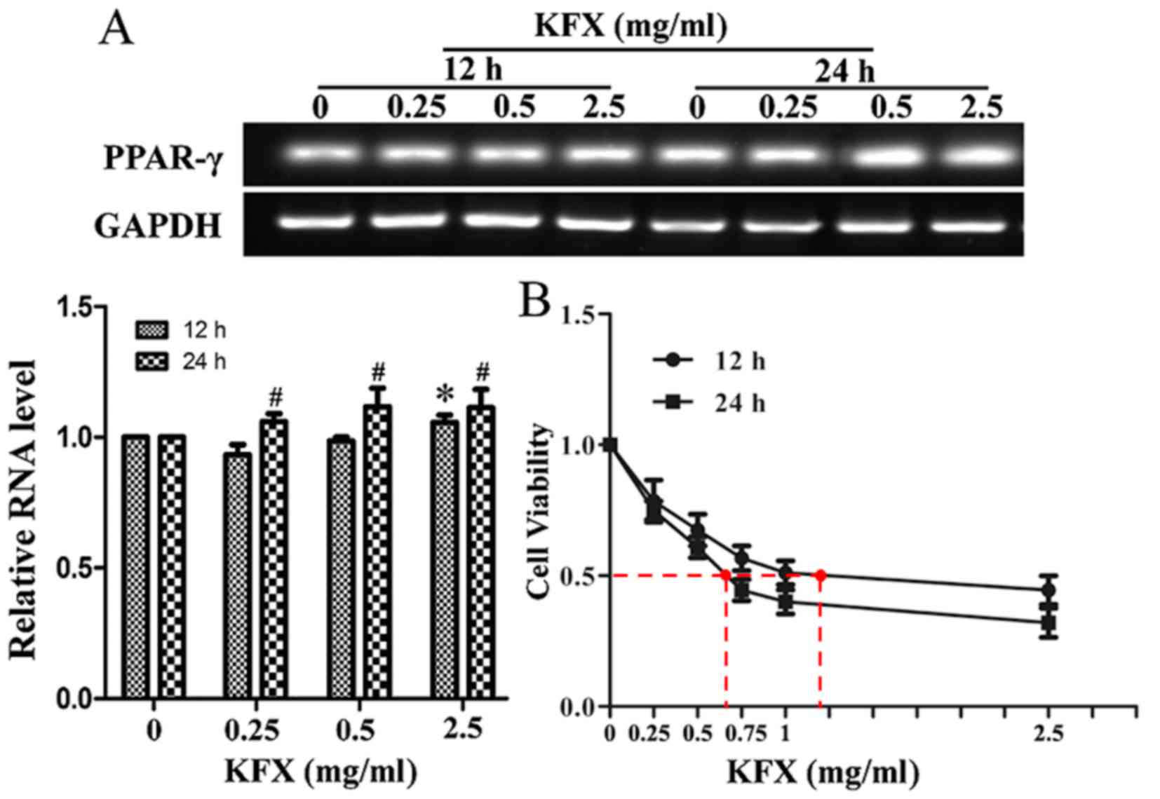 | Figure 1.KFX increases PPAR-γ mRNA expression
and decreases the viability of SGC-7901 cells. (A) SGC-7901 cells
were treated with KFX at different concentrations (0, 0.25, 0.5 and
2.5 mg/ml) for 12 and 24 h, and the PPAR-γ mRNA level was analyzed
by reverse transcription-polymerase chain reaction. The histogram
presents the quantitative analysis of PPAR-γ mRNA level, and the
data are expressed as fold over control group; n=6 for each group.
Data are presented as the mean ± standard error of the mean.
*P<0.05 compared with KFX (0 mg/ml) treatment for 12 h;
#P<0.05 compared with KFX (0 mg/ml) treatment for 24
h. (B) The anti-proliferative effect of KFX was investigated using
an MTT assay. SGC-7901 cells were treated with different dosages of
KFX (0, 0.05, 0.15, 0.25, 0.5 and 2.0 mg/ml) for 12 and 24 h; n=6
for each group. Data are presented as the mean ± standard error of
the mean. *P<0.05 compared with KFX (0 mg/ml) treatment for 12
h; #P<0.05 compared with KFX (0 mg/ml) treatment for
24 h. KFX, Kangfuxin; PPAR-γ, peroxisome proliferator-activated
receptor γ. |
KFX induces apoptosis and decreases
inflammation in SGC-7901 cells
To investigate the hypothesis that KFX is capable of
inducing apoptosis in SGC-7901 cells, caspase-3 activity was
detected using western blot analysis following treatment with 0,
0.25 or 0.50 mg/ml KFX for 12 h. Activated caspase-3, denoted by
increased expression of cleaved caspase-3, was detected in the
SGC-7901 cells following KFX treatment (Fig. 2A); this difference was significant
compared with the negative control (P<0.05). Furthermore, to
demonstrate the capability of KFX to induce apoptosis in SGC-7901
cells, the pro-survival protein Bcl-2 and the pro-apoptotic protein
Bax were evaluated using western blot analysis. A decrease in the
expression of Bcl-2 was revealed with KFX treatment; however, Bax
expression appeared to remain unaffected. Additionally, the
expression levels of p53, an initiator of cellular apoptosis, were
upregulated in SGC-7901 cells following KFX treatment (Fig. 2A). These results suggested that the
decrease in cell viability observed was due to cell apoptosis
induced by KFX. Furthermore, a TUNEL assay was performed to detect
the pro-apoptotic effect of KFX on SGC-7901 cells. Compared with
the untreated control, the number of apoptosis cells was
significantly increased following KFX treatment, in a
dose-dependent manner (Fig. 2B).
 | Figure 2.KFX induces cell apoptosis, inhibits
the inflammation response and prevents ERK phosphorylation.
SGC-7901 cells were treated with KFX at different dosages (0, 0.25
and 0.5 mg/ml) for 12 h and (A) the protein levels of c-caspase-3,
Bax/Bcl-2 and p53 were detected using western blot analysis. The
histogram is the quantitative analysis of the corresponding
immunoblots and the data are expressed as fold over control group;
n=6 for each group. Data are presented as the mean ± standard error
of the mean. *P<0.05 compared with KFX (0 mg/ml). (B) DAPI (top)
staining and TUNEL assay (bottom) of the SGC-7901 cell treated with
KFX at different dosages (0, 0.25 and 0.5 mg/ml) for 12 h. The
histogram is the quantitative analysis of TUNEL + cells in at least
6 separate fields; n=6 for each group. Scale bar, 100 µm.
*P<0.05 compared with control. Western blot assays of (C) IL-6,
IL-1β and TNF-α, and (D) p-ERK/ERK levels in SGC-7901 cell treated
with KFX at different dosages (0, 0.25 and 0.5 mg/ml) for 12 h. The
histogram is the quantitative analysis of the corresponding
immunoblots and the data are expressed as fold over control group;
n=6 for each group. Data are presented as the mean ± standard error
of the mean. *P<0.05 compared with KFX (0 mg/ml). KFX,
Kangfuxin; TUNEL, terminal deoxynucleotidyl transferase
dUTP-mediated nick-end labeling; p-ERK, phosphorylated
extracellular signal-regulated kinase; c-caspcase-3, cleaved
caspase-3; Bcl-2, B-cell lymphoma 2; Bax, Bcl-2 associated X; IL,
interleukin; TNF, tumor necrosis factor; con, control. |
The magnitude of inflammation is often augmented
during aging and age, in turn, is a major risk factor for
developing oncological diseases (24). Previous studies have demonstrated that
interleukin (IL)-1β, IL-6 and tumor necrosis factor (TNF)-α recruit
immune cells into the site of a developing tumor or tumor
microenvironment, thereby enhancing inflammation (25,26). To
investigate the ability of KFX to abate inflammation in the
microenvironment of SGC-7901 cells, the expression of IL-1β, IL-6
and TNF-α were detected with western blot analysis in the present
study. Fig. 2C shows that IL-1β, IL-6
and TNF-α protein levels significantly decreased following KFX
treatment compared with the levels in the untreated cells. These
results indicated that KFX may alleviate the production of
inflammatory cytokines in SGC-7901 cells.
KFX promotes SGC-7901 cell apoptosis
through the ERK pathway
Mitogen-activated protein kinase kinase (MAPKK) is
involved in a number of cellular biological functions, including
proliferation, differentiation, motility and death (27–29). ERKs
are the main members of the MAPKK signaling pathway, and the
activation of ERK1/2 is an anticancer target (30,31).
Therefore, to clarify whether the ERK signaling pathway is
activated in SGC-7901 cells by KFX, cells were treated with 0, 0.25
and 0.5 mg/ml KFX for 12 h in the present study. The results
demonstrated that phosphorylated (p)-ERK1/2 was significantly
decreased following KFX treatment, in a dose-depended manner,
whereas total ERK expression remained consistent (Fig. 2D).
To further investigate the role of p-ERK1/2 in
KFX-mediated SGC-7901 cell apoptosis, PMA, a specific activator of
protein kinase C, NF-κB and ERK, was introduced (32). Following cell treatment with PMA, ERK
phosphorylation was markedly increased (Fig. 3A). Additionally, pro-survival protein
Bcl-2, inflammatory cytokines IL-1β, IL-6 and TNF-α, along with the
downregulated p53 and cleaved caspase-3, were significantly
increased, whereas the pro-survival protein Bax remained unchanged
(Fig. 3B and C). As expected, KFX
exhibited an inhibitory effect on PMA-induced anti-apoptosis via
the ERK signaling pathway, as demonstrated by the decreased
expression following use of KFX and PMA together.
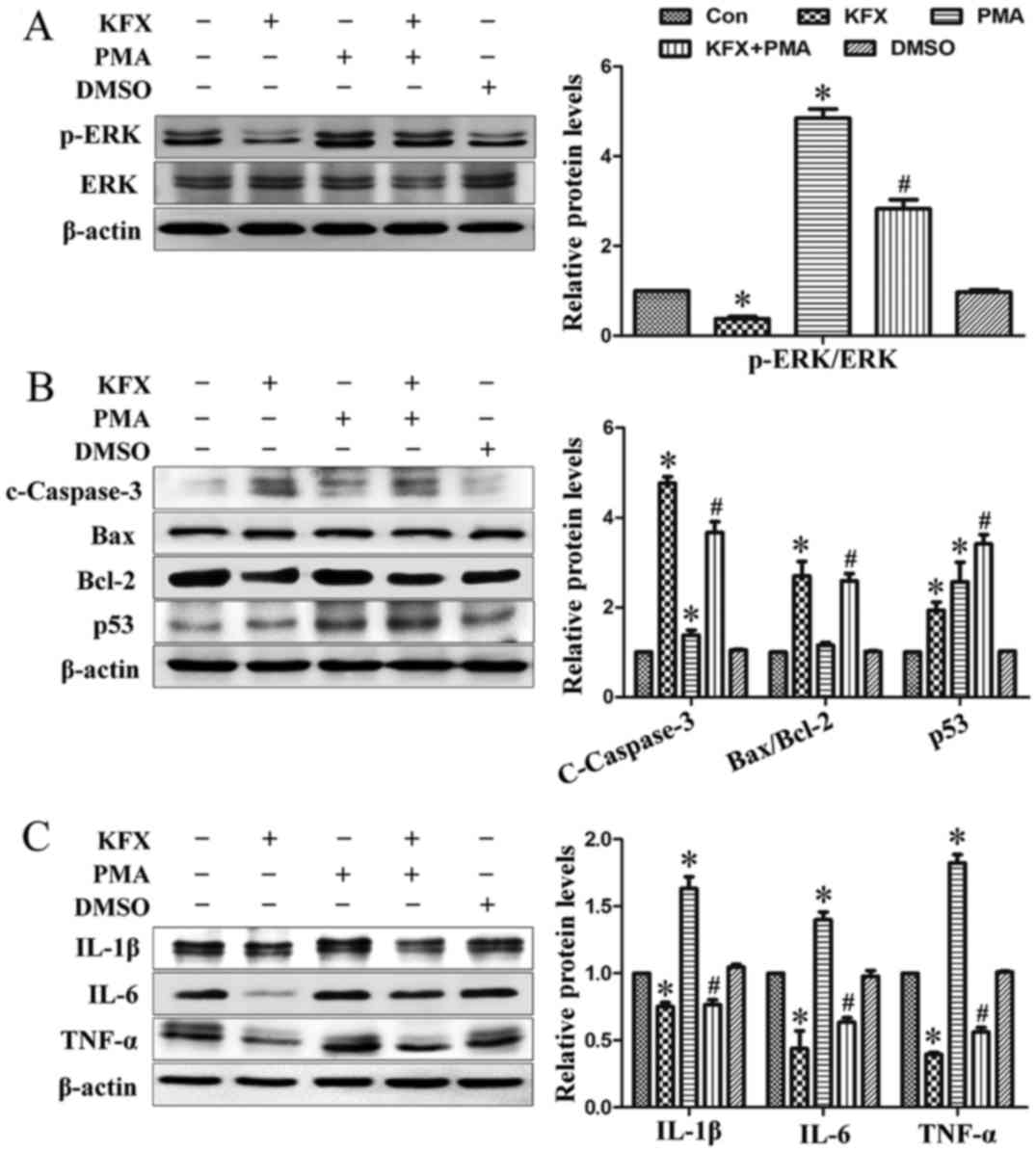 | Figure 3.KFX prevents ERK
phosphorylation-mediated anti-apoptotic and inflammatory responses.
SGC-7901 cells were cultured to near (80–90%) confluence and then
administrated with 0.5 mg/ml KFX in the presence or absence of 3
nmol/l PMA for 12 h. Immunoblotting assays of (A) p-ERK/ERK; (B)
c-caspase-3, Bax/Bcl-2 and p53; (C) IL-6, IL-1β and TNF-α in
SGC-7901 cells. The histogram is the quantitative analysis of the
corresponding immunoblots and the data are expressed as fold over
control group; n=6 for each group. Data are presented as the mean ±
standard error of the mean. *P<0.05 compared with control;
#P<0.05 compared with PMA. KFX, Kangfuxin; p-ERK,
phosphorylated extracellular signal-regulated kinase; c-caspcase-3,
cleaved caspase-3; Bcl-2, B-cell lymphoma 2; Bax, Bcl-2 associated
X; IL, interleukin; p53, tumor protein p53; TNF, tumor necrosis
factor; PMA, phorbol 12-myristate 13-acetate; DMSO, dimethyl
sulfoxide; con, control. |
By contrast, U0126, an inhibitor of p-ERK1/2, was
used to inhibit p-ERK1/2 expression and to mimic the function of
KFX on SGC-7901 cells. Following treatment with U0126, ERK
phosphorylation was blocked and cleaved-caspase 3 expression was
increased (Fig. 4A and B). Consistent
with the results obtained by KFX treatment, U0126 treatment also
exhibited a significant pro-apoptotic and anti-inflammatory effect
on SGC-7901 cells (Fig. 4C), which
suggested that KFX may inhibit phosphorylation in the ERK1/2
signaling pathway, blocking cell proliferation similar to U0126. In
addition, the role of ERK-mediated SGC-7901 cell apoptosis and
anti-proliferation induced by KFX was further investigated with an
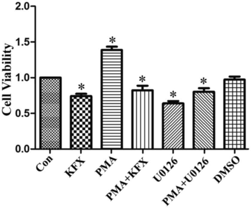
MTT assay. Cell viability of SGC-7901 cells incubated with PMA was
significantly increased compared with that of the control, but
largely abolished by KFX or U0126 (Fig.
5).
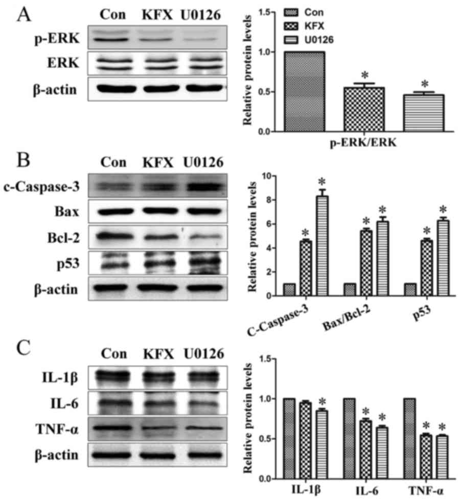 | Figure 4.KFX inhibits ERK1/2 pathway
phosphorylation similar to U0126. SGC-7901 cells were cultured to
near (80–90%) confluence and then administered with 0.5 mg/ml KFX
or 0.2 µmol/l U0126 for 12 h. Immunoblotting assays of (A)
p-ERK/ERK; (B) c-caspase-3, Bax/Bcl-2 and p53; (C) IL-6, IL-1β and
TNF-α levels in SGC-7901 cells. The histogram is the quantitative
analysis of the corresponding immunoblots and the data are
expressed as fold over control group; n=6 for each group. Data are
presented as the mean ± standard error of the mean. *P<0.05
compared with Con. KFX, Kangfuxin; p-ERK, phosphorylated
extracellular signal-regulated kinase; p53, tumor protein p53;
c-caspcase-3, cleaved caspase-3; Bcl-2, B-cell lymphoma 2; Bax,
Bcl-2 associated X; IL, interleukin; TNF, tumor necrosis factor;
con, control. |
Discussion
Gastric cancer is the fourth most common cause of
cancer-associated mortality worldwide, and remains difficult to
treat, primarily due to the majority of patients presenting with
advanced disease (1). In the USA,
stomach malignancy is currently the 15th most common type of cancer
(33,34). Although the majority of
chemotherapeutic regimens utilize antineoplastic agents as the
clinical standard of care for gastric cancer, patients commonly
experience a limited response to this therapy (35–37).
Therefore, investigation into more effective therapeutic
interventions for gastric cancer is important. Over the last 50
years, emerging evidence has suggested that a number of natural
extracts from plants and animals exhibit beneficial effects in the
prevention of cancer (16,38,39). The
present study demonstrated that KFX, an aqueous extract from P.
americana, exhibited potential anticancer effects in a gastric
cancer cell line through inhibiting cell proliferation and inducing
apoptosis; potentially via the ERK signaling pathway.
The results of the present study demonstrated that
KFX markedly inhibited SGC-7901 cell viability in a dose-dependent
manner, which indicated that KFX may exhibit a therapeutic effect
on gastric cancer. The induction of cellular apoptosis in malignant
cells is critical for the chemoprevention and chemotherapy of
cancer by natural product-derived anticancer agents (40,41); as
such, the inhibition of SGC-7901 cell viability following KFX
treatment may be as a result of apoptosis. Therefore, in order to
clarify the underlying molecular mechanism, the effect of KFX
treatment on SGC-7901 cell apoptosis was investigated. In general,
apoptosis is a type of organized cell self-destruction by a series
of signal cascades that include numerous gene products and
cytokines. Caspase-3 activation serves an important role in
apoptosis, whilst the imbalance between Bcl-2 (anti-apoptotic) and
Bax (pro-apoptotic) has been recognized as a signature of the
acquisition of apoptosis resistance in cancer cells (42,43). In
addition, p53, acting as a transcriptional factor, serves an
important role in promoting apoptosis in response to various
cellular stressors, including oncogene activation (44). The results of the present study
demonstrated that KFX may induce gastric cancer cell apoptosis,
supported by the observed caspase-3 activation, p53 upregulation
and Bcl-2 downregulation alongside unchanged Bax expression.
ERK is involved in a number of cellular programs,
and the activation of ERK through phosphorylation is a potential
anticancer target (45). In the
present study, it was revealed that KFX possessed anticancer
potential in SGC-7901 cells through inhibiting cell proliferation
and inducing apoptosis, potentially via the ERK signaling pathway.
KFX exhibited an inhibitory effect on protein kinases involved in
the phosphorylation of ERK, thereby leading to a decrease in p-ERK
protein. Furthermore, following incubation with an ERK activator,
PMA, the decrease observed in cleaved caspase-3 and p53, and the
increase in Bcl-2, inflammatory cytokines and cell proliferation,
suggested that KFX-induced apoptosis may occur via an ERK-mediated
signaling pathway. Referring to the results of the present study
and the relevant information available in the literature, a
proposed scheme presenting a potential explanation regarding the
pro-apoptotic effect of KFX on SGC-7901 cells was created (Fig. 6).
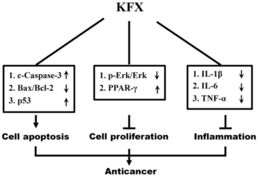 | Figure 6.Schematic diagram illustrating the
mechanism of KFX pro-apoptotic effects on SGC-7901 cells through
inhibition of the MEK/ERK signaling pathway. MEK, mitogen-activated
protein kinase/extracellular-signal-regulated kinase kinase; KFX,
Kangfuxin; p-ERK, phosphorylated extracellular signal-regulated
kinase; c-caspcase-3, cleaved caspase-3; p53, tumor protein p53;
Bcl-2, B-cell lymphoma 2; Bax, Bcl-2 associated X; IL, interleukin;
TNF, tumor necrosis factor; PPAR-γ, peroxisome
proliferator-activated receptor. |
In conclusion, to the best of our knowledge, the
results of the present study demonstrated for the first time that
KFX may potentially inhibit SGC-7901 gastric cancer cell
proliferation and induce apoptosis through modulation of the ERK
signaling pathway, thus suggesting the novel therapeutic potential
of KFX for gastric cancer treatment.
Acknowledgements
Not applicable.
Funding
The present study was supported by the National
Nature Science Foundation of China (grant no. 81570368), Technology
Program of Wenzhou (grant no. Y20140737) and Wenzhou Medical
University Crossing Research Program (grant no. KJHX1504).
Availability of data and materials
The datasets used and/or analyzed during the current
study are available from the corresponding author on reasonable
request.
Authors' contributions
WC and FG conceived the present study. XM and JS
wrote the manuscript and performed the cell cultures. WY performed
the western blot analysis. YH conducted the cell apoptosis assays.
CS performed and analyzed the PCR. YT and TW performed the western
blot analysis and MTT assays. All authors read and approved the
final manuscript.
Ethics approval and consent to
participate
Not applicable.
Consent for publication
Not applicable.
Competing interests
The authors declare that they have no competing
interests.
References
|
1
|
Yu J, Wang X, Li Y and Tang B: Tanshinone
IIA suppresses gastric cancer cell proliferation and migration by
downregulation of FOXM1. Oncol Rep. 37:1394–1400. 2017. View Article : Google Scholar : PubMed/NCBI
|
|
2
|
de Martel C, Eorman D and Plummer M:
Gastric cancer: Epidemiology and risk factors. Gastroenterol Clin
North Am. 42:219–240. 2013. View Article : Google Scholar : PubMed/NCBI
|
|
3
|
Zheng HC, Zheng YS, Li XH, Takahashi H,
Hara T, Masuda S, Yang XH, Guan YF and Takano Y: Arp2/3
overexpression contributed to pathogenesis, growth and invasion of
gastric carcinoma. Anticancer Res. 28:2225–2232. 2008.PubMed/NCBI
|
|
4
|
Sherr CJ: Principles of tumor suppression.
Cell. 116:235–246. 2004. View Article : Google Scholar : PubMed/NCBI
|
|
5
|
André T, Boni C, Navarro M, Tabernero J,
Hickish T, Topham C, Bonetti A, Clingan P, Bridgewater J, Rivera F
and de Gramont A: Improved overall survival with oxaliplatin,
fluorouracil, and leucovorin as adjuvant treatment in stage II or
III colon cancer in the MOSAIC trial. J Clin Oncol. 27:3109–3116.
2009. View Article : Google Scholar : PubMed/NCBI
|
|
6
|
Chinese Materia Medica Editoral Committee:
Chinese Materia Medica. 9. Shanghai Scientific and Technical
Publisher; Shanghai: pp. 149–151. 1999
|
|
7
|
Sun XY: Sheng Nong's herbal classic.
Beijing Commercial Press; Beijing: pp. 90–91. 1955
|
|
8
|
Jiang Y, Wang X, Jin C, Chen X, Li J, Wu
Z, Liu G and Li S: Inhibitory effect of Periplaneta
Americana extract on 3LL lung cancer in mice. Zhongguo Fei Ai
Za Zhi. 9:488–491. 2006.(In Chinese). PubMed/NCBI
|
|
9
|
Li W, Duan LF, He GQ, Shen ZQ, Yang HQ and
Liang YP: Periplaneta americana extract effects on
experimental liver fibrosis. Lishizhen Med Mater Med Res.
21:1137–1138. 2010.
|
|
10
|
He ZC, Peng F, Song LY, Wang XY, Hu MH,
Zhao Y and Liu GM: Review on investigations related to chemical
constituents and biological activities of Periplaneta
americana. Zhongguo Zhong Yao Za Zhi. 32:2326–2331. 2007.(In
Chinese). PubMed/NCBI
|
|
11
|
Sun YN: Apoptosis pathway for targets of
anti-tumor treatment research progress. Foreign Med Sci (Sect
Pharmarcy). 33:321–324. 2006.
|
|
12
|
Chen P, Shen Y, Shi H, Ma X, Lin B, Xiao
T, Wu F, Zhu J, Li Z, Xiao J, et al: Gastroprotective effects of
Kangfuxin-against ethanol-induced gastric ulcer via attenuating
oxidative stress and ER stress in mice. Chem Biol Interact. Oct
28–2016.(Epub ahead of print). View Article : Google Scholar
|
|
13
|
Luo SL, Huang XJ, Wang Y, Jiang RW, Wang
L, Bai LL, Peng QL, Song CL, Zhang DM and Ye WC: Isocoumarins from
American cockroach (Periplaneta americana) and their
cytotoxic activities. Fitoterapia. 95:115–120. 2014. View Article : Google Scholar : PubMed/NCBI
|
|
14
|
Zhang H, Wei L, Zhang Z, Liu S, Zhao G,
Zhang J and Hu Y: Protective effect of Periplaneta americana
extract on intestinal mucosal barrier function in patients with
sepsis. J Tradit Chin Med. 33:70–73. 2013. View Article : Google Scholar : PubMed/NCBI
|
|
15
|
Yuan F, Liu J, Qiao T, Li T, Shen Q and
Peng F: The effects and mechanisms of Periplaneta americana
extract reversal of multi-drug resistance in BEL-7402/5-FU cells.
Molecules. 21:pii: E852. 2016. View Article : Google Scholar
|
|
16
|
Chen PP, Ma XY, Lin Q, Xu HL, Shi HX,
Zhang HY, Xiao J, Geng FN and Zhao YZ: Kangfuxin promotes apoptosis
of gastric cancer cells through activating ER-stress and autophagy.
Mol Med Rep. 16:9043–9050. 2017. View Article : Google Scholar : PubMed/NCBI
|
|
17
|
Kim S, Lee JJ and Heo DS: PPARγ ligands
induce growth inhibition and apoptosis through p63 and p73 in human
ovarian cancer cells. Biochem Biophys Res Commun. 406:389–395.
2011. View Article : Google Scholar : PubMed/NCBI
|
|
18
|
Tsai CY, Wang CC, Lai TY, Tsu HN, Wang CH,
Liang HY and Kuo WW: Antioxidant effects of diallyl trisulfide on
high glucose-induced apoptosis are mediated by the
PI3K/Akt-dependent activation of Nrf2 in cardiomyocytes. Int J
Cardiol. 168:1286–1297. 2013. View Article : Google Scholar : PubMed/NCBI
|
|
19
|
Kersten S, Desvergne B and Wahli W: Roles
of PPARs in health and disease. Nature. 405:421–424. 2000.
View Article : Google Scholar : PubMed/NCBI
|
|
20
|
Brown CJ, Lain S, Verma CS, Fersht AR and
Lane DP: Awakening guardian angels: Drugging the p53 pathway. Nat
Rev Cancer. 9:862–873. 2009. View
Article : Google Scholar : PubMed/NCBI
|
|
21
|
Akinyeke TO and Stewart LV: Troglitazone
suppresses c-Myc levels in human prostate cancer cells via a
PPARγ-independent mechanism. Cancer Biol Ther. 11:1046–1058. 2011.
View Article : Google Scholar : PubMed/NCBI
|
|
22
|
Na HK and Surh YJ: Peroxisome
proliferator-activated receptor gamma (PPARgamma) ligands as
bifunctional regulators of cell proliferation. Biochem Pharmacol.
66:1381–1391. 2003. View Article : Google Scholar : PubMed/NCBI
|
|
23
|
Garcia-Bates TM, Lehmann GM,
Simpson-Haidaris PJ, Bernstein SH, Sime PJ and Phipps RP: Role of
peroxisome proliferator-activated receptor gamma and its ligands in
the treatment of hematological malignancies. PPAR Res.
2008:8346122008. View Article : Google Scholar : PubMed/NCBI
|
|
24
|
Dmitrieva OS, Shilovskiy IP, Khaitov MR
and Grivennikov SI: Interleukins 1 and 6 as main mediators of
inflammation and cancer. Biochemistry (Mosc). 81:80–90. 2016.
View Article : Google Scholar : PubMed/NCBI
|
|
25
|
Tawara K, Oxford JT and Jorcyk CL:
Clinical significance of interleukin (IL)-6 in cancer metastasis to
bone: potential of anti-IL-6 therapies. Cancer Manag Res.
3:177–189. 2011.PubMed/NCBI
|
|
26
|
Chang Q, Bournazou E, Sansone P, Berishaj
M, Gao SP, Daly L, Wels J, Theilen T, Granitto S, Zhang X, et al:
The IL-6/JAK/Stat3 feed-forward loop drives tumorigenesis and
metastasis. Neoplasia. 15:848–862. 2013. View Article : Google Scholar : PubMed/NCBI
|
|
27
|
Yang M and Huang CZ: Mitogen-activated
protein kinase signaling pathway and invasion and metastasis of
gastric cancer. World J Gastroenterol. 21:11673–11679. 2015.
View Article : Google Scholar : PubMed/NCBI
|
|
28
|
Ji CD, Wang YX, Xiang DF, Liu Q, Zhou ZH,
Qian F, Yang L, Ren Y, Cui W, Xu SL, et al: Kir2.1 interaction with
Stk38 promotes invasion and metastasis of human gastric cancer by
enhancing MEKK2-MEK1/2-ERK1/2 signaling. Cancer Res. Mar
16–2018.(Epub ahead of print). View Article : Google Scholar
|
|
29
|
Shin BA, Yoo HG, Kim HS, Kim MH, Hwang YS,
Chay KO, Lee KY, Ahn BW and Jung YD: P38 MAPK pathway is involved
in the urokinase plasminogen activator expression in human gastric
SNU-638 cells. Oncol Rep. 10:1467–1471. 2003.PubMed/NCBI
|
|
30
|
Rubinfeld H and Seger R: The ERK cascade:
A prototype of MAPK signaling. Mol Biotechnol. 31:151–174. 2005.
View Article : Google Scholar : PubMed/NCBI
|
|
31
|
Murphy LO and Blenis J: MARK signal
specificity: the right place at the right time. Trends Biochem Sci.
31:268–275. 2006. View Article : Google Scholar : PubMed/NCBI
|
|
32
|
Zhang L, Stuber F, Lippuner C, Schiff M
and Stamer UM: Phorbol-12-myristate-13-acetate induces nociceptin
in human Mono Mac 6 cells via multiple transduction signalling
pathways. Br J Anaesth. 117:250–257. 2016. View Article : Google Scholar : PubMed/NCBI
|
|
33
|
World Health Organization (WHO): Cancer:
Fact Sheet No 297. WHO; Geneva: 2015, http://www.who.int/mediacentre/factsheets/fs297/en/May
21–2015
|
|
34
|
National Cancer Institute (NCI): Cancer
Stat Facts: Stomach Cancer. NCI; Bethesda, MD: 2015, http://seer.cancer.gov/statfacts/html/stomach.htmlMay
21–2015
|
|
35
|
Nishikawa K, Tsuburaya A, Yoshikawa T,
Takahashi M, Tanabe K, Yamaguchi K, Yoshino S, Namikawa T, Aoyama
T, Rino Y, et al: A phase II trial of capecitabine plus cisplatin
(XP) for patients with advanced gastric cancer with early relapse
after S-1 adjuvant therapy: XParTS-I trial. Gastric Cancer. Feb
27–2018.(Epub ahead of print). View Article : Google Scholar
|
|
36
|
Abdel-Razeq H, Mansour A, Abdulelah H,
Al-Shwayat A, Makoseh M, Ibrahim M, Abunasser M, Rimawi D,
Al-Rabaiah A, Alfar R, et al: Thromboembolic events in cancer
patients on active treatment with cisplatin-based chemotherapy:
Another look! Thromb J. 16:22018. View Article : Google Scholar : PubMed/NCBI
|
|
37
|
Kim SM and Park SH: Chemotherapy beyond
second-line in advanced gastric cancer. World J Gastroenterol.
21:8811–8116. 2015. View Article : Google Scholar : PubMed/NCBI
|
|
38
|
Budchart P, Khamwut A, Sinthuvanich C,
Ratanapo S, Poovorawan Y and T-Thienprasert NP: Partially purified
gloriosa superba peptides inhibit colon cancer cell viability by
inducing apoptosis through p53 upregulation. Am J Med Sci.
354:423–429. 2017. View Article : Google Scholar : PubMed/NCBI
|
|
39
|
Lin SR, Fu YS, Tsai MJ, Cheng H and Weng
CF: Natural compounds from herbs that can potentially execute as
autophagy inducers for cancer therapy. Int J Mol. 18:pii: E1412.
2017.
|
|
40
|
Zhu X, Li Z, Li T, Long F, Lv Y, Liu L,
Liu X and Zhan Q: Osthole inhibits the PI3K/AKT signaling pathway
via activation of PTEN and induces cell cycle arrest and apoptosis
in esophageal squamous cell carcinoma. Biomed Pharmacother.
102:502–509. 2018. View Article : Google Scholar : PubMed/NCBI
|
|
41
|
Maryam A, Mehmood T, Yan Q, Li Y, Khan M
and Ma T: Proscillaridin a promotes oxidative stress and ER stress,
inhibits STAT3 activation and induces apoptosis in A549 lung
adenocarcinoma cells. Oxid Med Cell Longev. 2018:38534092018.
View Article : Google Scholar : PubMed/NCBI
|
|
42
|
Yu J, Peng H, Lin Y and Yi S: Effect of
moxibustion treatment on cell apoptosis and expressions of heat
shock protein and second mitochondrial activator of caspase in
acute gastric mucosal lesion of rats. J Tradit Chin Med.
33:258–261. 2013. View Article : Google Scholar : PubMed/NCBI
|
|
43
|
Zhang R, He Y, Zhang X, Xing B, Sheng Y,
Lu H and Wei Z: Estrogen receptor-regulated microRNAs contribute to
the BCL2/BAX imbalance in endometrial adenocarcinoma and
precancerous lesions. Cancer Lett. 314:155–165. 2012. View Article : Google Scholar : PubMed/NCBI
|
|
44
|
Harris BRE, Wang D, Zhang Y, Ferrari M,
Okon A, Cleary MP, Wagner CR and Yang DQ: Induction of the p53
tumor suppressor in cancer cells through inhibition of
cap-dependent translation. Mol Cell Biol. Feb 26–2018.(Epub ahead
of print). View Article : Google Scholar : PubMed/NCBI
|
|
45
|
Baillie TA: Metabolism and toxicity of
drugs. Two decades of progress in industrial drug metabolism. Chem
Res Toxicol. 21:129–137. 2008. View Article : Google Scholar : PubMed/NCBI
|