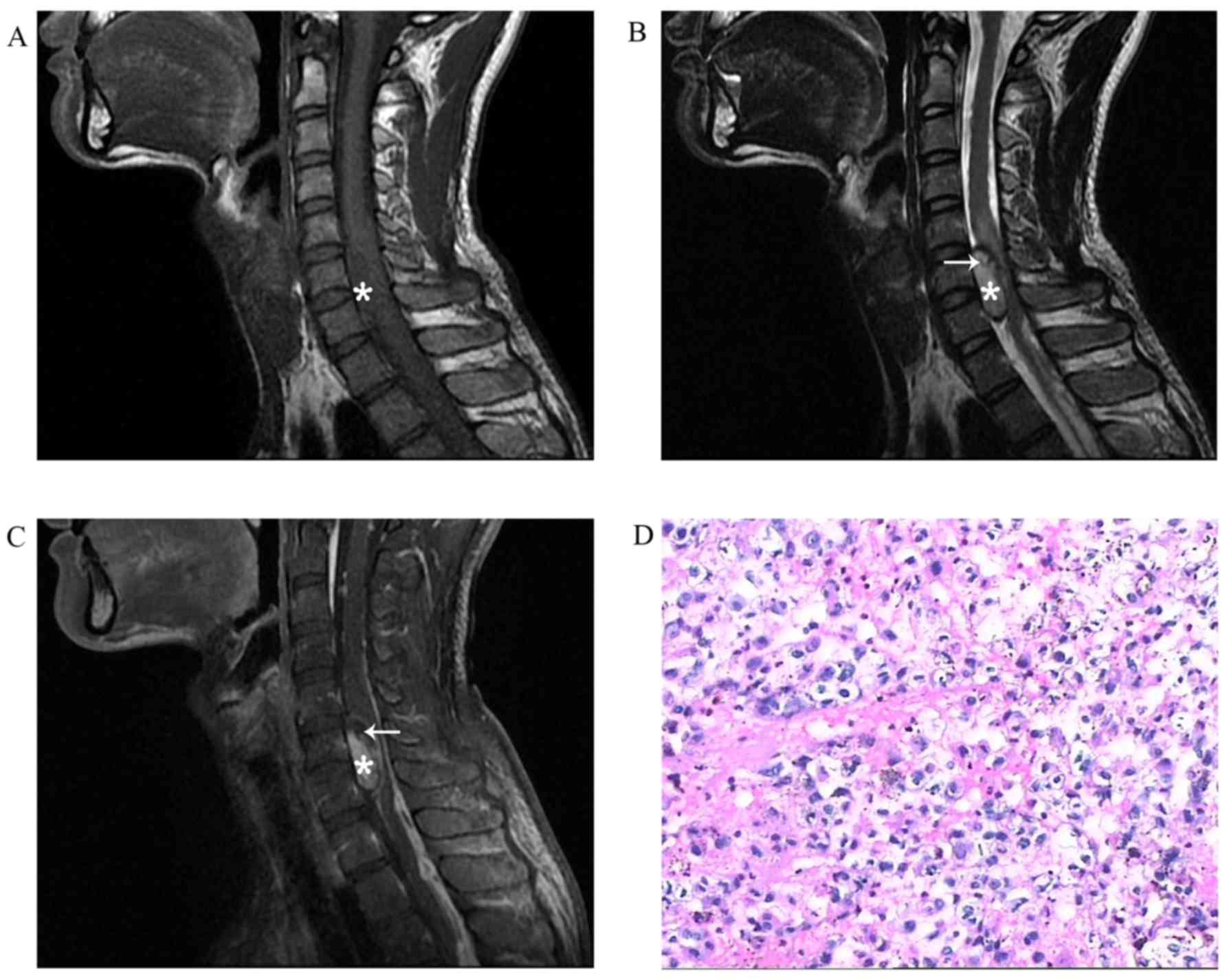
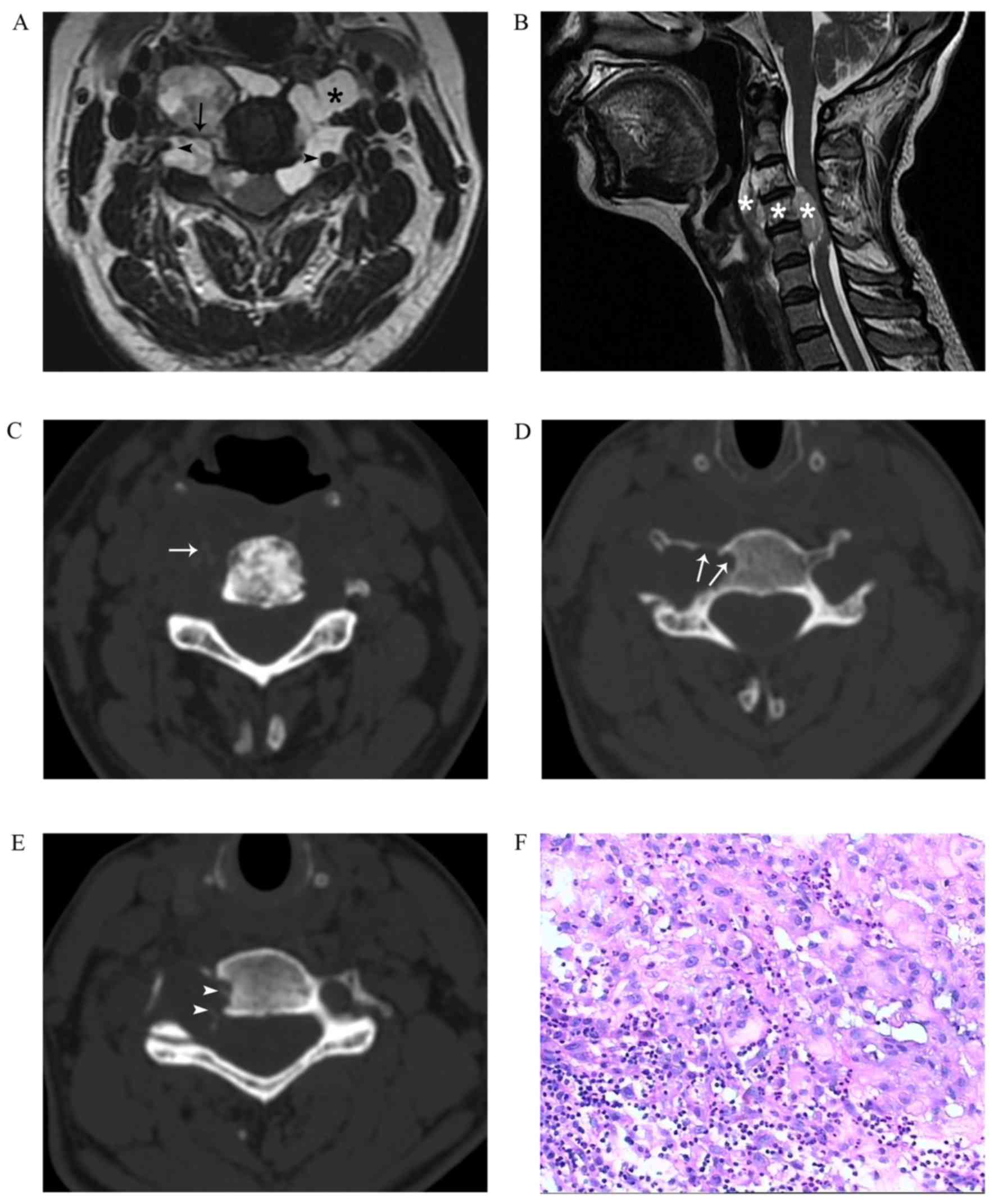
|
1
|
Christopher D, Fletcher JA and Krishnan U:
WHO classification of tumours of soft tissue and boneInternational
agency for research on cancer. 4th edition. WHO; Lyon: pp. 110–111.
2013
|
|
2
|
Wippold FJ II, Koeller KK and
Smirniotopoulos JG: Clinical and imaging features of cervical
chordoma. AJR Am J Roentgenol. 172:1423–1426. 1999. View Article : Google Scholar : PubMed/NCBI
|
|
3
|
Bjornsson J, Wold LE, Ebersold MJ and Laws
ER: Chordoma of the mobile spine. A clinicopathologic analysis of
40 patients. Cancer. 71:735–740. 1993. View Article : Google Scholar : PubMed/NCBI
|
|
4
|
Sundaresan N, Boriani S, Rothman A and
Holtzman R: Tumors of the osseous spine. J Neurooncol. 69:273–290.
2004. View Article : Google Scholar : PubMed/NCBI
|
|
5
|
Erdem E, Angtuaco EC, Van Hemert R, Park
JS and Al-Mefty O: Comprehensive review of intracranial chordoma.
Radiographics. 23:995–1009. 2003. View Article : Google Scholar : PubMed/NCBI
|
|
6
|
Yamaguchi T, Yamato M and Saotome K: First
histologically confirmed case of a classic chordoma arising in a
precursor benign notochordal lesion: Differential diagnosis of
benign and malignant notochordal lesions. Skeletal Radiol.
31:413–418. 2002. View Article : Google Scholar : PubMed/NCBI
|
|
7
|
Nishiguchi T, Mochizuki K, Tsujio T,
Nishita T and Inoue Y: Lumbar vertebral chordoma arising from an
intraosseous benign notochordal cell tumour: Radiological findings
and histopathological description with a good clinical outcome. Br
J Radiol. 83:e49–e53. 2010. View Article : Google Scholar : PubMed/NCBI
|
|
8
|
Deshpande V, Nielsen GP, Rosenthal DI and
Rosenberg AE: Intraosseous benign notochord cell tumors (BNCT):
Further evidence supporting a relationship to chordoma. Am J Surg
Pathol. 31:1573–1577. 2007. View Article : Google Scholar : PubMed/NCBI
|
|
9
|
Yamaguchi T, Watanabe-Ishiiwa H, Suzuki S,
Igarashi Y and Ueda Y: Incipient chordoma: A report of two cases of
early-stage chordoma arising from benign notochordal cell tumors.
Mod Pathol. 18:1005–1010. 2005. View Article : Google Scholar : PubMed/NCBI
|
|
10
|
D'Haen B, De Jaegere T, Goffin J, Dom R,
Demaerel P and Plets C: Chordoma of the lower cervical spine. Clin
Neurol Neurosurg. 97:245–248. 1995. View Article : Google Scholar : PubMed/NCBI
|
|
11
|
Ross JS and Moore KR: Diagnostic imaging:
spine. 3rd edition. Elsevier Health Sci; 2015
|
|
12
|
Murphey MD, Andrews CL, Flemming DJ,
Temple HT, Smith WS and Smirniotopoulos JG: From the archives of
the AFIP. Primary tumors of the spine: Radiologic pathologic
correlation. Radiographics. 16:1131–1158. 1996. View Article : Google Scholar : PubMed/NCBI
|
|
13
|
Darby AJ, Cassar-Pullicino VN, McCall IW
and Jaffray DC: Vertebral intra-osseous chordoma or giant
notochordal rest? Skeletal Radiol. 28:342–346. 1999. View Article : Google Scholar : PubMed/NCBI
|
|
14
|
Soo MY: Chordoma: Review of
clinicoradiological features and factors affecting survival.
Australas Radiol. 45:427–434. 2001. View Article : Google Scholar : PubMed/NCBI
|
|
15
|
Smolders D, Wang X, Drevelengas A,
Vanhoenacker F and De Schepper AM: Value of MRI in the diagnosis of
non-clival, non-sacral chordoma. Skeletal Radiol. 32:343–350. 2003.
View Article : Google Scholar : PubMed/NCBI
|
|
16
|
Vaz RM, Pereira JC, Ramos U and Cruz CR:
Intradural cervical chordoma without bone involvement. Case report.
J Neurosurg. 82:650–653. 1995. View Article : Google Scholar : PubMed/NCBI
|
|
17
|
Dow GR, Robson DK, Jaspan T and Punt JA:
Intradural cerebellar chordoma in a child: A case report and review
of the literature. Childs Nerv Syst. 19:188–191. 2003.PubMed/NCBI
|
|
18
|
Zhou H, Liu Z, Liu C, Ma Q, Liu X, Jiang L
and Wei F: Cervical chordoma in childhood without typical vertebral
bony destruction: Case report and review of the literature. Spine
(Phila Pa 1976). 34:E493–E497. 2009. View Article : Google Scholar : PubMed/NCBI
|
|
19
|
Firooznia H, Pinto RS, Lin JP, Baruch HH
and Zausner J: Chordoma: Radiologic evaluation of 20 cases. AJR Am
J Roentgenol. 127:797–805. 1976. View Article : Google Scholar : PubMed/NCBI
|
|
20
|
Mortelé B, Lemmerling M, Mortelé K,
Verstraete K, Defreyne L, Kunnen M and Vandekerckhove T: Cervical
chordoma with vertebral artery encasement mimicking neurofibroma:
MRI findings. Eur Radiol. 10:967–969. 2000. View Article : Google Scholar : PubMed/NCBI
|
|
21
|
Murphy JM, Wallis F, Toland J, Toner M and
Wilson GF: CT and MRI appearances of a thoracic chordoma. Eur
Radiol. 8:1677–1679. 1998. View Article : Google Scholar : PubMed/NCBI
|
|
22
|
Yamaguchi T, Suzuki S, Ishiiwa H, Shimizu
K and Ueda Y: Benign notochordal cell tumors: A comparative
histological study of benign notochordal cell tumors, classic
chordomas, and notochordal vestiges of fetal intervertebral discs.
Am J Surg Pathol. 28:756–761. 2004. View Article : Google Scholar : PubMed/NCBI
|
|
23
|
Yamaguchi T, Iwata J, Sugihara S, McCarthy
EF Jr, Karita M, Murakami H, Kawahara N, Tsuchiya H and Tomita K:
Distinguishing benign notochordal cell tumors from vertebral
chordoma. Skeletal Radiol. 37:291–299. 2008. View Article : Google Scholar : PubMed/NCBI
|
|
24
|
Nishiguchi T, Mochizuki K, Ohsawa M, Inoue
T, Kageyama K, Suzuki A, Takami T and Miki Y: Differentiating
benign notochordal cell tumors from chordomas: Radiographic
features on MRI, CT, and tomography. AJR Am J Roentgenol.
196:644–650. 2011. View Article : Google Scholar : PubMed/NCBI
|
|
25
|
Kyriakos M: Benign notochordal lesions of
the axial skeleton: A review and current appraisal. Skeletal
Radiol. 40:1141–1152. 2011. View Article : Google Scholar : PubMed/NCBI
|
|
26
|
Kreshak J, Larousserie F, Picci P, Boriani
S, Mirra J, Merlino B, Brunocilla E and Vanel D: Difficulty
distinguishing benign notochordal cell tumor from chordoma further
suggests a link between them. Cancer Imaging. 14:42014.PubMed/NCBI
|
|
27
|
Elefante A, Caranci F, Del Basso De Caro
ML, Peca C, Guadagno E, Severino R, Mariniello G and Maiuri F:
Paravertebral high cervical chordoma. A case report. Neuroradiol J.
26:227–232. 2013. View Article : Google Scholar : PubMed/NCBI
|
|
28
|
Karakida O, Aoki J, Seo GS, Ishii K, Sone
S, Nakakouji T and Otsuka K: Epidural dumbbell-shaped chordoma
mimicking a neurinoma. Pediatr Radiol. 26:62–64. 1996. View Article : Google Scholar : PubMed/NCBI
|