Introduction
Gallbladder cancer (GBC) has a high occurrence among
populations in the Andean area, Native Americans and Mexican
Americans (1). GBC is often diagnosed
at a late stage due to its unapparent symptoms at the early stage
(2). Surgery is currently the primary
option for GBC treatment alongside a combination of 5-fluorouracil
(5-FU) and cisplatin (CDDP), which is a common choice for advanced
GBC (3,4). Although chemotherapy exerts a
therapeutic effect in a number of patients, chemoresistance
eventually occurs in patients that receive chemotherapy (5).
CDDP is one of the most widely used cytotoxic
anticancer drugs (6–9). CDDP mainly reacts with the N7-position
of guanine, forming inter- and intra-strand DNA cross-links and
blocks replication and transcription, and may result in
replication-mediated double-strand breaks (DSBs) (10,11).
However, resistance to these drugs undermines their curative
potential. The resistance to CDDP and numerous other
chemotherapeutic agents is partially due to a wide range of genetic
and epigenetic alterations which result in abnormal cell survival
(12–14). In the present study, the expression of
a number of chemotherapy resistance-associated genes (DUSP1,
HIF-1α, MDR1, MRP1) was compared between CDDP-resistant SGC996 and
GBC-SD cells and normal SGC996 and GBC-SD cells. Notably, one gene
(dual-specificity phosphatase 1 (DUSP1)) expression was markedly
increased in the established CDDP-resistant cells compared with the
normal cells. Using an in vivo assay, DUSP1 expression in
subcutaneous tumors was also elevated following CDDP treatment.
DUSP1 is one member of the DUSP family, which
consists of a total of 25 members. The expression of DUSP1 is
cancer-dependent (15). In a range of
epithelial tumor types including pancreatic ductal adenocarcinoma
(PDAC), non-small-cell lung cancer, breast, ovarian, gastric and
early-stage prostate cancer, DUSP1 was revealed to be
overexpressed, however it was decreased in hepatocellular carcinoma
(16–19). The DUSP family are specific inhibitory
molecules which target mitogen-activated protein kinases (MAPKs)
(20). By inhibiting p38 and c-Jun
N-terminal kinase (JNK) activity, DUSP1 enhances resistance to
doxorubicin or paclitaxel in breast cancer, osteosarcoma and
non-small cell lung carcinoma cell lines (17,21–24).
However, there are few studies on the association between DUSP1
expression and chemoresistance in GBC.
The present study examined the expression of DUSP1
in two GBC cell lines: SGC996 and GBC-SD. DUSP1 expression was
revealed to be relatively low in GBC cells and was overexpressed in
the two cell lines. An MTS assay revealed that DUSP1 overexpressing
GBC cells had better survival and lower apoptosis following CDDP
treatment compared with untreated control cells. DUSP1
overexpression was verified to inhibit p38 MAPK activity and
decrease apoptosis compared with control cells. Further experiments
indicated that fewer DSB were formed in DUSP1 overexpressing cells
compared with control cells. Therefore, DUSP1 may be a potential
therapeutic target to enhance the efficiency of chemotherapy for
GBC.
Materials and methods
Cell culture
Human GBC cell line GBC-SD and SGC996 cells were
obtained from the Type Culture Collection of the Chinese Academy of
Sciences (Shanghai, China). GBC-SD and SGC996 were cultured in
RPMI-1640 (Gibco; Thermo Fisher Scientific, Inc, Waltham, MA, USA)
containing penicillin (100 IU/ml) and streptomycin (100 µg/ml),
supplemented with 10% fetal bovine serum which was diluted in PBS
(Gibco; Thermo Fisher Scientific, Inc.). All cell lines were
cultured in a 5% (v/v) CO2 humidified incubator at
37°C.
Construction of stable expression GBC
cell lines
DUSP1 expression plasmid was generated by cloning
DUSP1 cDNA into the basic retroviral transfer plasmid Pwpi
(Biovector Science Lab, Inc., Beijing, China) to generate the
plasmid pWPI-DUSP1. To generate DUSP1 overexpressing cells, GBC-SD
and SGC996 cells were transfected with lentiviral vectors
pWPI-DUSP1 or pWPI-Vec, the psAX2 packaging plasmid and the pMD2G
envelope plasmid were used to obtain the lentivirus at 37°C. This
was then collected and frozen at −80°C until use. The cells were
transfected using Lipofectamine® 2000 (Invitrogen;
Thermo Fisher Scientific, Inc.). Lentiviral supernatants were then
collected to infect GBC cells. Following viral infection, the media
was replaced with normal RPMI-1640 culture media. The stable cells
were selected and examined using reverse transcription-quantitative
polymerase chain reaction (RT-qPCR) and western blot analysis.
RT-qPCR
For RNA extraction, total RNA was isolated using
Trizol reagent (Invitrogen; Thermo Fisher Scientific, Inc.). A
total of 1–2 µg of total RNA was subject to RT using Superscript
III transcriptase (Invitrogen; Thermo Fisher Scientific, Inc.).
RT-qPCR was conducted using a Bio-Rad CFX96 system (Bio-Rad,
Laboratories, Inc., Hercules, CA, USA) with SYBR green to determine
the mRNA expression level of a gene of interest such as DUSP1,
HIF-1α, MDR1 and MRP1. Expression levels were normalized to the
expression of GAPDH mRNA. In brief, 50 ng small RNA was processed
for poly A addition by adding 1 unit of polymerase with 1 mM ATP in
1×RT buffer (Invitrogen; Thermo Fisher Scientific, Inc.) at 37°C
for 10 min in a 10 µl volume, and then heat inactivated at 95°C for
2 min, and then 50 pmol anchor primer (DUSP1, HIF-1α, MDR1 and
MRP1) were added to 12.5 µl volume total PCR mix, incubated at 65°C
for 5 min, and the last step was cDNA synthesis, with the addition
of 2 µl 5X RT buffer, 2 µl 10 mM dNTP and 1 µl reverse
transcriptase to a total volume of 20 µl and incubated at 42°C for
1 h 25 min. The sequences of the GAPDH primers are as follows:
Forward, 5′-GGAGTCAACGGATTTGGT-3′ and reverse,
5′-GTGATGGGATTTCCATTGAT-3′. The sequences of DUSP1 are as follows:
Forward, 5′-CCTGACAGCGCGGAATCT-3′ and reverse,
5′-GATTTCCACCGGGCCAC-3′. Analysis of relative gene expression was
quantified with the 2−ΔΔCq method (25).
Western blot analysis
Cells were lysed in RIPA buffer (Beyotime Institute
of Biotechnology, Haimen, China) and proteins [20-50 µg, determined
by a BCA Protein Assay kit (Thermo Fisher Scientific, Inc.)] were
separated on a 10% gel using SDS-PAGE and then transferred onto
polyvinylidene fluoride membranes (EMD Millipore, Billerica, MA,
USA). Membranes were blocked with 5% bovine serum albumin at room
temperature for 2 h and incubated with specific primary antibodies
at room temperature for 1 h (1:1,000 in 0.5% FBS) against GAPDH
(cat no. G8795; Sigma-Aldrich, Merck KGaA, Darmstadt, Germany),
Caspase-3 (active; cat no. 1476-1; Epitomics; Abcam, Cambridge,
UK), poly (ADP-ribose) polymerase (PARP; cat no. 9542; Cell
Signaling Technology, Inc., Danvers, MA, USA), phosphorylated-H2A
histone family (γH2AX; cat no. JBW301; EMD Millipore) and phospho
(p)-p38 (cat no. 4822; Abcam). The blots were incubated with
horseradish peroxidase-conjugated secondary antibodies at room
temperature for 1 h (including goat anti-rabbit IgG (cat no. 7054;
dilution, 1:10,000; Cell Signaling Technology, Inc., Danvers, MA,
USA) and goat anti-mouse IgG (cat no. 7056; dilution, 1:10,000;
Cell Signaling Technology, Inc.) and visualized using the
electrochemiluminesence system Image Lab V4.0 (Bio-Rad
Laboratories, Inc.).
MTS assay
Stable transfected cells (5×10³) were
seeded on a 96-well plate with 3 replicate wells. Following cell
adhesion, various concentrations of CDDP (Sigma-Aldrich, Merck
KGaA) diluted in DMSO (1, 1.5, 2, 2.5, 3, 3.5 µg/ml) were added.
Following incubation for 1 h at 37°C, cell viability was assessed
every 48 h utilizing the tetrazolium-based MTT colorimetric assay
(CellTiter 96 cell proliferation assay kit; Promega Corporation,
Madison, WI, USA) according to the manufacturer's protocol. All
experiments were performed at least in triplicate on three separate
occasions. A dose-response curve was plotted.
Apoptosis assay
Cell apoptosis was evaluated using a flow cytometry
assay. Briefly, (1×10⁵ SGC996 cells and 2×10⁴
GBC-SD cells) Vector and OE cells seeded in 6-well plates were
harvested and washed twice using phosphate buffered saline, stained
with propidium iodide (PI) in binding buffer (BD Biosciences, San
Jose, CA, USA) subsequent to 15 min incubation at room temperature
in darkness, and detected by Becton-Dickinson FACS Calibur FCM
using the software within the system (BD Biosciences).
Subcutaneous xenograft model
All experimental procedures were conducted in
accordance with the institutional guidelines for the care and use
of laboratory animals (26), and
ethical approval was provided by the Institutional Animal Care and
Use Committee of Zhejiang Medical College, Zhejiang University
(Zhejiang, China) for all animal experiments. Animals were
subjected to isoflurane anesthesia. Animal studies were conducted
using female 5-week-old nude mouse (20–30 g) from Silaike
Experimental Animal Co. Ltd. (Shanghai China). A total of 8 mice
were housed in a specific pathogen-free laboratory, airconditioned,
with a 12/12 h light/dark cycle. Subcutaneous implantation was
performed as previously described (27) where mice were injected subcutaneously
with DUSP1-Vector or DUSP1-OE SGC996 cells. A total of
1×10⁶ SGC996 stable cells (mixed with Matrigel in a
ratio of 1:1) were injected subcutaneously. After 1 week, mice were
randomized into two groups: Mice treated with CDDP solution at a
dose of 0.5 mg/kg in the treatment group (the CDDP(+) group) every
4 days for 3 weeks and the control group [CDDP(−) group treated
with DMSO]. The mice were sacrificed under the influence of
Fluothane on the 21st day before they lost 40% weight and tumor
tissue was used to extract total tissue protein. DUSP1 expression
was detected using western blot analysis.
Statistical analysis
Data are expressed as mean ± standard error of the
mean of at least 3 independent experiments. Statistical analyses
were performed using a paired Student's t-test using SPSS 17.0
(SPSS, Inc., Chicago, IL, USA). P<0.01 was considered to
indicate a statistically significant difference.
Results
DUSP1 expression was markedly
increased in the CDDP-resistant SGC996 cell lines and CDDP-treated
subcutaneous tumor types
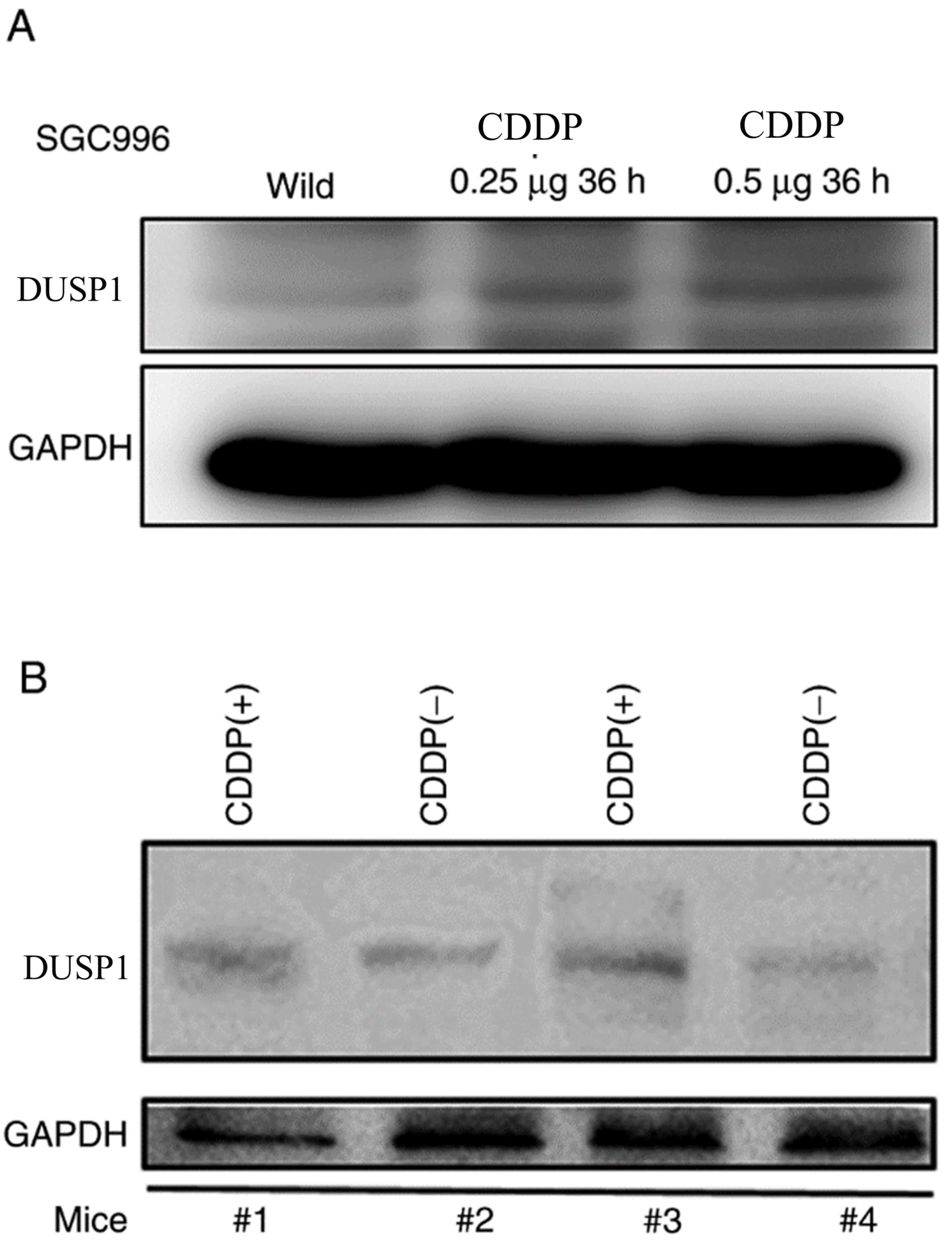
In order to explore the genes involved in GBC
chemoresistance, a CDDP-resistant SGC996 cell line was established.
Genes such as BCRP, MRP1, MDR1 and HIF1 were previously reported to
be associated with drug-resistance in various cancer types were
detected (28–30). DUSP1 expression was substantially
elevated in the CDDP-resistant SGC996 cells compared with the
normal SGC996 cells (Fig. 1A). DUSP1
was reported to be involved in gemcitabine-resistance in PDAC
(31). However, the association
between the induction of DUSP1 and CDDP-resistance in GBC remains
unclear. In order to further confirm the DUSP1 induction resulting
from CDDP treatment in vivo, subcutaneous xenograft GBC
mouse models were generated by transplanting normal SGC996 cells
into nude mice. The nude mice were divided into two groups: A
control tumor group and a CDDP-treated group. As presented in
Fig. 1B, DUSP1 expression was
increased in the CDDP-treated group compared with the control
group.
Altogether, the results presented in Fig. 1A and B reveal that DUSP1 expression is
higher in CDDP-resistant cell lines and CDDP-treated tumors,
suggesting that DUSP1 expression may be associated with
CDDP-resistance.
DUSP1 overexpression resulted in
enhanced chemoresistance in SGC996 and GBC-SD cells
To further confirm that DUSP1 expression is
associated with CDDP-resistance, DUSP1 was overexpressed using the
addition of DUSP1-cDNA (Fig. 2A;
protein level) in SGC996 and GBC-SD cells, and then these cells
were treated with CDDP or 5-FU. An MTT assay was applied to analyze
the survival rate of these cells. Furthermore, single staining for
PI was performed to evaluate the cytotoxicity of these cells
subsequent to CDDP treatment. SGC996 cells with DUSP1
overexpression (SGC996 OE) were less sensitive to CDDP and 5-FU
treatment compared with the control cells (SGC996-vector) (Fig. 2A). Fewer PI positive cells were
observed in SGC996 OE cells compared with the control cells, and
SGC996 OE cells presented a significantly higher cell viability
compared with the control cells (P<0.05) which indicated a lower
apoptosis rate (Fig. 2B). Similarly,
GBC-SD DUSP1 overexpressing (GBC-SD OE) cells were less sensitive
to CDDP and 5-FU treatment compared with the control cells
(Fig. 2C). Additionally, there was a
significantly higher cell viability of GBC-SD OE cells compared
with the control cells (P<0.01; Fig.
2D). Altogether, the evidence indicates that DUSP1 enhances
chemoresistance in GBC.
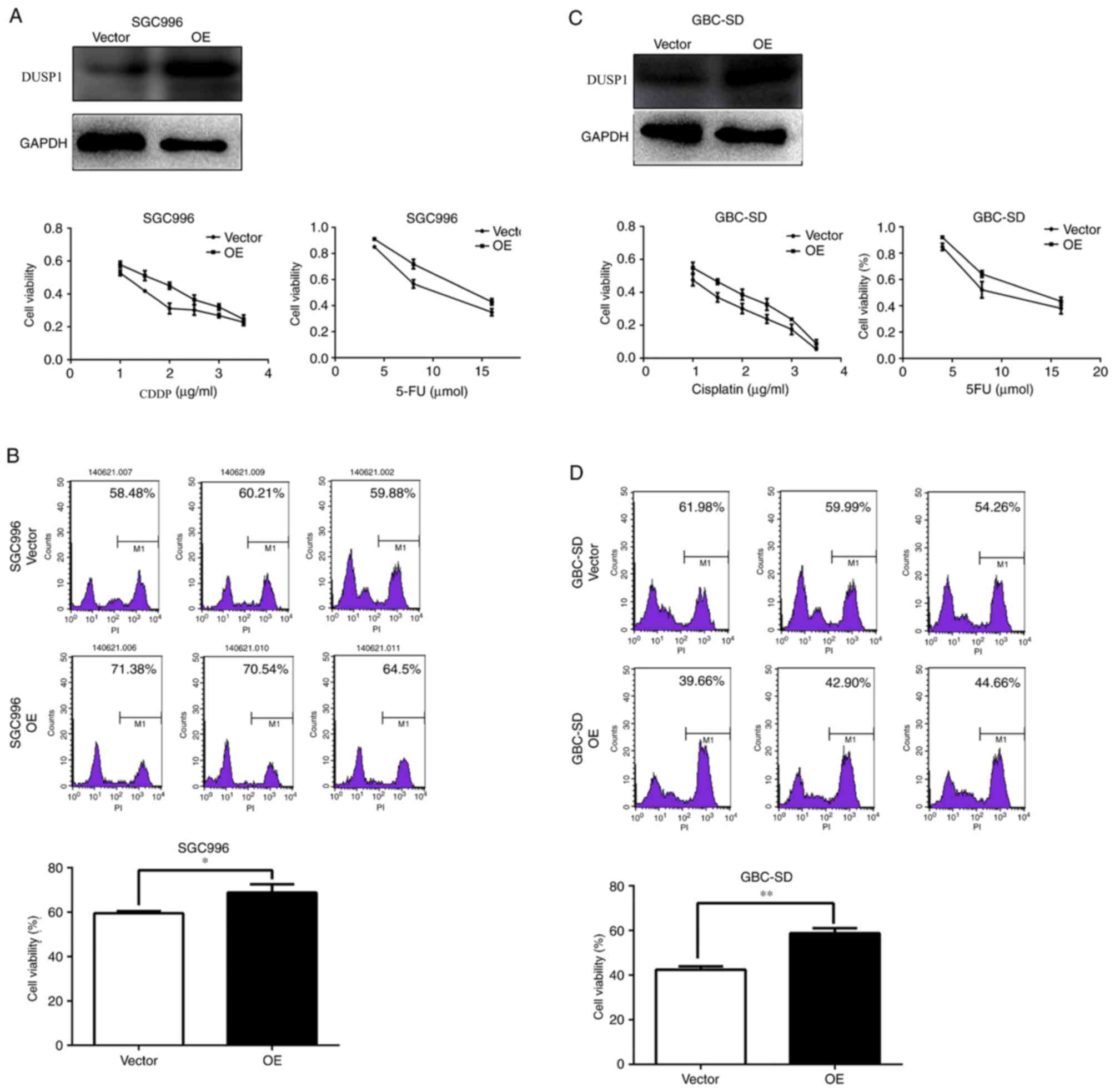 | Figure 2.DUSP1 may decrease CDDP and 5-FU
chemotherapy efficiency in GBC cells. (A) SGC996 cells that were
DUSP1 OE or normal (vector) were seeded in 96-well plates, and
treated with different concentrations of CDDP (1, 1.5, 2, 2.5, 3
and 3.5 µg/ml) or 5-FU (4, 8 and 16 µmol) for 48 h, and then MTT
assays for cell viability were performed. (B) SGC996 cells that
were DUSP1 OE or normal (vector) were seeded in 6-well plates,
treated with CDDP for 48 h, harvested and washed twice with PBS,
stained with PI in the binding buffer, and detected using flow
cytometry. (C) GBC-SD cells that were DUSP1 OE or normal (vector)
were seeded in 96-well plates, and treated with different
concentrations of CDDP (1, 1.5, 2, 2.5, 3 and 3.5 µg/ml) or 5-FU
(4, 8 and 16 µmol) for 48 h, and then MTT cell viability assays
were performed. (D) GBC-SD cells that were DUSP1 OE or normal
(vector) were seeded in 6-well plates, treated with CDDP for 48 h,
harvested and washed twice with PBS, stained with PI in the binding
buffer, and detected using flow cytometry. *P<0.05 and
**P<0.01 with comparisons shown by lines. DUSP, dual-specificity
phosphatase; CDDP, cisplatin; GBC, gallbladder cancer; OE,
overexpression; 5-FU, fluorouracil; PI, propidium iodide. |
DUSP1 overexpression resulted in
decreased p38 MAPK apoptosis and decreased DNA damage
DNA damage is a major factor resulting in normal
cell death and the DNA damage response is crucial for cell
survival. Once DNA damage occurs, the foci of phosphorylated H2AX
(γH2AX) will be rapidly formed in order to recruit repair factors
(32,33). Thus, γH2AX staining usually serves as
a marker for DNA damage in previous studies (34). DNA damage in SGC996-OE cells was
detected to be significantly decreased by immunofluorescent
staining compared with SGC996-Vector cells (P<0.01; Fig. 3A). This was further verified using a
western blot assay (Fig. 3B).
CDDP-induced p-p38 MAPK additionally decreased in GBC-SD-OE cells
compared with the control cells, which resulted in reduced
CDDP-induced apoptosis, evidenced by the relatively decreased
expression of cleaved PARP and cleaved caspase 3 in GBC-SD-OE cells
compared with the control cells (Fig.
3B).
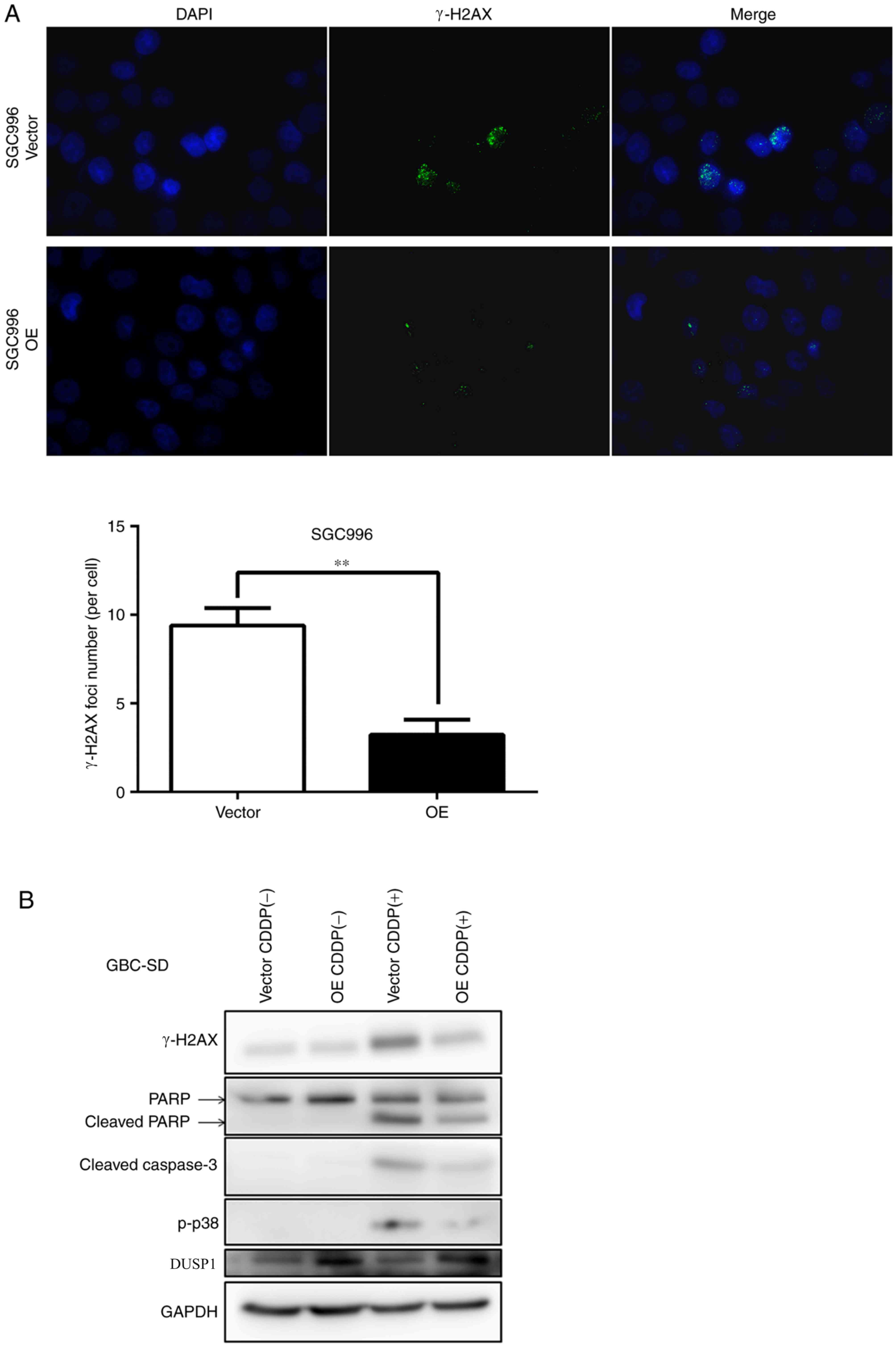 | Figure 3.DUSP1 decreases p-p38 and γH2AX
protein expression levels. Cells were treated with 4 µg/ml CDDP for
24 h. (A) Immunocytochemical detection of γH2AX-foci revealed a
significant decrease DNA double-strand breaks in SGC996 OE cells
compared with the control cells (vector). (B) Western blots
revealed that CDDP treatment increased cleaved PARP, cleaved
caspase 3, p-p38 and γH2AX in GBC-SD cells, and that in comparison
an overexpression of DUSP1 decreased the levels of these proteins.
**P<0.01 with comparisons shown by lines. OE, overexpression;
γH2AX, phosphorylated-H2A histone family, member X; CDDP,
cisplatin; CDDP(−), non-cisplatin treatment; CDDP(+), cisplatin
treatment; PARP, poly (ADP-ribose) polymerase; p-, phosphorylated;
DUSP, dual-specificity phosphatase. |
Discussion
DUSP1 is induced in response to oxidative stress,
hypoxia, and a number of other factors including nutritional
deprivation by the regulation of tumor protein p53, transcription
factor E2F1, c-Jun and activating transcription factor 2 (15,35,36). An
increased expression of DUSP1 was detected in PDAC following
gemcitabine treatment and served a function in the chemoresistance
of gemcitabine (31). Similarly, the
elevated expression of DUSP1 in CDDP-resistant GBC cells was
detected in the present study. CDDP, as a normal chemotherapy drug,
triggers DNA damage response and p38 MAPK activation, resulting in
cell death (13,37). A number of studies have indicated that
CDDP activates p38 MAPK, thereby inducing apoptosis in cells and
that the inhibition of p38 MAPK activation may be associated with
CDDP-resistance in ovarian cancer (38–40).
Additionally, CDDP inducing DNA cross links and DSBs contributes to
cancer treatment (41,42). However, the effectiveness of CDDP in
various cancer types such as pancreatic, breast and lung cancer
(43–45) is hampered by the development of drug
resistance over time.
In the present study, DUSP1 additionally enhanced
chemoresistance in GBC. Overexpressing DUSP1 may attenuate the
activation of p38 MAPK, thereby resulting in a lower apoptotic rate
as evidenced by the decreased cleaved PARP expression and activated
caspase 3 protein expression. Furthermore, fewer γH2AX foci were
formed in the SGC996-OE cells compared with the control cells. In
this sense, DUSP1 may serve a function in reducing DNA damage and
protecting GBC from cell death. Targeting DUSP1 may improve the
efficiency of chemotherapy in GBC.
To conclude, the results of the present study
demonstrated that DUSP1 may function through the downregulation of
p38 MAPK and DNA damage to influence the efficiency of GBC
chemotherapy. Previous finding have revealed that DUSP1 may
additionally function through JNK-MAPK signaling to reduce the
cytotoxicity caused by gemcitabine in pancreatic cancer (31). However, gemcitabine is widely applied
in GBC treatment (46). Novel small
molecules may be developed in the near future that target DUSP1 in
order to suppress GBC progression more effectively.
Acknowledgements
Not applicable.
Funding
No funding was received.
Availability of data and materials
The data generated in the present study are
available on reasonable request from the corresponding author.
Authors' contributions
JF and YW contributed to the research design and
operation, data analysis and manuscript preparation. The other
authors contributed to the research. ZY, FG, MY and QL contributed
to the cell research. JL, ZW and YX contributed to the animal
research.
Ethics approval and consent to
participate
Ethical approval was provided by the Institutional
Animal Care and Use Committee of Zhejiang Medical College, Zhejiang
University (Zhejiang, China) for all animal experiments.
Consent for publication
Not applicable.
Competing interests
The authors declare that they have no competing
interests.
Glossary
Abbreviations
Abbreviations:
|
GBC
|
gallbladder cancer
|
|
DUSP1
|
dual-specificity phosphatase 1
|
|
CDDP
|
cisplatin
|
|
DSB
|
double-strand breaks
|
References
|
1
|
Lazcano-Ponce EC, Miquel JF, Muñoz N,
Herrero R, Ferrecio C, Wistuba II, Alonso de Ruiz P, Aristi Urista
G and Nervi F: Epidemiology and molecular pathology of gallbladder
cancer. CA Cancer J Clin. 51:349–364. 2001. View Article : Google Scholar : PubMed/NCBI
|
|
2
|
Gourgiotis S, Kocher HM, Solaini L,
Yarollahi A, Tsiambas E and Salemis NS: Gallbladder cancer. Am J
Surg. 196:252–264. 2008. View Article : Google Scholar : PubMed/NCBI
|
|
3
|
Malik IA, Aziz Z, Zaidi SH and Sethuraman
G: Gemcitabine and Cisplatin is a highly effective combination
chemotherapy in patients with advanced cancer of the gallbladder.
Am J Clin Oncol. 26:174–177. 2003. View Article : Google Scholar : PubMed/NCBI
|
|
4
|
Chatni SS, Sainani RS, Mehta SA and
Mohandas KM: Infusion chemotherapy with cisplatinum and
fluorouracil in the treatment of locally-advanced and metastatic
gallbladder cancer. J Cancer Res Ther. 4:151–155. 2008. View Article : Google Scholar : PubMed/NCBI
|
|
5
|
Reddy N and Czuczman MS: Enhancing
activity and overcoming chemoresistance in hematologic malignancies
with bortezomib: Preclinical mechanistic studies. Ann Oncol.
21:1756–1764. 2010. View Article : Google Scholar : PubMed/NCBI
|
|
6
|
Peiffert D, Seitz JF, Rougier P, François
E, Cvitkovic F, Mirabel X, Nasca S, Ducreux M, Hannoun-Levi JM,
Lusinchi A, et al: Preliminary results of a phase II study of
high-dose radiation therapy and neoadjuvant plus concomitant
5-fluorouracil with CDDP chemotherapy for patients with anal canal
cancer: A French cooperative study. Ann Oncol. 8:575–581. 1997.
View Article : Google Scholar : PubMed/NCBI
|
|
7
|
Doval DC, Sekhon JS, Gupta SK, Fuloria J,
Shukla VK, Gupta S and Awasthy BS: A phase II study of gemcitabine
and cisplatin in chemotherapy-naive, unresectable gall bladder
cancer. Br J Cancer. 90:1516–1520. 2004. View Article : Google Scholar : PubMed/NCBI
|
|
8
|
Bleiberg H, Conroy T, Paillot B, Lacave
AJ, Blijham G, Jacob JH, Bedenne L, Namer M, de Besi P, Gay F, et
al: Randomised phase II study of cisplatin and 5-fluorouracil
(5-FU) versus cisplatin alone in advanced squamous cell oesophageal
cancer. Eur J Cancer. 33:1216–1220. 1997. View Article : Google Scholar : PubMed/NCBI
|
|
9
|
Sohn BS, Yuh YJ, Kim KH, Jeon TJ, Kim NS
and Kim SR: Phase II trial of combination chemotherapy with
gemcitabine, 5-fluorouracil and cisplatin for advanced cancers of
the bile duct, gallbladder, and ampulla of Vater. Tumori.
99:139–144. 2013. View Article : Google Scholar : PubMed/NCBI
|
|
10
|
Roos WP and Kaina B: DNA damage-induced
cell death: From specific DNA lesions to the DNA damage response
and apoptosis. Cancer Lett. 332:237–248. 2013. View Article : Google Scholar : PubMed/NCBI
|
|
11
|
Roos WP and Kaina B: DNA damage-induced
cell death by apoptosis. Trends Mol Med. 12:440–450. 2006.
View Article : Google Scholar : PubMed/NCBI
|
|
12
|
Jordan P and Carmo-Fonseca M: Molecular
mechanisms involved in cisplatin cytotoxicity. Cell Mol Life Sci.
57:1229–1235. 2000. View Article : Google Scholar : PubMed/NCBI
|
|
13
|
Kartalou M and Essigmann JM: Mechanisms of
resistance to cisplatin. Mutat Res. 478:23–43. 2001. View Article : Google Scholar : PubMed/NCBI
|
|
14
|
Gosland M, Lum B, Schimmelpfennig J, Baker
J and Doukas M: Insights into mechanisms of cisplatin resistance
and potential for its clinical reversal. Pharmacotherapy. 16:16–39.
1996.PubMed/NCBI
|
|
15
|
Liu YX, Wang J, Guo J, Wu J, Lieberman HB
and Yin Y: DUSP1 is controlled by p53 during the cellular response
to oxidative stress. Mol Cancer Res. 6:624–633. 2008. View Article : Google Scholar : PubMed/NCBI
|
|
16
|
Bang YJ, Kwon JH, Kang SH, Kim JW and Yang
YC: Increased MAPK activity and MKP-1 overexpression in human
gastric adenocarcinoma. Biochem Biophys Res Commun. 250:43–47.
1998. View Article : Google Scholar : PubMed/NCBI
|
|
17
|
Wang HY, Cheng Z and Malbon CC:
Overexpression of mitogen-activated protein kinase phosphatases
MKP1, MKP2 in human breast cancer. Cancer Lett. 191:229–237. 2003.
View Article : Google Scholar : PubMed/NCBI
|
|
18
|
Vicent S, Garayoa M, López-Picazo JM,
Lozano MD, Toledo G, Thunnissen FB, Manzano RG and Montuenga LM:
Mitogen-activated protein kinase phosphatase-1 is overexpressed in
non-small cell lung cancer and is an independent predictor of
outcome in patients. Clin Cancer Res. 10:3639–3649. 2004.
View Article : Google Scholar : PubMed/NCBI
|
|
19
|
Denkert C, Schmitt WD, Berger S, Reles A,
Pest S, Siegert A, Lichtenegger W, Dietel M and Hauptmann S:
Expression of mitogen-activated protein kinase phosphatase-1
(MKP-1) in primary human ovarian carcinoma. Int J Cancer.
102:507–513. 2002. View Article : Google Scholar : PubMed/NCBI
|
|
20
|
Liu C, Shi Y, Du Y, Ning X, Liu N, Huang
D, Liang J, Xue Y and Fan D: Dual-specificity phosphatase DUSP1
protects overactivation of hypoxia-inducible factor 1 through
inactivating ERK MAPK. Exp Cell Res. 309:410–418. 2005. View Article : Google Scholar : PubMed/NCBI
|
|
21
|
Cortes-Sempere M, Chattopadhyay S, Rovira
A, Rodriguez-Fanjul V, Belda-Iniesta C, Tapia M, Cejas P,
Machado-Pinilla R, Manguan-García C, Sánchez-Pérez I, et al: MKP1
repression is required for the chemosensitizing effects of
NF-kappaB and PI3K inhibitors to cisplatin in non-small cell lung
cancer. Cancer Lett. 286:206–216. 2009. View Article : Google Scholar : PubMed/NCBI
|
|
22
|
Small GW, Shi YY, Higgins LS and Orlowski
RZ: Mitogen-activated protein kinase phosphatase-1 is a mediator of
breast cancer chemoresistance. Cancer Res. 67:4459–4466. 2007.
View Article : Google Scholar : PubMed/NCBI
|
|
23
|
Sánchez-Pérez I, Martínez-Gomariz M,
Williams D, Keyse SM and Perona R: CL100/MKP-1 modulates JNK
activation and apoptosis in response to cisplatin. Oncogene.
19:5142–5152. 2000. View Article : Google Scholar : PubMed/NCBI
|
|
24
|
Wang Z, Zhou JY, Kanakapalli D, Buck S, Wu
GS and Ravindranath Y: High level of mitogen-activated protein
kinase phosphatase-1 expression is associated with cisplatin
resistance in osteosarcoma. Pediatr Blood Cancer. 51:754–759. 2008.
View Article : Google Scholar : PubMed/NCBI
|
|
25
|
Livak KJ and Schmittgen TD: Analysis of
relative gene expression data using real-time quantitative PCR and
the 2(-Delta Delta C(T)) method. Methods. 25:402–408. 2001.
View Article : Google Scholar : PubMed/NCBI
|
|
26
|
Norton JN, Reynolds RP, Chan C, Valdivia
RH and Staats HF: Assessing the satisfaction and burden within an
academic animal care and use program. FASEB J. 31:3913–3921. 2017.
View Article : Google Scholar : PubMed/NCBI
|
|
27
|
Lin SJ, Lee SO, Lee YF, Miyamoto H, Yang
DR, Li G and Chang C: TR4 nuclear receptor functions as a tumor
suppressor for prostate tumorigenesis via modulation of DNA
damage/repair system. Carcinogenesis. 35:1399–1406. 2014.
View Article : Google Scholar : PubMed/NCBI
|
|
28
|
Cai BL, Xu XF, Fu SM, Shen LL, Zhang J,
Guan SM and Wu JZ: Nuclear translocation of MRP1 contributes to
multidrug resistance of mucoepidermoid carcinoma. Oral Oncol.
47:1134–1140. 2011. View Article : Google Scholar : PubMed/NCBI
|
|
29
|
Stein U, Lage H, Jordan A, Walther W,
Bates SE, Litman T, Hohenberger P and Dietel M: Impact of BCRP/MXR,
MRP1 and MDR1/P-Glycoprotein on thermoresistant variants of
atypical and classical multidrug resistant cancer cells. Int J
Cancer. 97:751–760. 2002. View Article : Google Scholar : PubMed/NCBI
|
|
30
|
Tong Y, Li QG, Xing TY, Zhang M, Zhang JJ
and Xia Q: HIF1 regulates WSB-1 expression to promote
hypoxia-induced chemoresistance in hepatocellular carcinoma cells.
FEBS Lett. 587:2530–2535. 2013. View Article : Google Scholar : PubMed/NCBI
|
|
31
|
Liu F, Gore AJ, Wilson JL and Korc M:
DUSP1 is a novel target for enhancing pancreatic cancer cell
sensitivity to gemcitabine. PLoS One. 9:e849822014. View Article : Google Scholar : PubMed/NCBI
|
|
32
|
Redon C, Pilch D, Rogakou E, Sedelnikova
O, Newrock K and Bonner W: Histone H2A variants H2AX and H2AZ. Curr
Opin Genet Dev. 12:162–169. 2002. View Article : Google Scholar : PubMed/NCBI
|
|
33
|
Celeste A, Petersen S, Romanienko PJ,
Fernandez-Capetillo O, Chen HT, Sedelnikova OA, Reina-San-Martin B,
Coppola V, Meffre E, Difilippantonio MJ, et al: Genomic instability
in mice lacking histone H2AX. Science. 296:922–927. 2002.
View Article : Google Scholar : PubMed/NCBI
|
|
34
|
Svetlova MP, Solovjeva LV and Tomilin NV:
Mechanism of elimination of phosphorylated histone H2AX from
chromatin after repair of DNA double-strand breaks. Mutat Res.
685:54–60. 2010. View Article : Google Scholar : PubMed/NCBI
|
|
35
|
Wang J, Yin DP, Liu YX, Baer R and Yin Y:
Dual specificity phosphatase 1/CL100 is a direct transcriptional
target of E2F-1 in the apoptotic response to oxidative stress.
Cancer Res. 67:6737–6744. 2007. View Article : Google Scholar : PubMed/NCBI
|
|
36
|
Breitwieser W, Lyons S, Flenniken AM,
Ashton G, Bruder G, Willington M, Lacaud G, Kouskoff V and Jones N:
Feedback regulation of p38 activity via ATF2 is essential for
survival of embryonic liver cells. Genes Dev. 21:2069–2082. 2007.
View Article : Google Scholar : PubMed/NCBI
|
|
37
|
Hernández Losa J, Parada Cobo C, Guinea
Viniegra J, Sánchez-Arevalo Lobo VJ, Ramón y Cajal S and
Sánchez-Prieto R: Role of the p38 MAPK pathway in cisplatin-based
therapy. Oncogene. 22:3998–4006. 2003. View Article : Google Scholar : PubMed/NCBI
|
|
38
|
Weir NM, Selvendiran K, Kutala VK, Tong L,
Vishwanath S, Rajaram M, Tridandapani S, Anant S and Kuppusamy P:
Curcumin induces G2/M arrest and apoptosis in cisplatin-resistant
human ovarian cancer cells by modulating Akt and p38 MAPK. Cancer
Biol Ther. 6:178–184. 2007. View Article : Google Scholar : PubMed/NCBI
|
|
39
|
Mansouri A, Ridgway LD, Korapati AL, Zhang
Q, Tian L, Wang Y, Siddik ZH, Mills GB and Claret FX: Sustained
activation of JNK/p38 MAPK pathways in response to cisplatin leads
to Fas ligand induction and cell death in ovarian carcinoma cells.
J Biol Chem. 278:19245–19256. 2003. View Article : Google Scholar : PubMed/NCBI
|
|
40
|
Yuan ZQ, Feldman RI, Sussman GE, Coppola
D, Nicosia SV and Cheng JQ: AKT2 inhibition of cisplatin-induced
JNK/p38 and Bax activation by phosphorylation of ASK1: Implication
of AKT2 in chemoresistance. J Biol Chem. 278:23432–23440. 2003.
View Article : Google Scholar : PubMed/NCBI
|
|
41
|
Jamieson ER and Lippard SJ: Structure,
recognition, and processing of cisplatin-DNA adducts. Chem Rev.
99:2467–2498. 1999. View Article : Google Scholar : PubMed/NCBI
|
|
42
|
Gong JG, Costanzo A, Yang HQ, Melino G,
Kaelin WG Jr, Levrero M and Wang JY: The tyrosine kinase c-Abl
regulates p73 in apoptotic response to cisplatin-induced DNA
damage. Nature. 399:806–809. 1999. View
Article : Google Scholar : PubMed/NCBI
|
|
43
|
Danilov AV, Neupane D, Nagaraja AS,
Feofanova EV, Humphries LA, DiRenzo J and Korc M:
DeltaNp63alpha-mediated induction of epidermal growth factor
receptor promotes pancreatic cancer cell growth and
chemoresistance. PLoS One. 6:e268152011. View Article : Google Scholar : PubMed/NCBI
|
|
44
|
Lapensee EW, Tuttle TR, Fox SR and
Ben-Jonathan N: Bisphenol A at low nanomolar doses confers
chemoresistance in estrogen receptor-alpha-positive and negative
breast cancer cells. Environ Health Perspect. 117:175–180. 2009.
View Article : Google Scholar : PubMed/NCBI
|
|
45
|
Sève P and Dumontet C: Chemoresistance in
non-small cell lung cancer. Curr Med Chem Anticancer Agents.
5:73–88. 2005. View Article : Google Scholar : PubMed/NCBI
|
|
46
|
Nakamura M, Nakashima H, Abe T, Ensako T,
Yoshida K and Hino K: Gemcitabine-based adjuvant chemotherapy for
patients with advanced gallbladder cancer. Anticancer Res.
34:3125–3129. 2014.PubMed/NCBI
|