Introduction
Gastric cancer (GC) is the most prevalent digestive
system carcinoma (1). In 2012, it was
diagnosed in 950,000 patients and caused 723,000 mortalities
(1). Its malignance is due to its
capacity for rapid proliferation and resistance to
chemoradiotherapy. While the biological events and key signaling
pathway disruptions that drive its growth are being studied at
present, the underlying mechanisms of resistance to conventional
DNA-damaging agents and ionizing radiation (IR) remain largely
unknown.
Histidine triad nucleotide binding protein 1 (Hint1)
is a haploinsufficient tumor suppressor gene which contributes to
intercellular communication, helps to regulate cell proliferation
and survival, and is frequently underexpressed in the early stages
of oncogenesis (2–5). Hint1 knockout mice demonstrated a marked
increase in susceptibility to colorectal, mammary and ovarian
tumors (2,3). In 2011, our team first reported that
Hint1 was underexpressed in human GC tissues at the protein and
gene level. Its downregulation was associated with poor tumor cell
differentiation and bacterial or viral infection, including
infection by Helicobacter pylori or the Epstein-Barr virus,
suggesting patients with Hint1 underexpression may present with
biologically aggressive tumors and poor prognosis (6).
The tumor suppressive effects of Hint1 protein
primarily serve an inhibitory function in a number of gene
transcription control pathways. For instance, Hint1 promotes
apoptosis via upregulation of p53 and downregulation of B cell
lymphoma-2 in the SW480 human colon cancer cell line and the MCF7
breast cancer cell line (7). Upon
association with the plenty of SH3 domains protein and mitogen
activated protein kinase 9 complex, Hint1 inhibits activity of the
activator protein-1 transcription factor responsible for the
proliferation and angiogenesis of colon cancer cells (8). In addition, Hint1 enhances cellular
responses to DNA damage by regulating the functions of γ-H2A
histone family member X and ATM serine/threonine kinase (ATM) in
normal cells (9). However, little is
known regarding the effect of Hint1 on radiotherapy in cancer, even
though a relationship between Hint1 and DNA damage repair was
reported previously (9).
The present study analyzed the tumor suppressive
effects of Hint1 in the SGC7901 gastric cancer cell line. Its
inhibition of cell viability in this cell line was demonstrated,
and the involved signaling cascades were investigated. Hint1 may
negatively regulate extracellular signal-regulated kinase (ERK),
which is involved in gastric carcinogenesis. In addition, Hint1
prevented IR-induced DNA damage repair in SGC7901 cells via the
repression of Cyclin D1-dependent retinoblastoma protein (Rb
protein) phosphorylation, which induced G1 arrest and cell
death.
Materials and methods
Cell culture and treatment
The SGC7901 and AGS human gastric cancer cell lines
were obtained from the Cell Bank of Type Culture Collection of the
Chinese Academy of Sciences (Shanghai, China). Cells were
maintained in RPMI-1640 medium (Invitrogen; Thermo Fisher
Scientific, Inc., Waltham, MA, USA), supplemented with 10% fetal
bovine serum (Gibco; Thermo Fisher Scientific, Inc.), and incubated
in a 100% moist incubator with 5% CO2 at 37°C. A Varian
medical linear accelerator (Varian Medical Systems, Palo Alto, CA,
USA), offered by the Department of Oncology, Nanjing First Hospital
(Nanjing, China) was used to treat the cells. Gastric cancer cells
were plated at a density of 1×10⁴ cells/well into
96-well plates, and were incubated overnight. Then, cells were
treated with 0, 1, 2, 4 or 6 Gy X-ray or 50 µM PD98059 for 24 h at
37°C (Sigma-Aldrich; Merck KGaA, Darmstadt, Germany), a specific
ERK inhibitor (ERKi), for further experiments.
RNA interference
RNA interference was used to selectively knock down
Hint1 in SGC7901 cells. The sequence of pGPU6/green fluorescent
protein/Neo-short hairpin RNA (shRNA)-Hint1 (Sangon Biotech Co.,
Ltd., Shanghai, China) was
5′-CCGGCGACACGATCTTTGGGAAGATCTCGAGATCTTCCCAAAGATCGTGTCGTTTTTG-3′.
Cells were transfected with the aforementioned shRNA using
Lipofectamine 2000 (Invitrogen; Thermo Fisher Scientific, Inc.) for
24–72 h. Then cells were re-seeded in RPMI-1640 medium containing
G418 (400 µg/ml; Gibco; Thermo Fisher Scientific, Inc.) to enrich
the culture for cells that were successfully transfected. Following
120 h transfection, the cells were harvested to determine knockdown
efficiency by reverse transcription-quantitative polymerase chain
reaction (RT-qPCR) and western blot analysis. A non-targeting shRNA
vector (cat. no. E-07/F-07; Shanghai GenePharma Co., Ltd.,
Shanghai, China.) was used as a negative control for all
experiments.
Colony survival assay
A colony survival assay was performed to determine
the influence of Hint1 on SGC7901 cell proliferation. Exponentially
growing cells were seeded at a low density in a 6-well plate (80
cells/well plate; Corning Incorporated, Corning, NY, USA) and
allowed to grow for 7–10 days in RPMI-1640 medium. The media was
then removed and replaced with 0.1% crystal violet dye. The size of
live colonies which contained >50 cells was evaluated using a
fluorescence microscope (Olympus IX71; Olympus Corporation, Tokyo,
Japan) at magnification, −200. The number of colonies were then
counted and the proliferation ratio was calculated as the ratio of
the number of colonies in Hint1 silenced cells formed to those
formed by the vector control group.
MTT assay
SGC7901 and AGS cells (1×10⁴) were seeded
in 96-well plates and then treated with 0, 1, 2, 4 or 6 Gy X-ray or
PD98059 (50 µm). MTT reagent (10 µl/well; Trivegen, Gaithersburg,
MD, USA) was added and cells were left inside the incubator for a
further 4 h at 37°C, followed by the addition of 100 µl detergent
reagent (Trivegen) according to the manufacturer's protocol.
Absorbance of the colored solution was measured by a fully
automated multi-detection microplate reader (POLARstar Optima; BMG
Labtech GmbH, Ortenberg, Germany) at 570 nm.
Comet assay
The alkaline comet assay was performed in SGC7901
cells either directly following irradiation or following a recovery
of 24 h in RPMI-1640 medium supplemented with 10% fetal bovine
serum using the OxiSelect Comet Assay kit (Cell Biolabs, Inc., San
Diego, CA, USA). All steps were performed on ice or in the cold to
minimize repair processes. Images were recorded using a
fluorescence microscope (Olympus IX71; Olympus Corporation, Tokyo,
Japan) at magnification, −200. For each data point, 2–3 areas on
parallel slides were scored with 51–60 cells each, and DNA in tail
(%) was calculated for each cell using the image-analysis software
CaspLab (version 1.2.3 beta1; http://casplab.com/download). The median of DNA in
tail (%) was calculated for each area, and the presented values are
the means of the medians of each data point.
Protein extraction and western
blot
A total of 1×10⁷ SGC7901 or AGS cells
were collected and lysed using ice-cold lysis buffer (50 mM Tris,
pH 7.4, 150 mM NaCl, 1% SDS, 1 mM EDTA, 1% NP-40) containing 1 mM
protein inhibitor and 1 mM phenylmethylsulfonyl fluoride for 30 min
on ice. The lysates were centrifuged at 10,000 × g at 4°C for 10
min and the supernatants were collected. Protein concentration was
measured using a bicinchoninic acid protein assay (Pierce; Thermo
Fisher Scientific, Inc.). Equal amounts of 30 µg total protein for
each sample was loaded and separated by 7.5–12.5% SDS-PAGE and then
transferred onto polyvinylidene difluoride membranes (Amersham
Biosciences). Membranes were blocked with 5% skim milk in TBST (20
mM Tris-HCl, pH 7.4, 150 mM NaCl, 0.1% Tween-20) for 1 h and
incubated with specific antibodies diluted as needed at 4°C
overnight. β-actin was used as the loading control. Then, membranes
were incubated with goat anti-rabbit IgG (cat. no. sc-2040; 1:400)
or goat anti-mouse IgG (cat no. sc-2039; 1:400; both Santa Cruz
Biotechnology, Inc., Dallas, TX, USA) secondary antibodies for 1 h
at room temperature. Target proteins were visualized with an
Amersham Enhanced Chemiluminesence Western Blotting Detection kit
following the manufacturer's protocol. (cat. no. RPN2108, GE
Healthcare Life Sciences, Chalfont, UK). The primary antibodies
included anti-Hint1 (1:500; ab64071; Abcam, Cambridge, UK),
anti-phosphorylated (p-)ERK1/2 (1:250; cat. no. 4370), anti-ERK1/2
(1:1,000; cat. no. 9107; Cell Signaling Technology, Inc., Danvers,
MA, USA), anti-p-IκB kinase (1:500; IκB; Ser32; ab92700),
anti-nuclear factor-κB (NF-κB) p65 (1:1,000; Ser536; cat no.
ab86299), anti-ATM (1:300; cat no. ab17995; all Abcam, Cambridge,
UK), anti-p-ATM (1:1,000; Ser1981; cat. no. 13050), anti-p-ATR
serine/threonine protein kinase (1:500; ATR; Ser428; cat. no.
2853), anti-ATR (1:500; cat. no. 2790; all Cell Signaling
Technology, Inc.), anti-Cyclin D1 (1:500; ab137875; Abcam),
anti-p-Rb (1:1,000; Ser780; cat. no. 5225), anti-BCL2 associated X,
apoptosis regulator (1:1,000; Bax; cat. no. 2772; both Cell
Signaling Technology, Inc.) and anti-β-actin antibody (1:1,000;
A1978, Sigma-Aldrich; Merck KGaA).
RT-qPCR
Hint1 mRNA levels were determined in SGC7901 cells
using RT-qPCR. Total RNA was extracted from cells using an RNeasy
kit (Qiagen, Inc., Valencia, CA, USA) and RT-qPCR was performed
using a TaqMan reverse transcription kit (Thermo Fisher Scientific,
Inc.) according to the manufacturer's protocol. The PCR primers
were as follows: Human Hint1 forward, 5′-ATTTCCCCTCAAGCACCAACA-3′
and reverse, 5′-ATCAGCAGCACATTTCTTGCC-3′; β-actin forward,
5′-CCCATCTATGAGGGTTACGC-3′ and reverse, 5′-TTTAATGTCACGCACGATTTC-3′
(Invitrogen; Thermo Fisher Scientific, Inc.). The thermocycler
conditions were as follows: Initial denaturation at 95°C for 15
sec, then 40 cycles at 95°C for 5 sec and 60°C for 30 sec. Relative
expression to the control gene was determined using the ΔΔCq method
(10). The experiments were repeated
three times.
NF-κB transcription factor DNA binding
activity
SGC7901 cells were treated as described previously.
Nuclear extracts were prepared by a nuclear extract kit (Active
Motif, Carlsbad, CA, USA) and subsequently analyzed for NF-κB
activity using the NF-κB p65 Transcription Factor Assay kit (Cayman
Chemical Company, Ann Arbor, MI, USA) according to the
manufacturer's protocol. Briefly, cellular nuclear extracts were
purified and plated in a 96-well plate. Then, samples were
incubated with NF-κB (p65) primary antibody and secondary antibody
successively. Absorbance of each well measured by a fully automated
multi-detection microplate reader (POLARstar OPTIMA) at 450 nm.
Flow cytometric analysis of the cell
cycle
SGC7901 cells (2×10⁵) were seeded in
6-well plates with RPMI-1640 medium supplemented with 10% fetal
bovine serum and then irradiated with X-rays (6 Gy). The cells were
harvested either prior to irradiation or following a recovery
period of 24 h. Then, cells were washed three times with cold PBS
and resuspended in 1 ml staining solution (50 µg/ml propidium
iodide, 20 µg/ml RNase A). Following incubation for 30 min at room
temperature, samples were analyzed using a fluorescence-activated
cell sorting flow cytometer (BD Biosciences, Franklin Lakes, NJ,
USA). The percentage of cells in G0/G1, S, and G2-M phases was
calculated and compared with ModFit LT software (version 3.2;
Verity Software House, Topsham, ME, USA).
Statistical analysis
All experiments were performed at least in
triplicate on separate experimental days. Statistical differences
between the values obtained in different experimental settings were
evaluated by the means of analysis of variance (two-way or one-way,
as appropriate) with post hoc Duncan's range tests, or unpaired
Student's t-tests, using IBM SPSS 21.0 software (IBM SPSS, Armonk,
NY, USA) for Windows. P<0.05 was considered to indicate a
statistically significant difference.
Results
Growth inhibitory effects of Hint1 in
SGC7901 cells
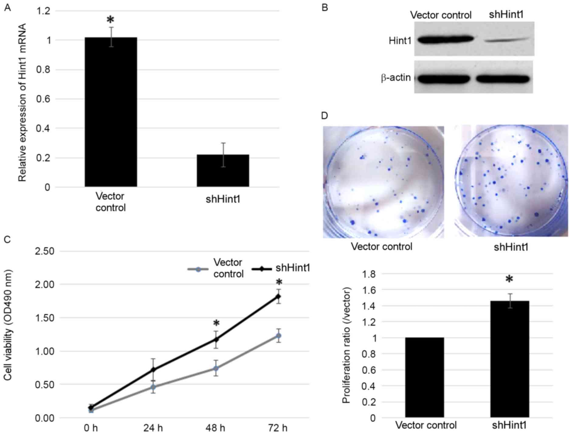
To analyze the involvement of Hint1 in gastric
cancer, a shRNA-Hint1 vector was transfected into SGC7901 gastric
cancer cells. Transfection with the shHint1 vector resulted in
significantly reduced Hint1 mRNA expression and visibly reduced
Hint1 protein expression (Fig. 1A and
B, respectively). In the subsequent MTT assay, cells
transfected with shHint1 demonstrated significantly increased
viability compared with the vector control group (Fig. 1C). Meanwhile, a colony survival assay
was performed to verify the results of this test. There was a ~1.5
fold increase in the number of colonies observed in the shHint1
group compared with the vector control under the same culture
conditions and time (Fig. 1D). These
data demonstrated an inhibitory function of Hint1 on gastric cancer
cell viability.
Suppression of ERK-dependent NF-κB
activation is involved in Hint1-mediated retardation of cell
viability
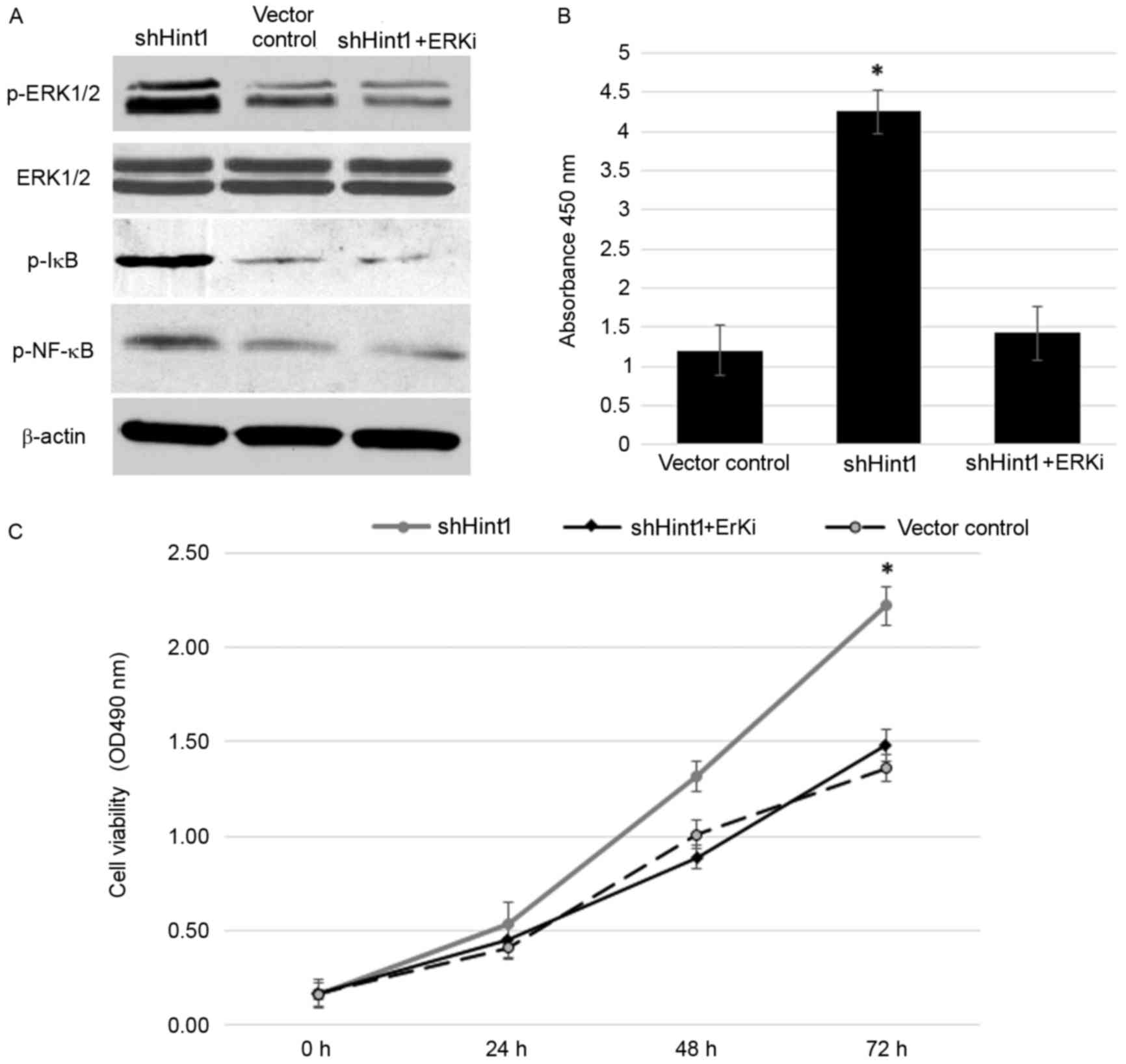
The ERK signaling cascade is a central
mitogen-activated protein kinase (MAPK) pathway that regulates
various oncogenic responses, including proliferation and survival
(11,12). To further investigate the mechanisms
by which Hint1 inhibited the viability of gastric cancer cells, the
interactions between Hint1 and ERK were determined. The results
revealed that ERK phosphorylation activity was significantly
increased following knockdown of Hint1 in SCG7901 cells (Fig. 2A). Activated ERK subsequently induced
IκB phosphorylation and activated the NF-κB p65 signal pathway when
lacking regulation from Hint1 (Fig. 2A
and B). These cascades were repressed by ERKi, demonstrating
that the activation of NF-κB p65 following Hint1 knockdown was ERK
dependent. To verify this result, the effect on cell viability in
the shHint1 group was analyzed following treatment with ERKi. ERKi
efficiently reversed the increased viability induced by Hint1
deficiency (Fig. 2C). These data
indicated that Hint1 may be a negative regulator of ERK, and
further prevent activation of its downstream signaling pathways,
including NF-κB, thus inhibiting cell viability.
Hint1 increased radiosensitivity in
gastric cancer cells
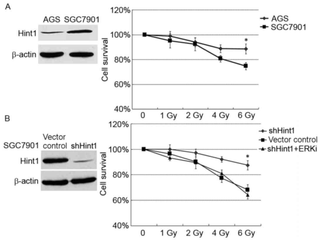
Due to the deleterious influence of Hint1 on cancer
cell viability, the present study investigated whether Hint1
modulated the radiation resistance of gastric cancer. Two human
gastric cancer cell lines with 0, 1, 2, 4 or 6 Gy X-ray. Prior to
the treatment, the protein expression of Hint1 was examined in
these cell lines (Fig. 3A). Notably,
differences in cell survival were observed between the gastric
cancer cells lines following radiation. SGC7901 cells, which
demonstrated higher Hint1 protein expression, exhibited higher
sensitivity to radiation, whereas AGS cells, with lower Hint1
protein expression, were relatively resistant to treatment
(Fig. 3A). shHint1-transfected
SGC7901 cells were then treated with radiation, with ERKi or
without. Radiation resistance was induced in the shHint1 SGC7901
cells, while ERKi overcame this (Fig.
3B).
Hint1 delayed radiation-induced DNA
damage repair in gastric cancer cells
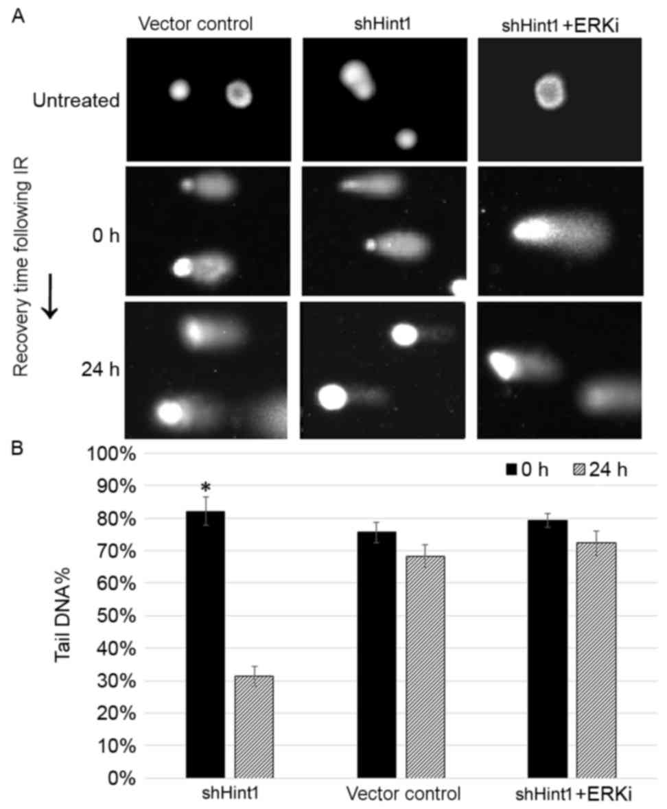
A previous study demonstrated that Hint1 is involved
in DNA damage repair in normal cells (9). The influence of Hint1 expression on DNA
damage, a hallmark of radiation-induced cytotoxicity, was explored
on SGC7901 cells using single cell gel electrophoresis, also known
as a comet assay. shHint1 cells demonstrated visibly smaller DNA
tails than the vector control 24 h following radiation treatment of
6 Gy, suggesting enhanced DNA repair in Hint1-deficient cells
(Fig. 4A). There was no significant
difference in the tail DNA fraction between 0 and 24 h in the
vector control and ERKi treatment group (Fig. 4B). In this experiment, ERKi treatment
did not affect the formation of DNA damage, instead retarding DNA
damage repair.
Hint1-mediated Cyclin D1 suppression
results in persistent G1 arrest in gastric cancer cells following
IR
The mechanisms underlying the involvement of Hint1
in the radiation response remain unclear. First, the effect of
Hint1 expression on the cellular DNA damage response signal pathway
was examined. As reported by a previous study, p-ATM levels
increase in SGC7901 cells following radiation treatment, but this
increase is partially suppressed in shHint1 cells (13). Notably, the activation of ATR, the
other important DNA damage sensor, did not seem to be altered by
Hint1 expression, but it was not possible to explain why Hint1
increased radiosensitivity with enhanced ATM activation (13). Similar results were observed in the
present study (Fig. 5A). Therefore,
the present study focused on the downstream effectors of the DNA
damage response. Cyclin D1, a cell-cycle regulator essential for
the G1 phase, was downregulated by Hint1 (Fig. 5B). During the G1 phase, Cyclin D1
increases DNA synthesis via Rb phosphorylation and promotes S phase
entry (14). Decreased Cyclin D1
levels in SGC7901 cells significantly inhibited p-Rb activation,
resulting in a persistent G1 arrest following IR treatment
(Fig. 5B and C). Accordingly, the
pro-apoptotic protein Bax was upregulated in vector control cells
following IR compared with shHint1 cells following IR (Fig. 5B).
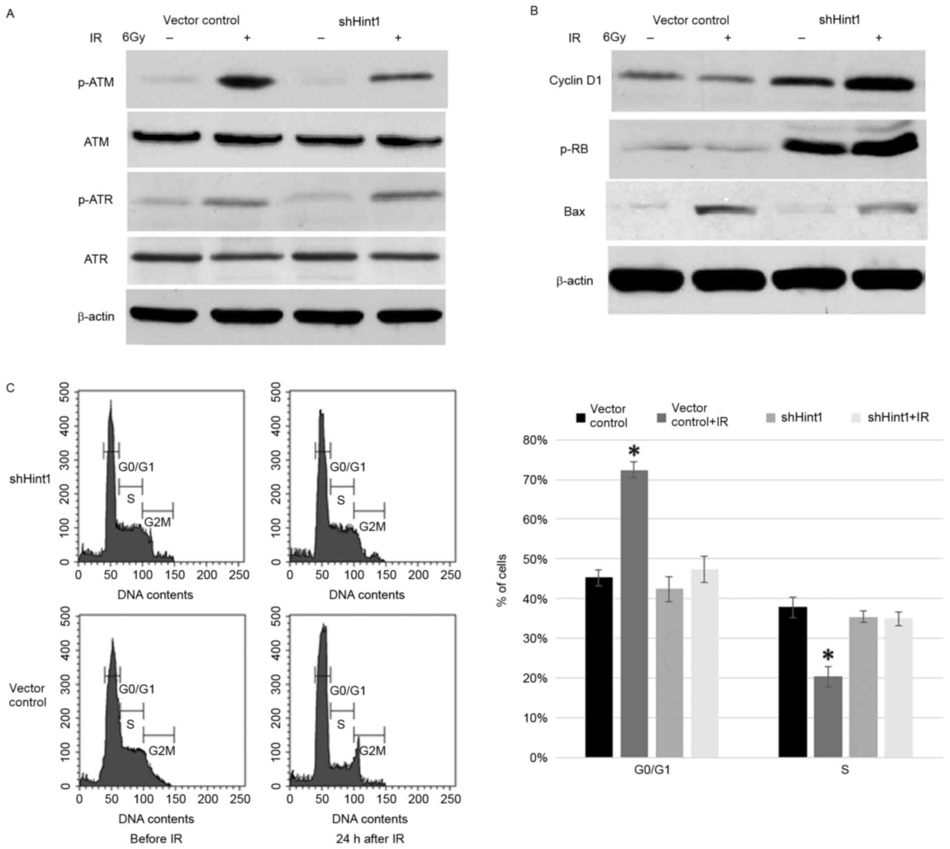 | Figure 5.(A) Expression of the DNA damage
response sensors, ATM and ATR, was assessed by Western blot
analysis. (B) Cyclin D1, p-Rb and Bax protein expression was
assessed by Western blot. (C) G1 arrest was observed via flow
cytometry in gastric cancer cells following IR. Data are presented
as the mean ± standard error of the mean. *P<0.05 vs. other
groups. ATM, ATM serine/threonine kinase; ATR, ATR serine/threonine
protein kinase; p-, phosphorylated; Rb, retinoblastoma protein;
Bax, BCL2 associated X, apoptosis regulator; IR, ionizing
radiation; si, small interfering RNA. |
Discussion
Previous investigations using genetically engineered
mice have demonstrated that Hint1 is a novel haploinsufficient
tumor suppressor gene (2,3). It demonstrates a strong analogy with
fragile histidine triad, a known tumor suppressor gene on common
fragile site fragile site, aphidicolin type, common, fra(3)(p14.2)
(2), but its functions are yet to be
completely understood. A previous study analyzed clinical samples
and reported a potential tumor suppressor role for Hint1 in human
gastric cancer (6). In the present
study, the function of Hint1 was analyzed in human gastric cancer
cells. The results obtained demonstrated, for the first time, that
Hint1 decreased gastric cancer cell proliferation in vitro
by decreasing cell viability. These observations were consistent
with the proposed tumor suppressor function of Hint1 and with
previous findings demonstrating that Hint1 inhibited human colon
and breast cancer cell growth (2,3,8).
Mechanistic studies subsequently demonstrated the
negative regulation of Hint1 on ERK signal pathway activation.
ERK1/2 are associated protein-serine/threonine kinases that
participate in the Ras-Raf-mitogen-activated protein kinase
kinase-ERK signal transduction cascade. This cascade participates
in the regulation of a number of processes, including cell
adhesion, cell cycle progression, cell migration, cell survival,
differentiation, metabolism, proliferation and transcription
(11,12). ERKs directly phosphorylate multiple
transcription factors, including the ETS transcription factor
family, c-Jun and c-Myc (15–17). ERK also phosphorylates and activates
the 90 kDa ribosomal S6 kinase (p90Rsk), which, in turn, leads to
the activation of the transcription factor cAMP response element
binding protein (18). Furthermore,
ERK results in activation of the NF-κB transcription factor by
phosphorylating and activating IκB kinase (19). Deregulated activation of NF-κB, which
induces uncontrolled cell growth, is a central signature of
multiple types of epithelial cancer. For instance, over-activation
of NF-κB, regulated by ERK, promoted malignant transformation in
endometrial epithelia cells (20). In
the present study, silencing Hint1 resulted in the activation of
the ERK-NF-κB signal pathway in SGC7901 cells. This demonstrated
that Hint1 acted as an ERK-control factor in stomach
carcinogenesis, resulting in tumor suppression.
As radiation therapy has become a pillar of the
therapeutic approach to gastric cancer, the study of the molecular
mechanisms underlying the radiation resistance of gastric cancer
cells has become important. A previous study indicated that
Hint1−/− mouse embryonic fibroblasts cells had defects
in the double-stranded break (DSB) repair pathway, including loss
of ATM activation and the presence of unrepaired DSB, while another
study demonstrated that Hint1-deficient cells exhibited resistance
to IR-induced apoptosis (2,9). Therefore, the exact function of Hint1 in
response to radiation in gastric cancer cells was worthy of
investigation. In the present study, Hint1-deficient SGC7901 cells
also exhibited resistance to IR. Although Hint1 deficiency affected
ATM activation, DNA damage repair was not retarded in
Hint1-deficient SGC7901 cells in the present study. On the
contrary, vector control cells demonstrated repressed DNA damage
repair functions. One reason for this may be that cancer cells have
more inordinate or defective DNA damage repair functions than
normal cells. The loss of critical tumor suppressor genes,
including Hint1, may activate other pro-survival signal pathways
which enhance DNA repair in cancer cells. In addition, ATM
activation is not completely inhibited following Hint1 silencing or
in Hint1−/- cells, as previously described (9). In addition, Hint1 deficiency did not
affect the activation of ATR, another important DNA damage sensor,
in the present study.
As Hint1 was demonstrated to inhibit ERK activation,
its downstream effectors, which may be involved in DNA repair, were
investigated. Cyclin D1, a component of the core cell cycle
machinery, was revealed to be significantly upregulated following
silencing of Hint1. Cyclin D1 phosphorylates and inactivates Rb
protein and promotes progression through the G1-S phase of the cell
cycle (21). A number of studies have
demonstrated that endogenous high levels of cyclin D1 promote
carcinogenesis and radioresistance in the majority of malignant
tumors (22–24). Downregulation of Cyclin D1 induces G1
arrest, followed by apoptosis, in human skin cancer A431 cells
following UV exposure (25). Cyclin
D1 overexpression perturbs DNA replication and induces
replication-associated DNA DSBs, which may gradually induce
formation of acquired radioresistance during long term fractionated
radiotherapy (26,27). In the present study, the negative
regulation of ERK mediated by Hint1 may suppress Cyclin D1
expression, which leads to persistent G1 arrest followed by
apoptosis in SGC7901 cells following IR exposure.
In summary, the results of the present study first
indicated that Hint1 was involved in the regulation of cell
proliferation and radioresponse via negative control of the ERK
signal pathway. These results demonstrated the critical function of
Hint1 in the biology of human gastric cancers. Acting as a tumor
growth suppressor and a radiosensitive agent, this protein is a
potential biomarker and an attractive target for specific
therapeutic interventions against gastric cancer.
Acknowledgements
The authors would like to thank Dr Hong Fan (Key
Laboratory of Developmental Genes and Human Diseases, Ministry of
Education, Southeast University, Nanjing, China) for her help and
advice.
Funding
The present study was supported by the National
Natural Science Foundation of China (grant no. 81201882), the
Natural Science Foundation of Jiangsu Province, China (grant no.
BK2012076) and the Key Project supported by the Medical Science and
Technology Development Foundation, Nanjing Department of Health,
China (grant no. ZKX14039).
Availability of data and materials
All data generated or analyzed during the present
study are included in this published article.
Authors' contributions
The present study was concieved and designed by XWW
and JFC. The methodology was developed by XMW, LZH and ZX. The data
was aquired by XWW, JZ, HYZ and XWW. Analysis and interpretation
was performed by XMW and XWW. The manuscript was written and
revised by XMW and JFC. Administrative and technical support was
provided by XWW and ZX. The study was overseen by JFC.
Ethics approval and consent to
participate
The present study was approved by Nanjing Medical
University (Nanjing, China) Ethics Committee in 2012.
Consent for publication
Not applicable.
Competing interests
The authors declare that they have no competing
interests.
References
|
1
|
Stewart BW and Wild CP: World cancer
report 2014. World Health Organization. Chapter 1.1 ISBN
9283204298. 2014
|
|
2
|
Su T, Suzui M, Wang L, Lin CS, Xing WQ and
Weinstein IB: Deletion of histidine triad nucleotide-binding
protein 1/PKC-interacting protein in mice enhances cell growth and
carcinogenesis. Proc Natl Acad Sci USA. 100:7824–7829. 2003.
View Article : Google Scholar : PubMed/NCBI
|
|
3
|
Li H, Zhang Y, Su T, Santella RM and
Weinstein IB: Hint1 is a haplo-insufficient tumor suppressor in
mice. Oncogene. 25:713–721. 2006. View Article : Google Scholar : PubMed/NCBI
|
|
4
|
Cen B, Li H and Weinstein IB: Histidine
triad nucleotide-binding protein 1 up-regulates cellular levels of
p27KIP1 by targeting ScfSKP2 ubiquitin ligase and Src. J Biol Chem.
284:5265–5276. 2009. View Article : Google Scholar : PubMed/NCBI
|
|
5
|
Weiske J and Huber O: The histidine triad
protein Hint1 interacts with pontin and reptin and inhibits
TCF-beta-catenin-mediated transcription. J Cell Sci. 118:3117–3129.
2005. View Article : Google Scholar : PubMed/NCBI
|
|
6
|
Huang H, Wei X, Su X, Qiao F, Xu Z, Gu D,
Fan H and Chen J: Clinical significance of expression of Hint1 and
potential epigenetic mechanism in gastric cancer. Int J Oncol.
38:1557–1564. 2011.PubMed/NCBI
|
|
7
|
Weiske J and Huber O: The histidine triad
protein Hint1 triggers apoptosis independent of its enzymatic
activity. J Biol Chem. 281:27356–27366. 2006. View Article : Google Scholar : PubMed/NCBI
|
|
8
|
Wang L, Zhang Y, Li H, Xu Z, Santella RM
and Weinstein IB: Hint1 inhibits growth and activator protein-1
activity in human colon cancer cells. Cancer Res. 67:4700–4708.
2007. View Article : Google Scholar : PubMed/NCBI
|
|
9
|
Li H, Balajee AS, Su T, Cen B, Hei TK and
Weinstein IB: The HINT1 tumor suppressor regulates both gamma-H2AX
and ATM in response to DNA damage. J Cell Biol. 183:253–265. 2008.
View Article : Google Scholar : PubMed/NCBI
|
|
10
|
Livak KJ and Schmittgen TD: Analysis of
relative gene expression data using real-time quantitative PCR and
the 2(-Delta Delta C(T)) method. Methods. 25:402–408. 2001.
View Article : Google Scholar : PubMed/NCBI
|
|
11
|
Samatar AA and Poulikakos PI: Targeting
RAS-ERK signalling in cancer: Promises and challenges. Nat Rev Drug
Discov. 13:928–942. 2014. View
Article : Google Scholar : PubMed/NCBI
|
|
12
|
Huang G, Tang B, Tang K, Dong X, Deng J,
Liao L, Liao Z, Yang H and He S: Isoquercitrin inhibits the
progression of liver cancer in vivo and in vitro via the MAPK
signalling pathway. Oncol Rep. 31:2377–2384. 2014. View Article : Google Scholar : PubMed/NCBI
|
|
13
|
Smith J, Tho LM, Xu N and Gillespie DA:
The ATM-Chk2 and ATR-Chk1 pathways in DNA damage signaling and
cancer. Adv Cancer Res. 108:73–112. 2010. View Article : Google Scholar : PubMed/NCBI
|
|
14
|
Cheng YH, Li LA, Lin P, Cheng LC, Hung CH,
Chang NW and Lin C: Baicalein induces G1 arrest in oral cancer
cells by enhancing the degradation of cyclin D1 and activating AhR
to decrease Rb phosphorylation. Toxicol Appl Pharmacol.
263:360–367. 2012. View Article : Google Scholar : PubMed/NCBI
|
|
15
|
Liu H, Duan Z, Zheng H, Hu D, Li M, Tao Y,
Bode AM, Dong Z and Cao Y: EBV-encoded LMP1 upregulates Igκ
3′enhancer activity and Igκ expression in nasopharyngeal cancer
cells by activating the Ets-1 through ERKs signaling. PLoS One.
7:e326242012. View Article : Google Scholar : PubMed/NCBI
|
|
16
|
Park KH, Shin KS, Zhao TT, Park HJ, Lee KE
and Lee MK: L-DOPA modulates cell viability through the ERK-c-Jun
system in PC12 and dopaminergic neuronal cells. Neuropharmacology.
101:87–97. 2016. View Article : Google Scholar : PubMed/NCBI
|
|
17
|
Kwon YW, Jang S, Paek JS, Lee JW, Cho HJ,
Yang HM and Kim HS: E-Ras improves the efficiency of reprogramming
by facilitating cell cycle progression through JNK-Sp1 pathway.
Stem Cell Res. 15:481–494. 2015. View Article : Google Scholar : PubMed/NCBI
|
|
18
|
Chuang JI, Huang JY, Tsai SJ, Sun HS, Yang
SH, Chuang PC, Huang BM and Ching CH: FGF9-induced changes in
cellular redox status and HO-1 upregulation are FGFR-dependent and
proceed through both ERK and AKT to induce CREB and Nrf2
activation. Free Radic Biol Med. 89:274–286. 2015. View Article : Google Scholar : PubMed/NCBI
|
|
19
|
Tsao GSW, Zhu DD, Zhang J and Deng W:
Abstract 1046: The role of NF-kB activation in the immortalization
of nasopharyngeal epithelial cells. Cancer Res. 75:2015. View Article : Google Scholar
|
|
20
|
Mizumoto Y, Kyo S, Kiyono T, Takakura M,
Nakamura M, Maida Y, Mori N, Bono Y, Sakurai H and Inoue M:
Activation of NF-kappaB is a novel target of KRAS-induced
endometrial carcinogenesis. Clin Cancer Res. 17:1341–1350. 2011.
View Article : Google Scholar : PubMed/NCBI
|
|
21
|
Lee Y, Dominy JE, Choi YJ, Jurczak M,
Tolliday N, Camporez JP, Chim H, Lim JH, Ruan HB, Yang X, et al:
Cyclin D1-Cdk4 controls glucose metabolism independently of cell
cycle progression. Nature. 510:547–551. 2014. View Article : Google Scholar : PubMed/NCBI
|
|
22
|
Musgrove EA, Caldon CE, Barraclough J,
Stone A and Sutherland RL: Cyclin D as a therapeutic target in
cancer. Nat Rev Cancer. 11:558–572. 2011. View Article : Google Scholar : PubMed/NCBI
|
|
23
|
Peurala E, Koivunen P, Haapasaari KM,
Bloigu R and Jukkola-Vuorinen A: The prognostic significance and
value of cyclin D1, CDK4 and p16 in human breast cancer. Breast
Cancer Res. 15:R52013. View
Article : Google Scholar : PubMed/NCBI
|
|
24
|
Choi YJ, Li X, Hydbring P, Sanda T,
Stefano J, Christie AL, Signoretti S, Look AT, Kung AL, von Boehmer
H and Sicinski P: The requirement for cyclin D function in tumor
maintenance. Cancer Cell. 22:438–451. 2012. View Article : Google Scholar : PubMed/NCBI
|
|
25
|
Kim AL, Athar M, Bickers DR and Gautier J:
Ultraviolet-B-induced G1 arrest is mediated by downregulation of
cyclin-dependent kinase 4 in transformed keratinocytes lacking
functional p53. J Invest Dermatol. 118:818–824. 2002. View Article : Google Scholar : PubMed/NCBI
|
|
26
|
Shimura T, Ochiai Y, Noma N, Oikawa T,
Sano Y and Fukumoto M: Cyclin D1 overexpression perturbs DNA
replication and induces replication-associated DNA double-strand
breaks in acquired radioresistant cells. Cell Cycle. 12:773–782.
2013. View
Article : Google Scholar : PubMed/NCBI
|
|
27
|
Shimura T, Fukumoto M and Kunugita N: The
role of cyclin D1 in response to long-term exposure to ionizing
radiation. Cell Cycle. 12:2738–2743. 2013. View Article : Google Scholar : PubMed/NCBI
|