Introduction
Osteosarcoma, a kind of lethal malignant bone tumor,
predominantly occurs among children as well as adolescents
(1). Accumulating studies have showed
that morbidity and mortality are both high in osteosarcoma.
Currently, the major therapies for osteosarcoma are chemotherapy
and surgery (2). With the development
of combination including neoadjuvant chemotherapy and limb salvage
surgery, the survival rates of osteosarcoma patients have
significantly increased (3,4). However, osteosarcoma patients with
metastasis often have a poor prognosis, in the meantime, overall
clinical outcomes of osteosarcoma remain unsatisfactory (5). Therefore, it is necessary to understand
the molecular mechanisms underlying the osteosarcoma development
and progression. Moreover, finding out useful novel biomarkers for
the diagnosis and treatment of osteosarcoma is one of the most
important clinical problems. In recent years, emerging studies have
indicated that miRNAs have important roles in different kinds of
tumors.
miRNA is a kind of small non-coding RNA and plays
crucial role in regulating gene expression, being considered as
cancer biomarkers (6,7). Studies have indicated that the
expressions of almost 30% human genes were controlled by different
miRNAs (8,9). Briefly, miRNAs can regulate the
expression of their mRNA targets by complete or partial binding to
the 3′untranslated regions (3′-UTRs) (10). Based on the characteristics of their
target genes, they can inhibit or promote multiple biological
processes of obvious cancers (11).
For instance, miR-130b suppresses epithelial ovarian carcinoma by
regulating runt-related transcription factor 3 (RUNX3) (12); miR-92b functions as an oncogene in
hepatocellular carcinoma via regulating Smad7 (13); miR-143 functions as a suppressor in
prostate cancer via targeting matrix metalloproteinase 13 (14). In addition, miR-210 has been reported
to modulate the development of many malignancies, including
glioblastoma (15), breast carcinoma
(16) and pancreatic carcinoma
(17). However, the function of
miR-210 in osteosarcoma needs to be fully elucidated.
Fibroblast growth factor receptor-like 1 (FGFRL1) is
one member of FGFR family and unlike the other members of FGFR,
FGFRL1 has no intracellular kinase domain, instead, it contains a
short intracellular domain (18,19).
Increasing evidence has shown that the FGFR family has vital
regulatory roles in proliferation, differentiation and migration of
various cancers (20). FGFRL1 has
also been reported to exert multiple biological functions in
different kinds of cancers. However, the specific functions of
FGFRL1 in osteosarcoma still remain unclear. Present study aimed to
investigate the correlation between miR-210 and FGFRL1 in
osteosarcoma cell migration and invasion.
Materials and methods
Osteosarcoma cell lines and tissue
specimens
Two osteosarcoma cell lines (MG63 and Saos-2) and
one human normal osteoblast cell line hFOB 1.19 were purchased from
Shanghai Institute for Biological Sciences (Shanghai, China). Both
the osteosarcoma cells and the hFOB 1.19 were maintained in DMEM
medium which contained penicillin-streptomycin and 10% FBS (all
from Invitrogen; Thermo Fisher Scientific, Inc., Waltham, MA, USA)
in an atmosphere with 5% CO2 at 37°C.
A total of 54 osteosarcoma tissue specimens and the
corresponding para-carcinoma tissues were collected from the
People's Hospital of Qingdao West Coast New District between 2015
and 2017 and snap-frozen at −80°C. The clinical characteristics of
osteosarcoma patients were shown in Table
I. All the patients involved in current study signed written
informed consent. The present study was approved by the Ethics
Committee of the People's Hospital of Qingdao West Coast New Area
(Shandong, China).
 | Table I.Correlation between miR-210 expression
and the clinicopathological characteristics of the osteosarcoma
patients. |
Table I.
Correlation between miR-210 expression
and the clinicopathological characteristics of the osteosarcoma
patients.
|
|
| miR-210a expression |
|
|---|
|
|
|
|
|
|---|
| Clinicopathological
features | Cases (n=54) | High (n=39) | Low (n=15) | P-value |
|---|
| Age (years) |
|
|
| 0.5768 |
|
>60 | 26 | 20 | 6 |
|
| ≤60 | 28 | 19 | 9 |
|
| Sex |
|
|
| 0.4937 |
|
Male | 28 | 18 | 8 |
|
|
Female | 26 | 21 | 7 |
|
| Tumor size
(cm) |
|
|
| 0.3864 |
|
≥5.0 | 30 | 25 | 8 |
|
|
<5.0 | 24 | 14 | 7 |
|
| TNM stage |
|
|
| 0.0066b |
|
I–II | 22 | 11 | 11 |
|
|
III | 32 | 28 | 4 |
|
| Lymph-node
metastasis |
|
|
| 0.0042b |
|
Yes | 33 | 31 | 4 |
|
| No | 21 | 8 | 11 |
|
| Distant
metastasis |
|
|
| 0.5204 |
|
Yes | 28 | 18 | 9 |
|
| No | 26 | 21 | 6 |
|
Cell transfection
Lipofectamine 2000 (Invitrogen; Thermo Fisher
Scientific, Inc.) was used to transfect miR-210 mimics, inhibitor
or scrambled negative miR-control (NC) as well as FGFRL1
overexpression plasmid and the negative control vector (GenePharma
Co., Ltd., Shanghai, China) into osteosarcoma cells. Plasmids were
transfected into osteosarcoma cell lines by X-treme GENE HP DNA
Transfection Reagent (Roche Diagnostics, Basel, Switzerland).
Reverse transcription-quantitative
polymerase chain reaction (RT-qPCR)
RT-qPCR was conducted to measure the miR-210
expressions and FGFRL1 expressions in osteosarcoma cell lines and
tissues. Firstly, the total RNA from osteosarcoma tissues and cells
as well as normal tissues was isolated using TRIzol reagent
(Invitrogen; Thermo Fisher Scientific, Inc.) in accordance with the
manufacturer's recommendation. Reverse transcription assay was
conducted to obtain the complementary DNA (cDNA) by PrimeScript RT
reagent kit (Takara Biotechnology Co., Ltd., Dalian, China) and
qPCR assay was conducted by SYBR® Premix Ex
Taq™ (Takara Biotechnology Co., Ltd.) on the system of
ABI 7900 Sequence Detection System (Applied Biosystems; Thermo
Fisher Scientific, Inc.). The miR-210 expression was normalized to
U6 while the FGFRL1 was normalized to GAPDH. The relative
expression levels of genes were calculated by the 2−ΔΔCq
method (21). The primers used were
shown in Table II.
| Table II.Primer sequences for reverse
transcription-quantitative polymerase chain reaction. |
Table II.
Primer sequences for reverse
transcription-quantitative polymerase chain reaction.
| Primer and
direction | Sequence |
|---|
| miR-210
forward |
5′-GTGCAGGGTCCGAGGT-3′ |
| miR-210
reverse |
5′-CTGTGCGTGTGACAGCGGCTGA-3′ |
| U6 forward |
5′-CTCGCTTCGGCAGCACA-3′ |
| U6 reverse |
5′-AACGCTTCACGAATTTGCGT-3′ |
| FGFRL1 forward |
5′-TGTGAACACAACGGTGGACT-3′ |
| FGFRL1 reverse |
5′-GGGCAACACCACAAACTTCT-3′ |
| GAPDH forward |
5′-TAATCTTCGCCTTAATACTT-3′ |
| GAPDH reverse |
5′-AGCCTTCATACATCTCAA-3′ |
Western blot analysis
The total proteins were extracted by lysing cells
with RIPA buffer which contained protease and phosphatase
inhibitors (Beyotime Institute of Biotechnology, Haimen, China).
Put the lysates onto ice for half an hour, then centrifuged them
for 20 min at 12,000 × g. The protein concentrations were
quantified with the BCA kit (Pierce; Thermo Fisher Scientific,
Inc.). The protein lysates were separated by SDS-PAGE and
subsequently transferred onto PVDF membrane which was blocked with
5% non-fat milk and incubated with the primary antibodies at 4°C
overnight. Then, a secondary incubation step was performed with
appropriate secondary antibody at room temperature for one hour.
The proteins were detected by Chemoluminescene method. GAPDH
(Abcam, Cambridge, UK) was used as an internal loading control.
Migration and invasion assays
The invasion and migration abilities of the treated
osteosarcoma cells were assessed by Transwell assays using
Transwell chambers (Corning Incorporated, Corning, NY, USA) with or
without Matrigel (Clontech Laboratories, Inc., Mountainview, CA,
USA). For the invasion assay, the Transwell chambers were
pretreated with Matrigel. Firstly, osteosarcoma cell transfections
were suspended in serum-free medium and seeded into the upper
chamber. At the same time, DMEM including 10% FBS was joined into
the lower chamber. Being cultivated for 48 h, cells on the upper
chambers were removed with cotton swabs carefully, while the
invasive cells on the lower chamber were subsequently fixed and
stained with 4% formaldehyde and 0.1% crystal violet, respectively.
The difference between the migration assay and the invasion assay
was that there was no Matrigel in the Transwell chambers. An
inverted microscope (Olympus Corporation, Tokyo, Japan) was used to
measure and count the invasive and migratory cells in five randomly
selected fields.
Luciferase reporter assay
The amplified FGFRL1-3′-UTR-WT and corresponding
FGFRL1-3′-UTR-MUT were respectively cloned into pGL3 luciferase
vector (Promega Corporation, Madison, WI, USA). Osteosarcoma cells
were added into a 96-well plate, and after 24 h, FGFRL1-3′UTR-WT
and miR-210 mimics or FGFRL1-3′UTR-MUT and miR-210 mimics were
transfected into the treated osteosarcoma cells using Lipofectamine
2000 (Invitrogen; Thermo Fisher Scientific, Inc.). Subsequently,
the Dual Luciferase Reporter Assay kit (Promega Corporation) was
used to detect the relative luciferase activities 48 h after the
transfections.
Statistical analysis
All the above assays were performed three times.
Statistical analysis was performed using SPSS statistical software,
version 17.0 (SPSS, Inc., Chicago, IL, USA) with Student's t-test
and one-way ANOVA with Scheffe's post hoc test. Correlation between
mRNA and miRNA were estimated using the Spearman's correlation
method. Kaplan-Meier method with log-rank test were applied to
estimate the survival rates and compare the survival curves
respectively. The data was indicated as means ± SD. P<0.05 was
considered to indicate a statistically significant difference.
Results
miR-210 expression is upregulated and
FGFRL1 expression is downregulated in osteosarcoma
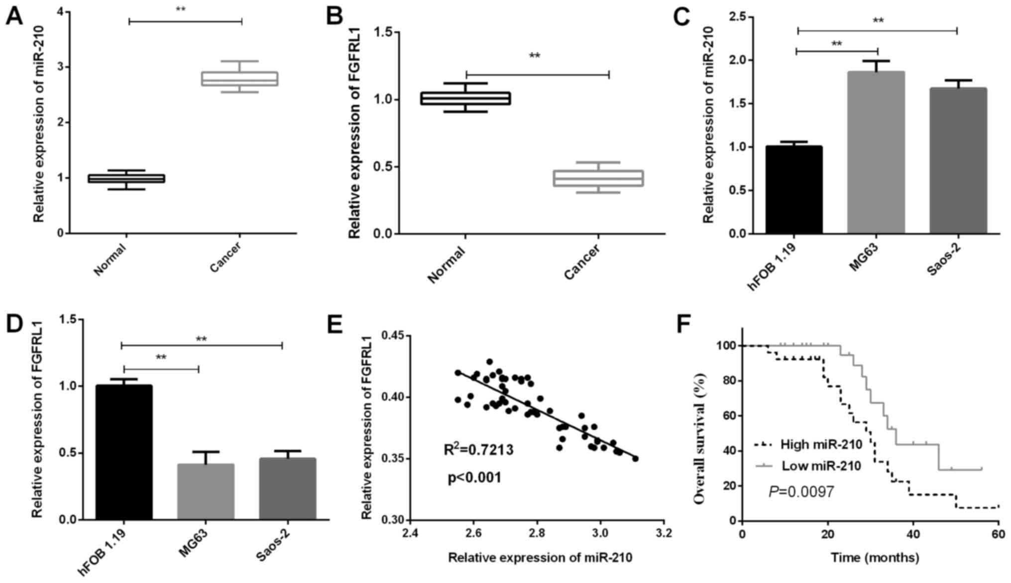
In this current study, the expressions of miR-210
and FGFRL1 in 54 paired osteosarcoma tissue specimens and cell
lines were measured. The results of RT-qPCR revealed that the
miR-210 expressions in osteosarcoma tissues were significantly
increased in contrast with that in the matched para-carcinoma
tissues (P=0.0015; Fig. 1A). On the
contrary, FGFRL1 expressions in osteosarcoma tissues were found to
be prominently downregulated (P=0.0023; Fig. 1B). Additionally, the same result was
also found in osteosarcoma cell lines. From the results of RT-qPCR,
we could also easily find a higher miR-210 expression both in MG63
(P=0.0064) and Saos-2 cells (P=0.0060) than that in hFOB 1.19
(P<0.01; Fig. 1C). Subsequently,
we also measured the FGFRL1 mRNA expression in osteosarcoma cell
lines, the results demonstrated a significant decrease in MG63
(P=0.0035) and Saos-2 (P=0.0039) cells compared to that in hFOB
1.19 (Fig. 1D). Moreover, to better
understand the relationship between miR-210 and FGFRL1, we analyzed
the correlation of miR-210 and FGFRL1 expression and found a
negative correlation between them (P<0.001; Fig. 1E). To address the clinical
significance of miR-210 in osteosarcoma, the mean expression level
of miR-210 was defined as cutoff value to divide osteosarcoma
patients into miR-210 low group and high group. Subsequently, the
results indicated that osteosarcoma patients who had low miR-210
expression levels showed higher overall survival rates than those
with high miR-210 expression levels (P=0.0097; Fig. 1F). Clinical association analysis
demonstrated that the high miR-210 expression was notably
correlated with advanced TNM stage (P=0.0066) and lymph node
metastasis (P=0.0042; Table I).
miR-210 accelerates osteosarcoma cell
invasion and migration
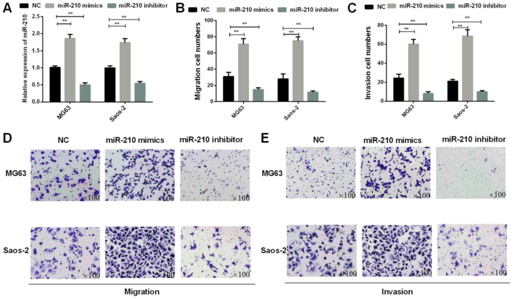
Subsequently, we investigated the invasion and
migration abilities of osteosarcoma cells which were transfected
with miR-210 mimics or inhibitor. The osteosarcoma cells (MG63 and
Saos-2) transfected with miR-210 mimics or inhibitor were used to
detect the roles of miR-210 in osteosarcoma cell invasion and
migration. The results of RT-qPCR assays demonstrated that the
expression of miR-210 mimics was high in MG63 (P=0.0035) and Saos-2
(P=0.0041) cells (P<0.01; Fig.
2A). The results of Transwell assays indicated that miR-210
overexpression facilitated migration ability of osteosarcoma cells
(P=0.0042 for MG63 cells and 0.0030 for Saos-2 cells). On the
contrary, the transwell results also demonstrated a significant
decrease in the migration of MG63 (P=0.0048) and Saos-2 (P=0.0035)
cells transfected with miR-210 inhibitor in contrast to the control
group (Fig. 2B and D). In addition,
according to the Transwell assays, the consequences also
demonstrated that miR-210 could promote osteosarcoma invasion
ability (P=0.0021 for MG63 cells and 0.0019 for Saos-2 cells)
(Fig. 2C and E).
miR-210 suppresses FGFRL1 gene
transcription in osteosarcoma by targeting its 3′-UTR
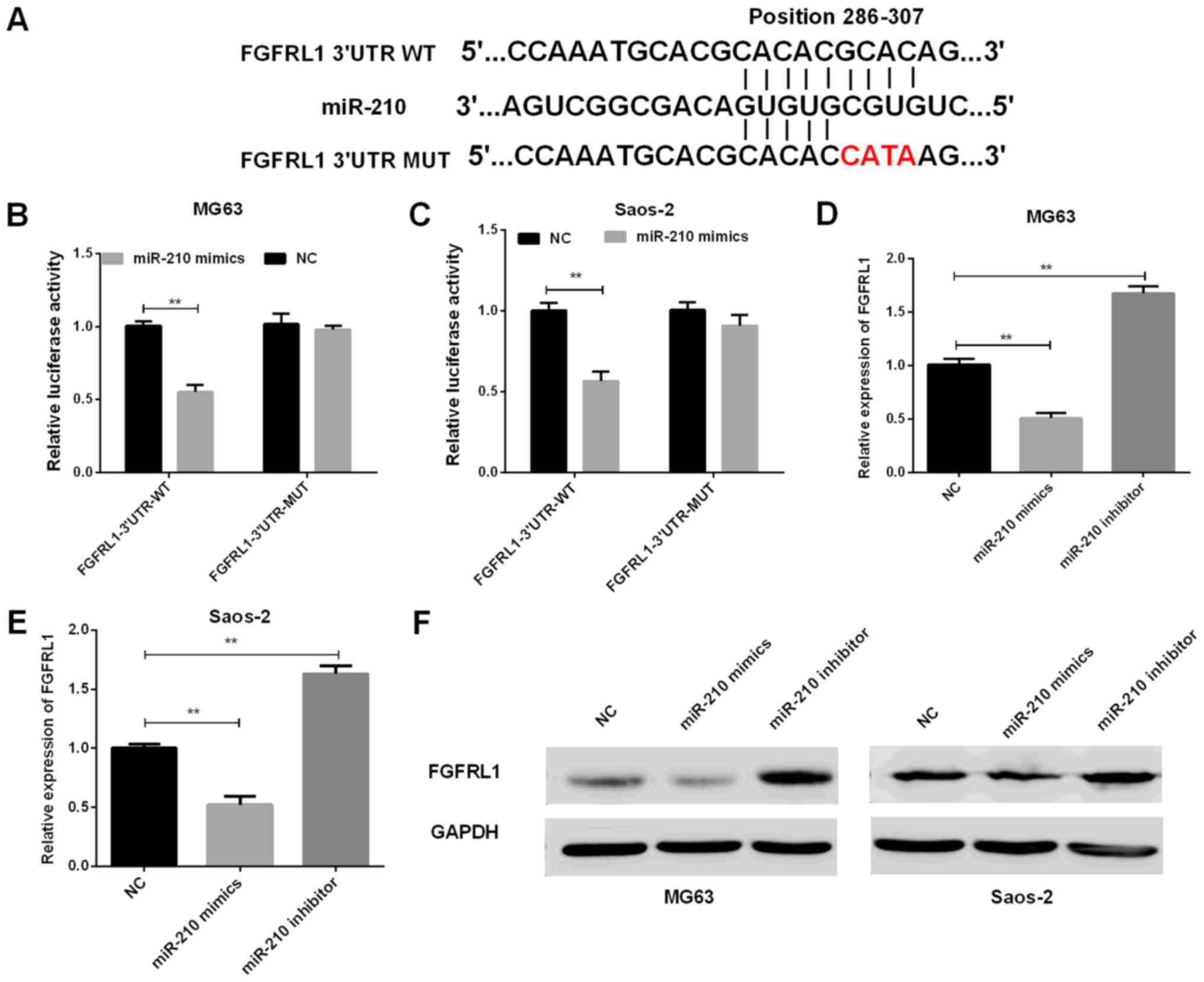
To investigate whether FGFRL1 expression was
associated with the expression of miR-210 and to better understand
the mechanisms of miR-210 in osteosarcoma, Target Scan was used to
find out the target sites in the FGFRL1 sequence of miR-210
(Fig. 3A). To confirm the results,
the luciferase reporter vector contained the wide-type (WT) or
mutant (Mut) FGFRL1 3′-UTR was constructed. Then, the luciferase
reporter assays were conducted in osteosarcoma cells. miR-210
mimics and FGFRL1-3′UTR-WT vector or FGFRL1-3′UTR-Mut vector were
cotransfected into osteosarcoma cells to investigate whether FGFRL1
was the target of miR-210. Then, we detected the roles of miR-210
in the regulation of FGFRL1 mRNA and protein expression. The
results of luciferase reporter assays indicated that miR-210
remarkably reduced the FGFRL1 3′-UTR-WT luciferase activities in
both MG63 and Saos-2 cells. However, miR-210 had no significant
influence on the FGFRL1 3′-UTR-Mut luciferase activities (P=0.0039
for MG63 cells and 0.0027 for Saos-2 cells) (Fig. 3B and C), suggesting that FGFRL1 was
direct target of miR-210. Furthermore, we also detected the FGFRL1
expressions at mRNA level as well as at protein level in
osteosarcoma cell lines transfected with miR-210 mimics or
inhibitor. The results of RT-qPCR and western blot both
demonstrated that miR-210 could inhibit the expression of FGFRL1 in
MG63 (P=0.0044 for miR-210 mimics and 0.0032 for miR-210 inhibitor)
and Saos-2 cells (P=0.0051 for miR-210 mimics and 0.0023 for
miR-210 inhibitor (Fig. 3D-F).
The roles of FGFRL1 in regulating
miR-210 effects in osteosarcoma cell migration and invasion
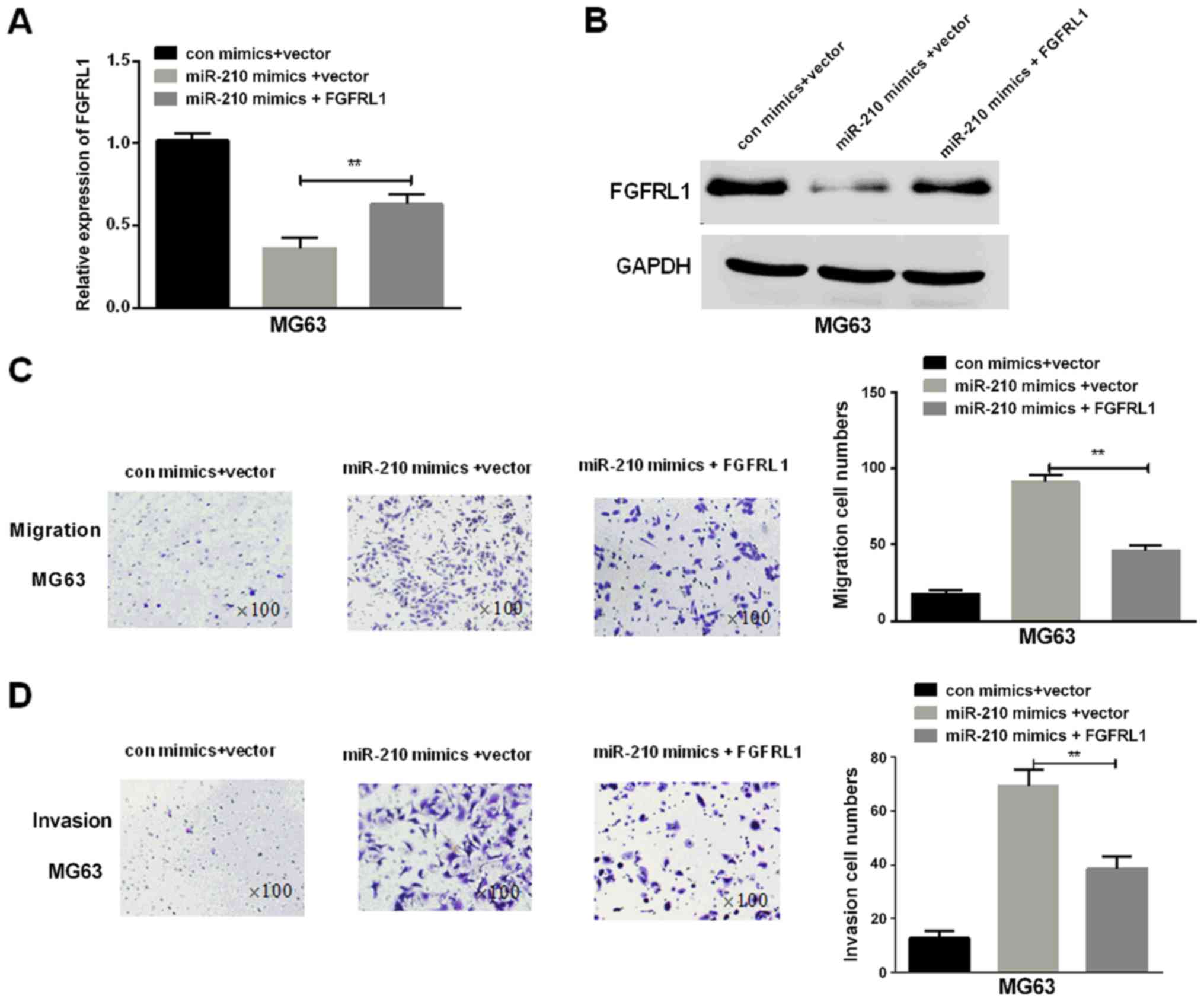
We continued to investigate whether FGFRL1 was
needed in regulation of the promoting functions mediated by miR-210
in osteosarcoma cell invasion and migration. Firstly, FGFRL1
overexpression vector and miR-210 mimics were transfected into
osteosarcoma cells. Then, RT-qPCR and western blotting were
performed to measure the mRNA and protein expression levels
respectively and the results indicated that the FGFRL1 expression
in osteosarcoma cells transfected with FGFRL1 was significantly
increased in contrast to the control group (P=0.0056; Fig. 4A and B). The results of Transwell
assays indicated that FGFRL1 deprived the promotion effect of
miR-210 on cell migration and invasion, suggesting that FGFRL1
played important roles in the miR-210-mediated biological functions
in osteosarcoma cells (P=0.0043 for migration and 0.0036 for
invasion) (Fig. 4C and D).
Discussion
Osteosarcoma is one common bone tumor which affects
more and more children globally. At present, the primary treatments
for osteosarcoma are radiotherapy, chemotherapy and surgical
resection (22). Although great
efforts have been made to explore the underlying mechanisms of
osteosarcoma carcinogenesis, the prognosis is still unsatisfied due
to extremely-high metastatic rate as well as aggressive invasion
into local tissues (23,24). Therefore, it is urgent to elucidate
the molecular mechanism of osteosarcoma to explore the novel
therapeutic approaches, such as diagnostic biomarkers and
therapeutic targets. During the past decades, increasing studies
have demonstrated that various miRNAs are involved in osteosarcoma
proliferation, apoptosis and metastasis (25).
Growing evidence has demonstrated that miR-210 is
aberrantly expressed in multiple tumors. Additionally, miR-210 can
modulate multiple physiological processes, including cell
differentiation, apoptosis and survival proliferation via
regulating its target genes (26).
For instance, miR-210 has been reported to accelerate
proliferation, autophagy and angiogenesis of schwannoma cells
(27); miR-210 has also been reported
to induce endothelial cell apoptosis by directly targeting PDK1 in
the setting of atherosclerosis (28);
in addition, miR-210-3p can inhibit the bladder cancer growth and
metastasis through regulating FGFRL1 (29). As we all know, miR-210 has been
demonstrated to play important roles in many tumors, but its role
in osteosaroma remains unclear. This study aimed to explore the
function of miR-210 in osteosaroma. The results in this study
revealed that miR-210 was significantly upregulated in osteosaroma.
Recent studies have indicated that hypoxia is one of the hallmarks
of cancer and cells within the tumor become hypoxic as the tumor
mass increases, resulting in activation of HIF-1α to induce various
malignant phenotypes (30,31). The potential mechanism underlying the
miR-210 overexpression in osteosaroma is that miR-210 is a hypoxia
regulated miRNA and the induction of miR-210 is a consistent
characteristic of the hypoxic response (32). FGFRL1, the fifth member of the FGFR
family, plays important roles in the development of virtually all
cell types such as the proliferation, differentiation, migration
and apoptosis (33). FGFRL1 is newly
described and usually expressed in adult pancreas and embryonic
bone (34). A study has demonstrated
that FGFRL1 overexpression in HEK 293 Tet-On cells suppresses the
development of tumor in nude mice (35). For another example, FGFRL1 can't
enhance cell proliferation but induce cell adhesion (36). However, little is known about the
potential functions of FGFRL1 in osteosaroma. In current study, we
investigated the relationship between miR-210 and FGFRL1 in
osteosaroma.
In conclusion, miR-210 was upregulated not only in
osteosaroma tissues but also in osteosaroma cells and its
expression was correlated with clinicopathological features. In
addition, we found that the expressions of miR-210 were negatively
correlated with the expression of FGFRL1 in osteosarcoma tissues.
miR-210 exerted tumor carcinogenic functions in osteosarcoma by
targeting FGFRL1 directly. In summary, all the results in this
study suggested that miR-210-FGFRL1 axis may be the novel biomarker
for the diagnosis and treatment of osteosarcoma.
Acknowledgements
Not applicable.
Funding
No funding was received.
Availability of data and materials
The datasets used and/or analyzed during the present
study are available from the corresponding author on reasonable
request.
Authors' contributions
XL and CZ perform the analysis with constructive
discussions. CW contributed to analysis and manuscript preparation.
JS performed the data analyses and wrote the manuscript. DW and YZ
contributed to the conception of the study. XX contributed to
manuscript preparation and the conception of the study. All authors
read and approved the final manuscript.
Ethics approval and consent to
participate
The present study obtained approval from the Ethics
Committee of the People's Hospital of Qingdao West Coast New Area
(Shandong, China). Signed written informed consents were obtained
from the patients.
Patient consent for publication
Not applicable.
Competing interests
The authors declare that they have no competing
interests.
References
|
1
|
Angulo P, Kaushik G, Subramaniam D,
Dandawate P, Neville K, Chastain K and Anant S: Natural compounds
targeting major cell signaling pathways: A novel paradigm for
osteosarcoma therapy. J Hematol Oncol. 10:102017. View Article : Google Scholar : PubMed/NCBI
|
|
2
|
Ferrari S and Serra M: An update on
chemotherapy for osteosarcoma. Expert Opin Pharmacother.
16:2727–2736. 2015. View Article : Google Scholar : PubMed/NCBI
|
|
3
|
Isakoff MS, Bielack SS, Meltzer P and
Gorlick R: Osteosarcoma: Current treatment and a collaborative
pathway to success. J Clin Oncol. 33:3029–3035. 2015. View Article : Google Scholar : PubMed/NCBI
|
|
4
|
Chen Y, Xu SF, Xu M and Yu XC: Intentional
marginal resection of periosteal osteosarcoma in combination with
neoadjuvant chemotherapy: A report of two cases and a review of the
literature. Oncol Lett. 13:1343–1347. 2017. View Article : Google Scholar : PubMed/NCBI
|
|
5
|
Hirotsu M, Setoguchi T, Sasaki H,
Matsunoshita Y, Gao H, Nagao H, Kunigou O and Komiya S: Smoothened
as a new therapeutic target for human osteosarcoma. Mol Cancer.
9:52010. View Article : Google Scholar : PubMed/NCBI
|
|
6
|
Margue C, Reinsbach S, Philippidou D,
Beaume N, Walters C, Schneider JG, Nashan D, Behrmann I and Kreis
S: Comparison of a healthy miRNome with melanoma patient miRNomes:
Are microRNAs suitable serum biomarkers for cancer? Oncotarget.
6:12110–12127. 2015. View Article : Google Scholar : PubMed/NCBI
|
|
7
|
Guo K, Liang Z, Li F and Wang H:
Comparison of miRNA and gene expression profiles between metastatic
and primary prostate cancer. Oncol Lett. 14:6085–6090.
2017.PubMed/NCBI
|
|
8
|
Sun B, Liu X, Gao Y, Li L and Dong Z:
Downregulation of miR-124 predicts poor prognosis in pancreatic
ductal adenocarcinoma patients. Br J Biomed Sci. 73:152–307. 2016.
View Article : Google Scholar : PubMed/NCBI
|
|
9
|
Ohzawa H, Miki A, Teratani T, Shiba S,
Sakuma Y, Nishimura W, Noda Y, Fukushima N, Fujii H, Hozumi Y, et
al: Usefulness of miRNA profiles for predicting pathological
responses to neoadjuvant chemotherapy in patients with human
epidermal growth factor receptor 2-positive breast cancer. Oncol
Lett. 13:1731–1740. 2017. View Article : Google Scholar : PubMed/NCBI
|
|
10
|
Gee HE, Ivan C, Calin GA and Ivan M:
HypoxamiRs and cancer: From biology to targeted therapy. Antioxid
Redox Signal. 21:1220–1238. 2014. View Article : Google Scholar : PubMed/NCBI
|
|
11
|
Manikandan J, Aarthi JJ, Kumar SD and
Pushparaj PN: Oncomirs: The potential role of non-coding microRNAs
in understanding cancer. Bioinformation. 2:330–334. 2008.
View Article : Google Scholar : PubMed/NCBI
|
|
12
|
Paudel D, Zhou W, Ouyang Y, Dong S, Huang
Q, Giri R, Wang J and Tong X: MicroRNA-130b functions as a tumor
suppressor by regulating RUNX3 in epithelial ovarian cancer. Gene.
586:48–55. 2016. View Article : Google Scholar : PubMed/NCBI
|
|
13
|
Zhuang LK, Yang YT, Ma X, Han B, Wang ZS,
Zhao QY, Wu LQ and Qu ZQ: MicroRNA-92b promotes hepatocellular
carcinoma progression by targeting Smad7 and is mediated by long
non-coding RNA XIST. Cell Death Dis. 7:e22032016. View Article : Google Scholar : PubMed/NCBI
|
|
14
|
Wu D, Huang P, Wang L, Zhou Y, Pan H and
Qu P: MicroRNA-143 inhibits cell migration and invasion by
targeting matrix metalloproteinase 13 in prostate cancer. Mol Med
Rep. 8:626–630. 2013. View Article : Google Scholar : PubMed/NCBI
|
|
15
|
Zhang S, Lai N, Liao K, Sun J and Lin Y:
MicroRNA-210 regulates cell proliferation and apoptosis by
targeting regulator of differentiation 1 in glioblastoma cells.
Folia Neuropathol. 53:236–244. 2015. View Article : Google Scholar : PubMed/NCBI
|
|
16
|
Liu D, Xia H, Wang F, Chen C and Long J:
MicroRNA-210 interacts with FBXO31 to regulate cancer proliferation
cell cycle and migration in human breast cancer. Onco Targets Ther.
9:5245–5255. 2016. View Article : Google Scholar : PubMed/NCBI
|
|
17
|
Amponsah PS, Fan P, Bauer N, Zhao Z,
Gladkich J, Fellenberg J and Herr I: microRNA-210 overexpression
inhibits tumor growth and potentially reverses gemcitabine
resistance in pancreatic cancer. Cancer Lett. 388:107–117. 2017.
View Article : Google Scholar : PubMed/NCBI
|
|
18
|
Trueb B: Biology of FGFRL1, the fifth
fibroblast growth factor receptor. Cell Mol Life Sci. 68:951–964.
2011. View Article : Google Scholar : PubMed/NCBI
|
|
19
|
Zhuang L, Karotki AV, Bruecker P and Trueb
B: Comparison of the receptor FGFRL1 from sea urchins and humans
illustrates evolution of a zinc binding motif in the intracellular
domain. BMC Biochem. 10:332009. View Article : Google Scholar : PubMed/NCBI
|
|
20
|
Kähkönen TE, Ivaska KK, Jian M, Büki KG,
Väänänen HK and Härkönen PL: Role of fibroblast growth factor
receptors (FGFR) and FGFR like-1 (FGFRL1) in mesenchymal stromal
cell differentiation to osteoblasts and adipocytes. Mol Cell
Endocrinol. 461:194–204. 2018. View Article : Google Scholar : PubMed/NCBI
|
|
21
|
Livak KJ and Schmittgen TD: Analysis of
relative gene expression data using real-time quantitative PCR and
the 2(-Delta Delta C(T)) method. Methods. 25:402–408. 2001.
View Article : Google Scholar : PubMed/NCBI
|
|
22
|
Sun Z, Liu Q, Hong H, Zhang H and Zhang T:
miR-19 promotes osteosarcoma progression by targeting SOCS6.
Biochem Biophys Res Commun. 495:1363–1369. 2018. View Article : Google Scholar : PubMed/NCBI
|
|
23
|
Urciuoli E, Coletta I, Rizzuto E, De Vito
R, Petrini S, D'Oria V, Pezzullo M, Milano GM, Cozza R, Locatelli F
and Peruzzi B: Src nuclear localization and its prognostic
relevance in human osteosarcoma. J Cell Physiol. 233:1658–1670.
2018. View Article : Google Scholar : PubMed/NCBI
|
|
24
|
Wang D, Song Z and Wang Z: Common
mechanism of pathogenesis in various types of metastatic
osteosarcoma. Oncol Lett. 14:6307–6313. 2017.PubMed/NCBI
|
|
25
|
Luo T, Yi X and Si W: Identification of
miRNA and genes involving in osteosarcoma by comprehensive analysis
of microRNA and copy number variation data. Oncol Lett.
14:5427–5433. 2017.PubMed/NCBI
|
|
26
|
Diao H, Liu B, Shi Y, Song C, Guo Z, Liu
N, Song X, Lu Y, Lin X and Li Z: MicroRNA-210 alleviates oxidative
stress-associated cardiomyocyte apoptosis by regulating BNIP3.
Biosci Biotechnol Biochem. 81:1712–1720. 2017. View Article : Google Scholar : PubMed/NCBI
|
|
27
|
Wang Z, Deng M, Liu Z and Wu S:
Hypoxia-induced miR-210 promoter demethylation enhances
proliferation, autophagy and angiogenesis of schwannoma cells.
Oncol Rep. 37:3010–3018. 2017. View Article : Google Scholar : PubMed/NCBI
|
|
28
|
Li Y, Yang C, Zhang L and Yang P:
MicroRNA-210 induces endothelial cell apoptosis by directly
targeting PDK1 in the setting of atherosclerosis. Cell Mol Biol
Lett. 22:32017. View Article : Google Scholar : PubMed/NCBI
|
|
29
|
Yang X, Shi L, Yi C, Yang Y, Chang L and
Song D: MiR-210-3p inhibits the tumor growth and metastasis of
bladder cancer via targeting fibroblast growth factor receptor-like
1. Am J Cancer Res. 7:1738–1753. 2017.PubMed/NCBI
|
|
30
|
Ruan K, Song G and Ouyang G: Role of
hypoxia in the hallmarks of human cancer. J Cell Biochem.
107:1053–1062. 2009. View Article : Google Scholar : PubMed/NCBI
|
|
31
|
Arvelo F and Cotte C: Hypoxia in cancer
malignity. Review. Invest Clin. 50:529–546. 2009.(In Spanish).
PubMed/NCBI
|
|
32
|
Qin Q, Furong W and Baosheng L: Multiple
functions of hypoxia-regulated miR-210 in cancer. J Exp Clin Cancer
Res. 33:502014. View Article : Google Scholar : PubMed/NCBI
|
|
33
|
Beenken A and Mohammadi M: The FGF family:
Biology, pathophysiology and therapy. Nat Rev Drug Discov.
8:235–253. 2009. View
Article : Google Scholar : PubMed/NCBI
|
|
34
|
Zhou WY, Zheng H, Du XL and Yang JL:
Characterization of FGFR signaling pathway as therapeutic targets
for sarcoma patients. Cancer Biol Med. 13:260–278. 2016. View Article : Google Scholar : PubMed/NCBI
|
|
35
|
Zhuang L, Steinberg F and Trueb B:
Receptor FGFRL1 acts as a tumor suppressor in nude mice when
overexpressed in HEK 293 Tet-On cells. Oncol Lett. 12:4524–4530.
2016. View Article : Google Scholar : PubMed/NCBI
|
|
36
|
Yang X, Steinberg F, Zhuang L, Bessey R
and Trueb B: Receptor FGFRL1 does not promote cell proliferation
but induces cell adhesion. Int J Mol Med. 38:30–38. 2016.
View Article : Google Scholar : PubMed/NCBI
|