Introduction
Bladder cancer is a common malignancy and a primary
cause of cancer-related morbidity and mortality in the urinary
system worldwide (1). In recent
years, the incidence of bladder cancer continues to rapidly
increase, with approximately 75–85% of the diagnosed tumours
non-muscle-invasive bladder cancer (2). With the development of detection and
diagnostic techniques, most patients can be detected in the early
stage and treated by the transurethral resection of tumours
combined with standard intravesical chemotherapy or immunotherapy
(3,4).
However, 50–70% of non-muscle-invasive tumours are susceptible to
recur and 10–20% of cases may rapidly progress to muscle-invasive
type (5,6). Muscle-invasive bladder cancer is usually
accompanied with pelvic lymph node or distant metastasis, leading
to poor therapeutic effect and prognosis (7,8). Thus,
intravesical chemotherapy or immunotherapy combined with surgery is
thus far the most effective treatment strategy for the prevention
of tumour recurrence and progress (9). Although these adjuvant drugs have
exhibited relatively acceptable effects, they are always associated
with multi-drug resistance and strong systemic toxicity. Therefore,
exploring more effective drugs with lower toxicity will be helpful
in preventing and treating the disease.
Bufalin is a major digoxin-like molecular with
immunoreactivity derived from Chan Su, a traditional Chinese
medicine extracted from the skin and parotid venom glands of the
toad (10). It can also induce a wide
range of pharmacological effects, including cardiotonic,
anaesthetic, antitumour, antimicrobial, respiration-improving and
so on (11,12). In particular, Bufalin has significant
antitumour activity in a wide spectrum of tumour models, such as
the inhibition of cell proliferation and angiogenesis, induction of
cell differentiation and apoptosis, disruption of the cell cycle,
reversal of multidrug resistance and modulation of the immune
response (13,14). Numerous studies have indicated that
NKA is a main target of Bufalin, and an aberrantly expressed NKA
subunit is tightly associated with cell apoptosis and proliferation
in several cancers (15,16). However, the effect of Bufalin on cell
proliferation and apoptosis of bladder cancer cells has not been
thoroughly clarified, and the underlying mediating mechanisms such
as antitumour effects remain to be elucidated.
The present study aimed to investigate the
antitumour effect of Bufalin on bladder cancer and to determine the
possible molecular mechanisms of bufalin mediated by
Na+-K+-ATPase (NKA), focusing on cell
apoptosis and proliferation.
Materials and methods
Cell lines and cell culture
Two bladder cancer cell lines, T24 and 5637, were
purchased from the American Type Culture Collection (ATCC,
Manassas, VA, USA). Cells were cultured in RPMI 1640 medium
supplemented with 10% foetal bovine serum (Gibco; Thermo Fisher
Scientific, Inc., Waltham, MA, USA) at 37°C with 5%
CO2.
Plasmid construction and cell
transfection
Human bladder cancer cell line T24 was used to
generate cells that express the NKA-α3 subunit. For construction of
the vectors, the green fluorescent protein (GFP) coding sequence
was inserted in the pIRES-puro vector using EcoRI and NotI
digestion enzymes. The coding sequences of the α3 subunit of NKA
were ligated into pIRES-puro in frame with GFP. Cell transfection
was performed using Lipofectamine 2000 (Invitrogen; Thermo Fisher
Scientific, Inc.), according to the manufacturer's protocol.
Positive GFP-fluorescent clones were observed under a fluorescence
microscope to examine GFP expression. After culture for 48 h, cells
were harvested, and total RNA was extracted. Conventional RT-PCR
and quantitative real-time PCR (qRT-PCR) were used to detect α3
subunit expression.
Cell proliferation assay
Cell proliferation was measured using a Cell
Counting kit-8 (Beyotime Institute of Biotechnology, Jiangsu,
China). After being inoculated into 96-well plates at a density of
2×10³ cells/well, cells were stained with 20 µl of CCK8
reagent 48 h after transfection. Two h after incubation, cell
viability was measured by detecting the absorbance of samples at
450 nm.
Cell apoptosis assay
Cell apoptosis was measured by Annexin V-fluorescein
isothiocyanate (FITC)/propidium iodide (PI) staining (BD
PharMingen, San Jose, CA, USA) following the manufacturer's
instructions. In brief, T24 cells were collected in 6-well plates
at a concentration of 105 cells/ml. Then, Annexin V-FITC (5 µl) and
PI (5 µl) were distributed to each well, and the cells were
incubated in the dark for 15 min to undergo flow cytometry (BD LSR
II; BD PharMingen).
Quantitative real-time PCR
Total RNA was extracted from cancer cells by using
TRIzol Reagent (Invitrogen; Thermo Fisher Scientific, Inc.)
according to the manufacturer's instructions. After that, all RNAs
were reversed transcribed into cDNA using a reverse transcription
reagent kit (Takara Biotechnology, Dalian, China). Real-time
quantitative PCR was performed via an Applied Biosystems SYBR-Green
mix kit and the ABI 7900 Real-Time PCR system (Applied Biosystems
Life Technologies, Foster City, CA, USA). Primer sequences are
shown in Table I. Relative mRNA
expression was normalized to GAPDH. The relative amount of mRNA was
calculated using the 2−∆∆Cq method (17). All primers are shown in Table I.
 | Table I.Primer sequences used in quantitative
polymerase chain reaction. |
Table I.
Primer sequences used in quantitative
polymerase chain reaction.
| Name | Primer | Sequence 5′ to
3′ |
|---|
| Na+/K+-ATPase a1 | Forward |
AGTACACGGCAGTGATCTAAAGG |
|
| Reverse |
CAGTCACAGCCACGATAGCAC |
| Na+/K+-ATPase a2 | Forward |
GGAGATGCAAGATGCCTTTCA |
|
| Reverse |
GCTCATCCGTGTCGAATTTGA |
| Na+/K+-ATPase a3 | Forward |
GACCTCATTTGACAAGAGTTCGC |
|
| Reverse |
GGGCAGACTCAGACGCATC |
| Bcl-2 | Forward |
TTTGATTTCTCCTGGCTGTCT |
|
| Reverse |
CTGATTTGACCATTTGCCTG |
| Caspase-3 | Forward |
GACAACAACGAAACCTCCG |
|
| Reverse |
AGGGTTAGCTGCATCGACA |
| GAPDH | Forward |
ACAGCAACAGGGTGGTGGAC |
|
| Reverse |
TTTGAGGGTGCAGCGAACTT |
Western blot analysis
Cells were collected and lysed using RIPA buffer
with PMSF (Beyotime Institute of Biotechnology) on ice. Protein
concentration was qualified using a bicinchoninic acid assay (BCA)
kit (Beyotime Institute of Biotechnology). Equivalent amounts of
protein samples were separated by 10% sodium dodecyl
sulfate-polyacrylamide gel electrophoresis (SDS-PAGE) gel
electrophoresis and subsequently transferred to polyvinylidene
fluoride (PVDF) membranes. Membranes were blocked in Tris-buffered
saline (TBS) containing 5% non-fat milk. After that, membranes were
incubated with primary antibody against subunit α1 (cat. no.
ab7671), α3 (cat. no. ab2826; both Abcam, Cambridge, UK) and GAPDH
(cat. no. 2118S; Cell Signaling Technology, Inc., Danvers, MA, USA)
at 4°C overnight, followed by incubation with secondary antibodies,
detected by enhanced chemiluminescent (ECL) and qualified using
ImageJ software (National Institutes of Health, Bethesda, MD,
USA).
Statistical analysis
All data are presented as the mean ± SD. Differences
were assessed by a two-tailed Student's t-test and one-way analysis
of variance (ANOVA), and the Student-Newman-Keuls test was used as
a post hoc test after ANOVA. P<0.05 was considered to indicate a
statistically significant difference. All experiments were
performed at least 3 times. Statistical analyses were carried out
using SPSS 19.0 (SPSS, Inc., Chicago, IL, USA).
Results
Bufalin inhibits cell viability and
induces cell apoptosis in bladder cancer cell lines
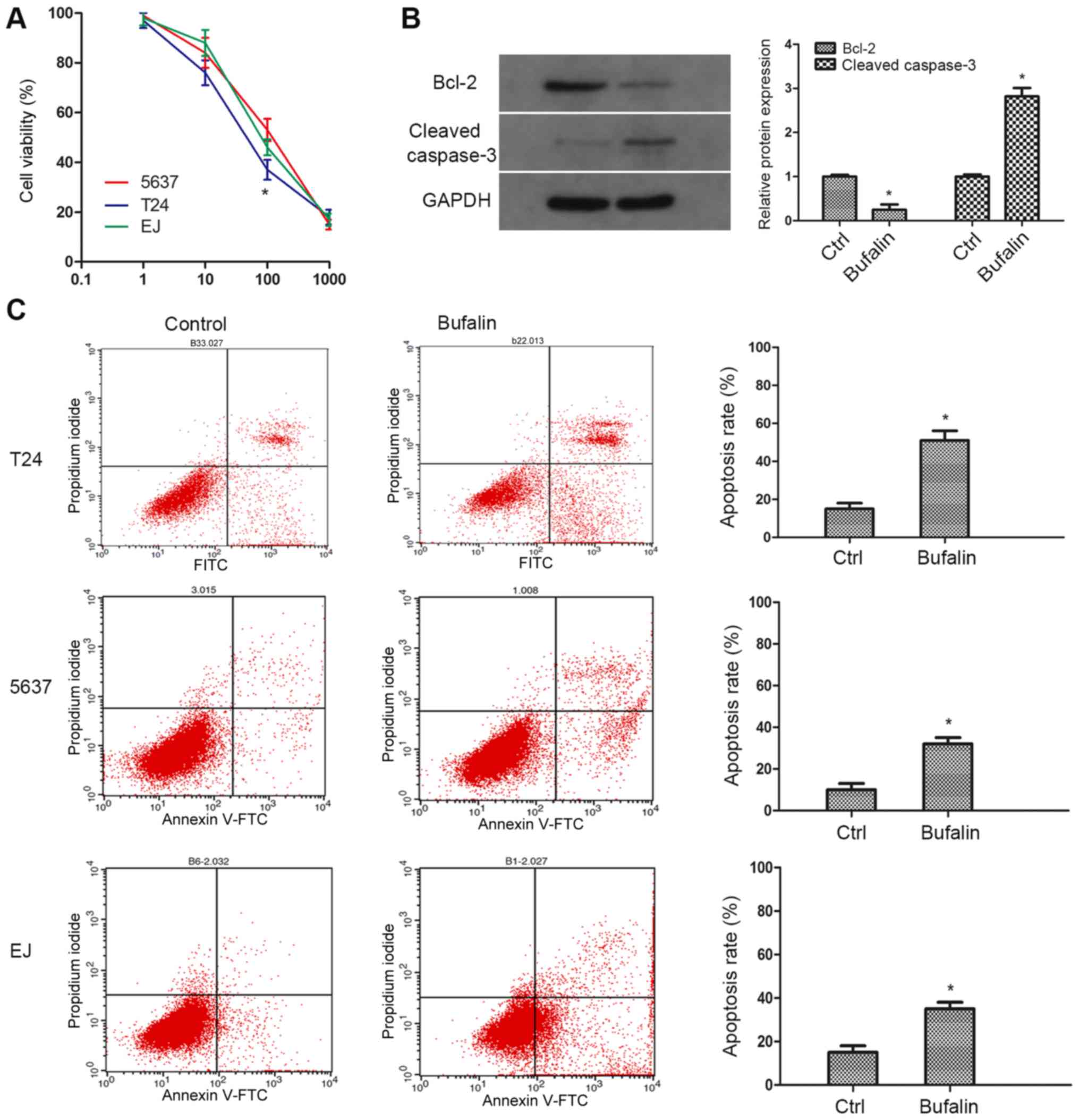
To investigate the pharmacological effect of Bufalin
on bladder cancer, two bladder cancer cell lines (T24, 5637) were
employed to simulate different stages of the tumour (Fig. 1). A CCK-8 assay was performed to
examine sensitivities to Bufalin in tumour cells. As the result
showed, compared with 5,637 cells, cell viability of T24 was
significantly inhibited by Bufalin at a concentration of 100 nM
(Fig. 1A). A cell apoptosis assay was
performed to determine the cytostatic effect of Bufalin. As the
result showed, compared with the control group, Bufalin induced
remarkable apoptosis in a total of three cell lines, and the
apoptosis rate in T24 cells was the most significant (Fig. 1C). Therefore, T24 cells and 100 nM
concentration of Bufalin were used for subsequent research. Western
blotting was additionally performed to explore the protein
expression of apoptotic phenotypes caspase-3 and Bcl-2. In
accordance with our expectation, the expression of caspase-3 was
markedly upregulated in contrast with the expression of Bcl-2
(Fig. 1B). These results demonstrated
that Bufalin inhibits tumour cell growth and promotes cell
apoptosis in bladder cancer.
Bufalin-induced apoptosis in bladder
cancer cells through inactivation of NKA
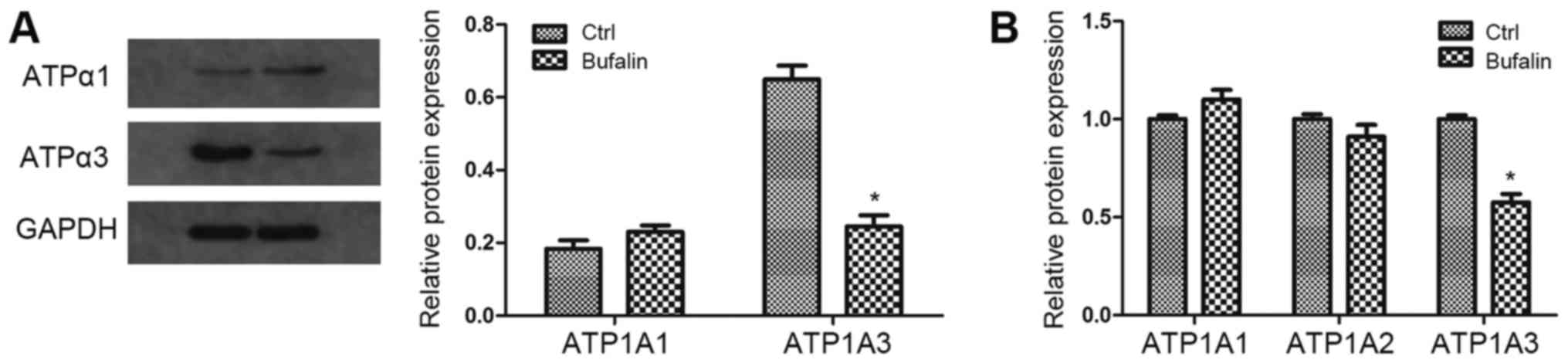
To determine whether ATPase is involved in
Bufalin-induced cell apoptosis, we examined the expression of three
subunits of ATPase (α1, α2 and α3) on protein or mRNA level in T24
cells. The results revealed that the expression of α3-ATPase was
significantly inhibited by Bufalin on both the protein and mRNA
level, while the expression of α1-ATPase and α2-ATPase was
moderately changed (Fig. 2). These
findings suggest that NKA is involved in the cell growth and
apoptosis of bladder cancer, and the α3 subunit of ATPase may play
an important role among the three subunits.
α3-NKA overexpression attenuated
Bufalin-induced apoptosis in bladder cancer cells
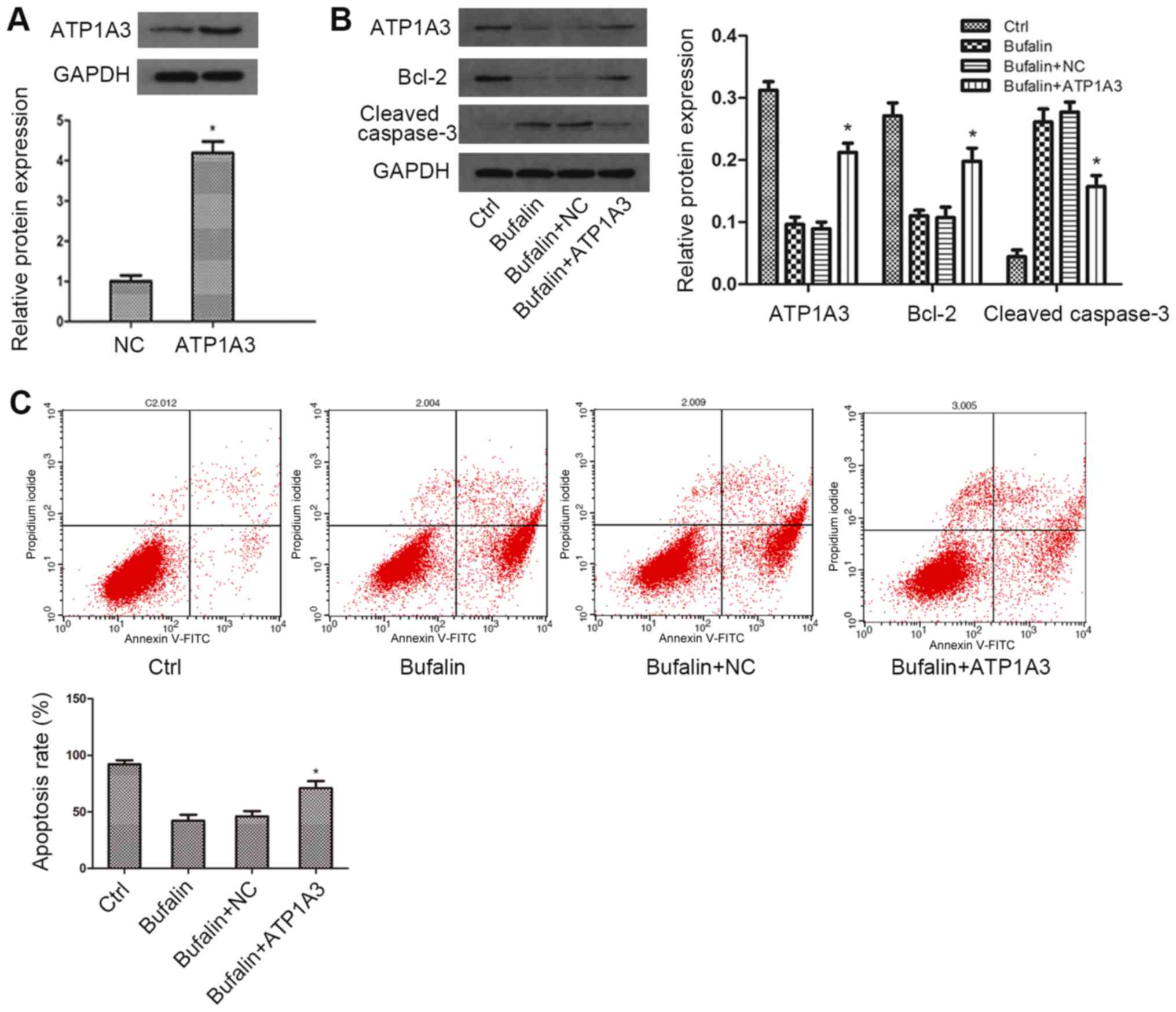
To further investigate the role of α3 subunit of NKA
in Bufalin-induced cytostatic effect in bladder cancer cells, we
constructed plasmids encoding α3 isoforms of ATPase to overexpress
α3-ATPase. The transfection efficiency was examined by western blot
analysis. As the western blot result showed, compared with the
control group, the expression of α3-ATPase was remarkably
upregulated on the protein level (Fig.
3A). Subsequently, we explored the expression of α3-ATPase,
caspase-3 and Bcl-2 under Bufalin treatment by western blotting. As
the data showed, compared with the Bufalin group, the expression of
α3-ATPase was moderately increased in the α3-isoform overexpression
group, while the expression change of caspase-3 and Bcl-2 induced
by Bufalin was significantly weakened in α3-isoform overexpressing
cells (Fig. 3B). The results from the
cell apoptosis assay showed that compared with the Bufalin group,
overexpression of α3-ATPase attenuated Bufalin-induced cell
apoptosis (Fig. 3C). Taken together,
these results confirmed that α3 subunit of NKA was the most
important subunit in cell growth and apoptosis of bladder
cancer.
Discussion
Bladder cancer is one of the most common urological
malignant tumours worldwide and is the 6th leading cause of new
cancer cases and the 9th leading cause of cancer-associated
mortality among all types of cancer (18,19). The
majority of non-muscle-invasive bladder cancers can be diagnosed
and treated early for their clinical symptoms and signs; however,
non-muscle-invasive bladder cancer is vulnerable to recur or
progress to invasive-muscle disease, which is considered an
aggressive and extremely virulent disease (20). Therefore, it is necessary to search
for more effective chemotherapy drugs to improve the prognosis and
survival of patients. Bufalin is a topoisomerase II inhibitor and
is involved in the regulation of the development process of
leukaemia, gastric, colon, breast, and ovarian cancer and other
malignant tumours (21,22). Based on these results, we aimed to
demonstrate that Bufalin may play an antitumour role in bladder
cancer cells by downregulation of NKA.
Apoptosis is a self-killing process of programmed
cell death that includes a range of cellular, morphological and
biochemical changes (23). It is
known that human mammalian cells exhibit two major pathways of
apoptosis: The intrinsic (or mitochondrial) and extrinsic (or death
receptor) signal transduction pathways (24). It has been demonstrated that the
mitochondrial pathway process of apoptosis is regulated by gene
expression and its activation may stimulate the degradation of
cellular substrates and participate in the pathogenesis of many
diseases (25). Bcl-2 may prevent the
release of cytochrome c from the mitochondria to inhibit
apoptosis, and caspase-3 is considered the convergence point of
multiple apoptosis-activating signals that determines the extent of
apoptosis. Its activation means an irreversible commitment to
cellular apoptosis (26). In previous
study, Qi et al (27) showed
that Bufalin can reduce the expression level of Bcl-2 and stimulate
the activation of caspase-3 to promote cell apoptosis through
mitochondria-mediated pathways in hepatocellular carcinoma cells.
One of the major features of Bufalin in the present study is the
inhibition of proliferation, a vital process that plays an
important role in maintaining normal tissue structure and functions
(28). Recent studies showed that
Bufalin induced cell apoptosis in non-small cell lung cancer,
choriocarcinoma and osteosarcoma cells (29–31). To
explore the effect of Bufalin on cell apoptosis and proliferation
in bladder cancer, we performed an MTT assay, cell apoptosis assay
and western blot. The results showed that the T24 cell line was
markedly inhibited and the most sensitive to Bufalin. Moreover,
Bufalin treatment resulted in the cleavage of caspase-3 activation
while blocking Bcl-2 expression. These results indicated that
Bufalin promotes apoptosis and inhibits proliferation in bladder
cancer.
NKA is a trans-membrane protein complex in mammals,
which pumps three Na+ ions out and two K+ ions into a
cell per molecule of hydrolysed ATP to regulate the intracellular
ion gradients (32). Apart from its
function as an ion pump, NKA is also a multifunctional protein in
signal transduction, cell junctions, adhesion and motility
(33,34). NKA contains four isoforms of the
α-subunit (α1, α2, α3 and α4) and three of the β-subunit (β1, β2
and β3) in vertebrates, and the α-subunit of NKA is the active
subunit participated in the binding of cardiac steroids and NKA
(16,35). Generally, the α1 subunit is widely
expressed in various cell types, and the α2 subunit is mostly
expressed in the heart muscle, skeletal muscle and brain. The α3
subunit is found in the central nervous system, ovaries and
placental tissues, and the α4 subunit is restricted to the testes
and is synthesized at the stage of spermatogonium (36,37). A
number of in vivo and in vitro studies have confirmed
that the α1 and α3 subunit is aberrantly expressed in a wide range
of tumours. For example, the expression level of NKA-α1 is
upregulated in glioblastoma, lung and skin cancers, while is
downregulated in bowel cancer. On the other hand, NKA-α3 is found
to be upregulated in rectal and colorectal cancers (38,39). Our
results also showed that the expression level of the α3 subunit in
bladder cancer cells was significantly upregulated among all α
subunits. Furthermore, previous studies have demonstrated that NKA
is a main target of Bufalin and is tightly associated with cell
apoptosis and proliferation in malignant tumour occurrence and
progression. For example, Bufalin induces apoptosis by
downregulating NKA in human lymphoblastic leukaemia cells (40), and Bufalin suppresses hepatocellular
carcinoma cells proliferation by negatively regulating NKA
(41). In our study, we found that
Bufalin significantly inhibited α3 subunit expression in T24 cells.
By transfection with α3 isoform overexpressing plasmids, we found
that compared with Bufalin groups, cell apoptosis was markedly
attenuated in NKA-α3 overexpressing cells. These results suggested
that the α3 isoform of NKA played a critical role in
Bufalin-induced cell apoptosis.
In conclusion, our study indicated that Bufalin can
promote cell apoptosis in bladder cancer, and this cytostatic
effect may be ascribed to the inactivation of NKA. These findings
implied that Bufalin has the potential to be applied as an
effective antitumour medicine in the treatment of bladder
cancer.
Acknowledgements
Not applicable.
Funding
No funding was received.
Availability of data and materials
The datasets during and/or analysed during the
current study available from the corresponding author on reasonable
request.
Authors' contributions
HH was involved in the conception and design of the
study, and manuscript writing. WZ was involved in the study design,
supervision of all phases of the study, data collection and data
analysis.
Ethics approval and consent to
participate
Not applicable.
Patient consent for publication
Not applicable.
Competing interests
The authors declare that they have no competing
interests.
References
|
1
|
Antoni S, Ferlay J, Soerjomataram I, Znaor
A, Jemal A and Bray F: Bladder cancer incidence and mortality: A
global overview and recent trends. Eur Urol. 71:96–108. 2017.
View Article : Google Scholar : PubMed/NCBI
|
|
2
|
Wirth M, Plattner VE and Gabor F:
Strategies to improve drug delivery in bladder cancer therapy.
Expert Opin Drug Deliv. 6:727–744. 2009. View Article : Google Scholar : PubMed/NCBI
|
|
3
|
Liu H, Chang JK, Hou JQ, Zhao ZH and Zhang
LD: Inhibition of miR-221 influences bladder cancer cell
proliferation and apoptosis. Eur Rev Med Pharmacol Sci.
21:3193–3199. 2017.PubMed/NCBI
|
|
4
|
Akagashi K, Tanda H, Kato S, Ohnishi S,
Nakajima H, Nanbu A, Nitta T, Koroku M, Sato Y and Hanzawa T:
Recurrence pattern for superficial bladder cancer. Int J Urol.
13:686–691. 2006. View Article : Google Scholar : PubMed/NCBI
|
|
5
|
Herr HW: High-risk superficial bladder
cancer: Transurethral resection alone in selected patients with T1
tumor. Semin Urol Oncol. 15:142–146. 1997.PubMed/NCBI
|
|
6
|
Pasin E, Josephson DY, Mitra AP, Cote RJ
and Stein JP: Superficial bladder cancer: An update on etiology,
molecular development, classification, and natural history. Rev
Urol. 10:31–43. 2008.PubMed/NCBI
|
|
7
|
Masson-Lecomte A, Rava M, Real FX,
Hartmann A, Allory Y and Malats N: Inflammatory biomarkers and
bladder cancer prognosis: A systematic review. Eur Urol.
66:1078–1091. 2014. View Article : Google Scholar : PubMed/NCBI
|
|
8
|
Rose TL, Deal AM, Nielsen ME, Smith AB and
Milowsky MI: Sex disparities in use of chemotherapy and survival in
patients with advanced bladder cancer. Cancer. 122:2012–2020. 2016.
View Article : Google Scholar : PubMed/NCBI
|
|
9
|
Yoshida T, Okuyama H, Nakayama M, Endo H,
Nonomura N, Nishimura K and Inoue M: High-dose chemotherapeutics of
intravesical chemotherapy rapidly induce mitochondrial dysfunction
in bladder cancer-derived spheroids. Cancer Sci. 106:69–77. 2015.
View Article : Google Scholar : PubMed/NCBI
|
|
10
|
Wang J, Chen C, Wang S, Zhang Y, Yin P,
Gao Z, Xu J, Feng D, Zuo Q, Zhao R and Chen T: Bufalin inhibits
HCT116 colon cancer cells and its orthotopic xenograft tumor in
mice model through genes related to apoptotic and PTEN/AKT
pathways. Gastroenterol Res Pract. 2015:4571932015. View Article : Google Scholar : PubMed/NCBI
|
|
11
|
Yin PH, Liu X, Qiu YY, Cai JF, Qin JM, Zhu
HR and Li Q: Anti-tumor activity and apoptosis-regulation
mechanisms of bufalin in various cancers: New hope for cancer
patients. Asian Pac J Cancer Prev. 13:5339–5343. 2012. View Article : Google Scholar : PubMed/NCBI
|
|
12
|
Yang LH, Zhang HZ, Zhang B, Chen F, Lai
ZH, Xu LF and Jin XQ: Studies on the chemical constituents from the
skin of Bufo bufo gargarizans Cantor. Yao Xue Xue Bao. 27:679–683.
1992.(In Chinese). PubMed/NCBI
|
|
13
|
Yin P, Wang Y, Qiu Y, Hou L, Liu X, Qin J,
Duan Y, Liu P, Qiu M and Li Q: Bufalin-loaded mPEG-PLGA-PLL-cRGD
nanoparticles: Preparation, cellular uptake, tissue distribution,
and anticancer activity. Int J Nanomedicine. 7:3961–3969.
2012.PubMed/NCBI
|
|
14
|
Han KQ, Huang G, Gu W, Su YH, Huang XQ and
Ling CQ: Anti-tumor activities and apoptosis-regulated mechanisms
of bufalin on the orthotopic transplantation tumor model of human
hepatocellular carcinoma in nude mice. World J Gastroenterol.
13:3374–3379. 2007. View Article : Google Scholar : PubMed/NCBI
|
|
15
|
Koh CH, Wu J, Chung YY, Liu Z, Zhang RR,
Chong K, Korzh V, Ting S, Oh S, Shim W, et al: Electronic
supplementary material identification of a Na+/K+-ATPase
inhibition-independent proarrhythmic ionic mechanisms of cardiac
glycosides. Sci Rep. 7:24652017. View Article : Google Scholar : PubMed/NCBI
|
|
16
|
Liu M, Feng LX, Sun P, Liu W, Wu WY, Jiang
BH, Yang M, Hu LH, Guo DA and Liu X: A novel bufalin derivative
exhibited stronger apoptosis-inducing effect than bufalin in A549
lung cancer cells and lower acute toxicity in mice. PLoS One.
11:e01597892016. View Article : Google Scholar : PubMed/NCBI
|
|
17
|
Rao X, Huang X, Zhou Z and Lin X: An
improvement of the 2^(-delta delta CT) method for quantitative
real-time polymerase chain reaction data analysis. Biostat
Bioinforma Biomath. 3:71–85. 2013.PubMed/NCBI
|
|
18
|
Fan B, Zhang X, Ma Y and Zhang A:
Fangchinoline induces apoptosis, autophagy and energetic impairment
in bladder cancer. Cell Physiol Biochem. 43:1003–1011. 2017.
View Article : Google Scholar : PubMed/NCBI
|
|
19
|
Xu H, Xie L, Liu X, Zhang Y, Shen Z, Chen
T, Qiu X, Sha N, Xing C, Wu Z, et al: Impact of squamous and/or
glandular differentiation on recurrence and progression following
transurethral resection for non-muscle invasive urothelial
carcinoma of bladder. Oncol Lett. 14:3522–3528. 2017. View Article : Google Scholar : PubMed/NCBI
|
|
20
|
Tran K and Severn M: Blue light cystoscopy
in patients with suspected non-muscle invasive bladder carcinoma: A
review of clinical utility [Internet]. Ottawa (ON): Canadian Agency
for Drugs and Technologies in Health; 2017
|
|
21
|
Kang KH, Han MH, Jeong JW, Park C, Lee SH,
Lee HW and Hong SH, Choi YH and Hong SH: Bufalin sensitizes human
bladder carcinoma cells to TRAIL-mediated apoptosis. Oncol Lett.
14:853–859. 2017. View Article : Google Scholar : PubMed/NCBI
|
|
22
|
Zhang N, Xie Y, Tai Y, Gao Y, Guo W, Yu W,
Li J, Feng X, Hao J, Gao Y, et al: Bufalin inhibits hTERT
expression and colorectal cancer cell growth by targeting CPSF4.
Cell Physiol Biochem. 40:1559–1569. 2016. View Article : Google Scholar : PubMed/NCBI
|
|
23
|
Vaux DL and Korsmeyer SJ: Cell death in
development. Cell. 96:245–254. 1999. View Article : Google Scholar : PubMed/NCBI
|
|
24
|
Kerr JF, Wyllie AH and Currie AR:
Apoptosis: A basic biological phenomenon with wide-ranging
implications in tissue kinetics. Br J Cancer. 26:239–257. 1972.
View Article : Google Scholar : PubMed/NCBI
|
|
25
|
Arnoult D, Parone P, Martinou JC,
Antonsson B, Estaquier J and Ameisen JC: Mitochondrial release of
apoptosis-inducing factor occurs downstream of cytochrome c release
in response to several proapoptotic stimuli. J Cell Biol.
159:923–929. 2002. View Article : Google Scholar : PubMed/NCBI
|
|
26
|
Liang H, Yu F, Tong Z, Yuan B and Wang C:
Effect of ischemia post-conditioning on skeletal muscle oxidative
injury, mTOR, Bax, Bcl-2 proteins expression, and HIF-1α/β-actin
mRNA, IL-6/β-actin mRNA and caveolin-3/β-actin mRNA expression in
ischemia-reperfusion rabbits. Mol Biol Rep. 40:507–514. 2013.
View Article : Google Scholar : PubMed/NCBI
|
|
27
|
Qi F, Inagaki Y, Gao B, Cui X, Xu H,
Kokudo N, Li A and Tang W: Bufalin and cinobufagin induce apoptosis
of human hepatocellular carcinoma cells via Fas- and
mitochondria-mediated pathways. Cancer Sci. 102:951–958. 2011.
View Article : Google Scholar : PubMed/NCBI
|
|
28
|
Hanahan D and Weinberg RA: Hallmarks of
cancer: The next generation. Cell. 144:646–674. 2011. View Article : Google Scholar : PubMed/NCBI
|
|
29
|
Jiang Y, Zhang Y, Luan J, Duan H, Zhang F,
Yagasaki K and Zhang G: Effects of bufalin on the proliferation of
human lung cancer cells and its molecular mechanisms of action.
Cytotechnology. 62:573–583. 2010. View Article : Google Scholar : PubMed/NCBI
|
|
30
|
Takai N, Ueda T, Ishii T, Kira N, Nishida
M, Nishida Y, Nasu K and Narahara H: Effects of bufalin on the
proliferation of human choriocarcinoma cells. Int J Gynecol Cancer.
21:1105–1109. 2011. View Article : Google Scholar : PubMed/NCBI
|
|
31
|
Zhang J, Sha J, Zhou Y, Han K, Wang Y, Su
Y, Yin X, Hu H and Yao Y: Bufalin inhibits proliferation and
induces apoptosis in osteosarcoma cells by downregulating
MicroRNA-221. Evid Based Complement Alternat Med. 2016:73194642016.
View Article : Google Scholar : PubMed/NCBI
|
|
32
|
Ren YP, Zhang MJ, Zhang T and Huang RW:
Dual effects of ouabain on the regulation of proliferation and
apoptosis in human umbilical vein endothelial cells: Involvement of
Na(+)-K(+)-ATPase α-subunits and NF-κB. Int J Clin Exp Med.
7:1214–1222. 2014.PubMed/NCBI
|
|
33
|
Kaplan JH: Biochemistry of Na,K-ATPase.
Annu Rev Biochem. 71:511–535. 2002. View Article : Google Scholar : PubMed/NCBI
|
|
34
|
Mobasheri A, Avila J, Cózar-Castellano I,
Brownleader MD, Trevan M, Francis MJ, Lamb JF and Martín-Vasallo P:
Na+, K+-ATPase isozyme diversity; comparative biochemistry and
physiological implications of novel functional interactions. Biosci
Rep. 20:51–91. 2000. View Article : Google Scholar : PubMed/NCBI
|
|
35
|
Nguyen AN, Jansson K, Sánchez G, Sharma M,
Reif GA, Wallace DP and Blanco G: Ouabain activates the Na-K-ATPase
signalosome to induce autosomal dominant polycystic kidney disease
cell proliferation. Am J Physiol Renal Physiol. 301:F897–F906.
2011. View Article : Google Scholar : PubMed/NCBI
|
|
36
|
Felippe Gonçalves-de-Albuquerque C, Silva
Ribeiro A, da Silva Ignácio C, Castro-Faria-Neto Caire H and Burth
P: Na/K pump and beyond: Na/K-ATPase as a modulator of apoptosis
and autophagy. Molecules. 22:pii: E578. 2017.
|
|
37
|
Dostanic-Larson I, Lorenz JN, Van Huysse
JW, Neumann JC, Moseley AE and Lingrel JB: Physiological role of
the alpha1- and alpha2-isoforms of the Na+-K+-ATPase and biological
significance of their cardiac glycoside binding site. Am J Physiol
Regul Integr Comp Physiol. 290:R524–R528. 2006. View Article : Google Scholar : PubMed/NCBI
|
|
38
|
Sakai H, Suzuki T, Maeda M, Takahashi Y,
Horikawa N, Minamimura T, Tsukada K and Takeguchi N: Up-regulation
of Na(+),K(+)-ATPase alpha 3-isoform and down-regulation of the
alpha1-isoform in human colorectal cancer. FEBS Lett. 563:151–154.
2004. View Article : Google Scholar : PubMed/NCBI
|
|
39
|
Mijatovic T, Ingrassia L, Facchini V and
Kiss R: Na+/K+-ATPase alpha subunits as new targets in anticancer
therapy. Expert Opin Ther Targets. 12:1403–1417. 2008. View Article : Google Scholar : PubMed/NCBI
|
|
40
|
Kawazoe N, Aiuchi T, Masuda Y, Nakajo S
and Nakaya K: Induction of apoptosis by bufalin in human tumor
cells is associated with a change of intracellular concentration of
Na+ ions. J Biochem. 126:278–286. 1999. View Article : Google Scholar : PubMed/NCBI
|
|
41
|
Xu Y, Liu X, Schwarz S, Hu L, Guo D, Gu Q
and Schwarz W: Inhibitory efficacy of bufadienolides on Na+,K+-pump
activity versus cell proliferation. Biochem Biophys Rep. 6:158–164.
2016.PubMed/NCBI
|