Introduction
Gastric cancer (GC) is the fourth most common
malignancy and causes the second most cancer-associated mortalities
globally in 2010, particularly in East Asia (1,2). The
majority of GC cases are diagnosed at an advanced stage, which
notably contributes to the poor prognosis associated with the
disease (3); therefore, it is
important to determine novel effective biomarkers, in order to
improve the diagnosis and medical treatment of GC.
Long non-coding RNAs (lncRNAs) are transcribed RNA
sequences with a length >200 nucleotides (nt) and no
protein-encoding ability (4). A
number of previous studies have reported that numerous lncRNAs are
dysregulated in a series of cancer types, including GC, and that
they serve as biomarkers for GC diagnosis and prognosis of patients
with GC (5–8). For example, HOX antisense intergenic RNA
(HOTAIR) was one of the earliest lncRNAs studied. It is upregulated
in GC, compared with normal adjacent tissues (NATs), and its
expression is a significant predictor of poor prognosis (9). Endo et al (10) reported similar results in diffuse-type
GC. The lncRNA H19 has attracted much attention in recent years and
the expression levels of H19 in GC tissues are significantly
increased compared with NATs, and its expression is notably
associated with poor prognosis of patients with GC (5,11–13). However, the role of lncRNAs in GC
remains unknown and further research is required.
In the present study, the focus was on the lncRNA
AB007962, which is located on chromosome 1 and has a transcript
length of 5,053 nt (http://genome.ucsc.edu/cgi-bin/hgGene?hgg_gene=uc001eso.1&db=hg19).
Its expression was analyzed in GC tissues, and the association
between its expression and the clinicopathological features and
prognosis of patients with GC was assessed. Furthermore, a cohort
of patients with GC from The First Hospital of China Medical
University (Shenyang, China) and a cohort from The Cancer Genome
Atlas (TCGA) were reviewed, in order to assess the clinical utility
of AB007962 expression levels.
Materials and methods
Specimens and clinical data
collection
All of the 96 paired human GC tissues and matched
NAT samples were obtained from patients who underwent radical
resection of GC at The First Hospital of China Medical University
between May 2009 and July 2010. The median age of the 96 patients
with GC was 62 years (range, 30-81 years), with 70 males accounting
for 72.9% and 26 females accounting for 27.1% of the cohort.
Furthermore, 77 patients (80.2%) had lymph node metastasis (pN1-N3)
and all of the 96 patients completed follow-up. The matched NAT
samples were collected from tissues that were located ≥5 cm from
the edge of the tumor. All of the samples were frozen immediately
in liquid nitrogen and stored at −80°C until use. Two pathologists
from The First Hospital of China Medical University (Shenyang,
China) histopathologically confirmed each sample taken for
diagnosis of GC. In addition, there was no radiotherapy or
chemotherapy prior to resection. The histological grade of the
samples was staged by following the standards set by the World
Health Organization (14). The tumor
stage was also evaluated on the basis of the 7th edition of the
Tumor-Node-Metastasis (TNM) staging system (15). Overall survival (OS) was defined as
the interval between the date of surgery and the date of mortality
or the last follow-up examination. Disease-free survival (DFS) was
defined as the interval between the date of surgery and the date of
recovery from disease or the last follow-up examination.
Ethical approval of the study
protocol
The present study was performed according to the
principles expressed in The Declaration of Helsinki. Tissue samples
were collected once written informed consent was obtained from all
96 patients, and following institutional ethical guidelines that
were reviewed and approved by the Research Ethics Committee of
China Medical University (Shenyang, China).
RNA isolation and reverse
transcription-quantitative polymerase chain reaction (RT-qPCR)
According to the manufacturer's protocols, total RNA
was isolated from frozen specimens of tumor and NATs using
TRIzol® (Invitrogen; Thermo Fisher Scientific, Inc.,
Waltham, MA, USA). The purity of the total RNA was verified by
ultraviolet (UV) spectrophotometry (2.0>A260/A280>1.8) using
a NanoPhotometer UV/Vis spectrophotometer (Implen GmbH, Münich,
Germany). Using a PrimeScript RT reagent kit (Takara Biotechnology
Co., Ltd., Dalian, China), RNA was reverse transcribed into cDNA
for qPCR according to the manufacturer's protocol. For qPCR,
according to the manufacturer's protocol, the 25 µl final volume
contained 12.5 µl 2× SYBR® Premix Ex Taq™ (Takara
Biotechnology Co., Ltd.), 0.5 µl forward and reverse primers each
(10 µM), 9.5 µl nuclease-free water, and 2 µl diluted of the
reverse transcribed product The primers (Sangon Biotech Co., Ltd.,
Shanghai, China) used in the present study were as follows: GAPDH
forward, 5′-CGGATTTGGTCGTATTGGG-3′ and reverse,
5′-CTGGAAGATGGTGATGGGATT-3′; AB007962 forward,
5′-TGTGCTCACGATGTTGAATG-3′ and reverse, 5′-AATGGAAATGGGACATGGAC-3′.
The reaction was incubated in a 96-well optical plate and amplified
for: 1 cycle of 95°C for 30 sec; and 45 cycles of 95°C for 5 sec
and 60°C for 30 sec. Relative expression of AB007962 was normalized
to GAPDH and calculated using the 2−ΔΔCq method
(16). ∆Cq is the
difference in the Cq value between the target and
endogenous reference (∆Cq=Cq lncRNA-Cq
GAPDH). ∆∆Cq is the difference in ∆Cq
values between the tumor cells and the control
(∆∆Cq=∆Cq tumor lncRNA-∆Cq NATs
lncRNA).
TCGA gene expression data
The expression of AB007962 in human gastric
specimens was analyzed using publicly available TCGA Illumina
RNA-Seq datasets (http://cancergenome.nih.gov) of 274 patients with GC,
including their clinical parameters and follow-up data. The present
study used the reads per kilobase of exon per million reads mapped
(RPKM) value to represent the expression levels of AB007962 for
each patient from TCGA datasets (17). Amongst these, matched tumor and
non-tumor specimens were available for 29 patients with GC.
Statistical analysis
Statistical analysis was performed using the SPSS
21.0 software package (IBM Corp., Armonk, NY, USA). A paired
Student's t-test was used to evaluate the difference in AB007962
expression between tumor tissues and NATs and the expression levels
from TCGA datasets between groups were calculated by a
nonparametric test (Wilcoxon test). The association between
AB007962 expression and clinicopathological factors was analyzed
using the Mann-Whitney U test for two groups and the Kruskal-Wallis
test for three or more groups. Survival curves were evaluated by
the Kaplan-Meier method and the log-rank test was used to determine
whether there was a statistically significant difference between
survival curves. Cox proportional hazards analysis was performed to
calculate the hazard ratio (HR) and the 95% confidence interval
(CI) to estimate the association between AB007962 expression level
and OS and DFS survival time. P<0.05 was considered to indicate
a statistically significant difference.
Results
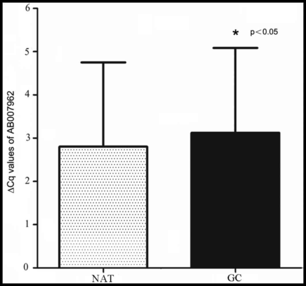
Expression of AB007962 is
downregulated in GC tissues
The expression level of AB007962 in 96 paired
cancerous and matched NATs was examined by RT-qPCR. The results
demonstrated that AB007962 expression levels were reduced in
cancerous tissues, compared with matched NATs (paired Student's
t-test; P<0.05; Fig. 1).
Furthermore, the AB007962 expression levels in 58/96 (60.4%) GC
tissues were downregulated, compared with matched NATs. The
AB007962 relative gene expression were further analyzed in patients
with GC from the TCGA gene expression datasets, and the results
demonstrated that there was no statistical differences of AB007962
expression in cancerous tissues, compared with NATs [read per
kilobase of transcript per million mapped reads
(RPKM)GC, 0.379±0.078; RPKMNATs, 0.400±0.095;
Wilcoxon test, P=0.633].
Association between AB007962 levels in
cancer tissues and clinicopathological factors in patients with
GC
The association between the expression level of
AB007962 and clinicopathological factors in 96 patients with GC was
investigated, including sex, age, tumor size, tumor location,
macroscopic type, histological grade and TNM stage. No significant
associations were identified between AB007962, and sex (P=0.154),
age (P=0.772), tumor location (P=0.515), tumor size (P=0.078)
macroscopic type (P=0.241), histological differentiation (P=0.492),
pathological (p)T stage (P=0.756), pN stage (P=0.070), pTNM stage
(P=0.978) or invasion into the lymphatic vessels (P=0.820).
Furthermore, through the use of TCGA gene expression datasets, no
significant associations were identified between AB007962
expression and clinicopathological factors, including sex
(P=0.726), Lauren type (P=0.152), histological differentiation
(P=0.532), pT stage (P=0.250), pN stage (P=0.472) or pTNM stage
(P=0.092).
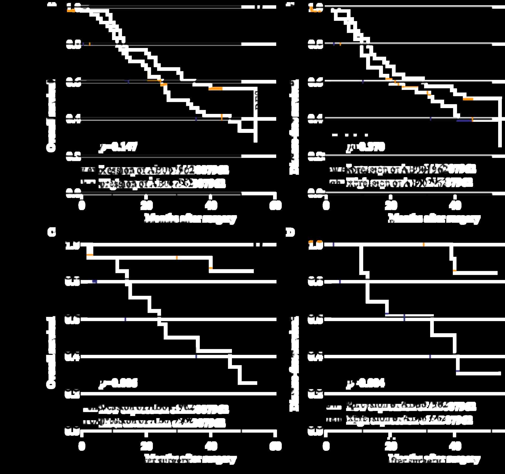
Association between AB007962
expression and patient survival time
In order to investigate whether the expression level
of AB007962 was associated with the prognosis of the 96 patients
with GC, survival data on the patients following gastrectomy was
collected. The median ratio of relative AB007962 expression in the
96 patients with GC was used as the cut-off value to divide the 96
patients into groups with high and low AB007962 expression.
Kaplan-Meier analysis and the log-rank test were used to evaluate
the association between AB007962 expression, and OS and DFS. The
results demonstrated that there was no significant difference in OS
(P=0.147; Fig. 2A) or DFS (P=0.370;
Fig. 2B) between the two groups.
However, among 28 patients with intestinal-type GC,
those with increased AB007962 expression had significantly
increased OS (P=0.006; Fig. 2C) and
DFS (P=0.004; Fig. 2D) compared with
the reduced expression group. The univariate survival analysis for
OS and DFS indicated that AB007962 expression (P=0.016) and tumor
location (P=0.026) were prognostic factors for OS, and AB007962
expression (P=0.015) and tumor size (P=0.033) were significant
prognostic factors for DFS (Table I).
P<0.05 was considered to indicate a statistically significant
difference for multivariate Cox analysis, and the multivariate
analysis confirmed that AB007962 expression levels were retained as
an independent prognostic indicator for OS (HR=6.185; 95% CI,
1.331-28.732; P=0.020) and DFS (HR=7.982; 95% CI, 1.638-38.894;
P=0.010) (Table II) of patients with
intestinal-type GC.
 | Table I.Association between the expression of
long non-coding RNA AB007962 and the clinicopathological features
in 96 patients with gastric cancer. |
Table I.
Association between the expression of
long non-coding RNA AB007962 and the clinicopathological features
in 96 patients with gastric cancer.
| Characteristics | No. of patients
(n=96) | Expression
levela | P-value |
|---|
| Age, years |
|
| 0.772 |
| ≤62 | 48 | 0.87 (0.49-1.32) |
|
|
>62 | 48 | 0.78 (0.44-1.18) |
|
| Sex |
|
| 0.151 |
|
Male | 70 | 0.71
(0.49-1.13) |
|
|
Female | 26 | 1.07
(0.48-1.49) |
|
| Tumor size, cm |
|
| 0.078 |
|
≤5.5 | 50 | 0.93
(0.49-1.49) |
|
|
>5.5 | 46 | 0.70
(0.50-1.04) |
|
| Tumor location |
|
| 0.515 |
| Upper
stomach | 5 | 0.41
(0.25-1.46) |
|
| Middle
stomach | 40 | 0.73
(0.49-1.17) |
|
| Lower
stomach | 47 | 0.92
(0.49-1.35) |
|
| Entire
stomach | 4 | 1.05
(0.63-1.48) |
|
| Macroscopic
typeb |
|
| 0.241 |
|
Borrmann I+II | 10 | 1.02
(0.79-1.37) |
|
|
Borrmann III+IV | 83 | 0.72
(0.48-1.20) |
|
| Histological
grade |
|
| 0.492 |
|
Good | 39 | 0.72
(0.48-1.18) |
|
|
Poor | 57 | 0.86
(0.49-1.32) |
|
| Lauren grade |
|
| 0.693 |
|
Intestinal type | 28 | 0.78
(0.52-1.40) |
|
| Diffuse
type | 68 | 0.83
(0.48-1.19) |
|
| pT stage |
|
| 0.953 |
|
T1+T2 | 13 | 0.73
(0.45-1.34) |
|
|
T3+
T4 | 83 | 0.83
(0.49-1.24) |
|
| pN stage |
|
| 0.070 |
| N0 | 19 | 0.71
(0.37-1.20) |
|
| N1 | 17 | 1.12
(0.87-1.86) |
|
| N2 | 23 | 0.67
(0.42-1.43) |
|
| N3 | 37 | 0.71
(0.49-1.05) |
|
| pTNM stage |
|
| 0.977 |
| I | 7 | 0.89
(0.35-1.20) |
|
| II | 19 | 0.73
(0.41-1.24) |
|
|
III | 70 | 0.82
(0.50-1.28) |
|
| Invasion into
lymphatic vessels |
|
| 0.817 |
|
Negative | 50 | 0.73
(0.49-1.21) |
|
|
Positive | 46 | 0.86
(0.49-1.41) |
|
| Table II.Univariate and multivariate Cox
regression analyses AB007962 for overall survival times or
Disease-free survival times of patients with intestinal-type
gastric cancer in the study cohort (n=28). |
Table II.
Univariate and multivariate Cox
regression analyses AB007962 for overall survival times or
Disease-free survival times of patients with intestinal-type
gastric cancer in the study cohort (n=28).
|
| OS | DFS |
|---|
|
|
|
|
|---|
| Variables | HR | 95% CI | P-value | HR | 95% CI | P-value |
|---|
| Univariate
analysis |
|
|
|
|
|
|
|
AB007962 expression (high vs.
low) | 6.540 | 1.419-30.134 | 0.016a | 7.044 | 1.469-33.784 | 0.015a |
| Tumor
size (≤5.5 vs. >5.5 cm) | 0.557 | 0.178-1.743 | 0.315 | 0.228 | 0.059-0.889 | 0.033a |
| Age
(≤62 vs. >62 years) | 1.048 | 0.331-3.318 | 0.936 | 1.271 | 0.367-4.396 | 0.705 |
| Sex
(male vs. female) | 0.883 | 0.192-4.054 | 0.872 | 0.595 | 0.075-4.708 | 0.623 |
| Tumor
location (middle + lower vs. upper) | 5.711 | 1.234-2.436 | 0.026a | 2.433 | 0.628-9.426 | 0.198 |
|
Macroscopic type (Borrmann
I+II vs. III+IV) | 0.689 | 0.143-3.329 | 0.643 | 0.464 | 0.059-3.676 | 0.467 |
|
Invasion depth (T1+T2 vs.
T3+T4) | 0.427 | 0.055-3.314 | 0.415 | 0.569 | 0.072-4.493 | 0.592 |
|
Regional lymph nodes (N0 vs.
N1+N2+N3) | 1.800 | 0.565-5.732 | 0.320 | 1.727 | 0.482-6.188 | 0.401 |
| TNM
stage (I+II vs. III+IV) | 1.340 | 0.430-4.177 | 0.614 | 0.885 | 0.249-3.143 | 0.850 |
|
Invasion into lymphatic
vessels (negative vs. positive) | 1.497 | 0.449-4.986 | 0.511 | 1.049 | 0.295-3.727 | 0.941 |
| Multivariate
analysis |
|
|
|
|
|
|
|
AB007962 expression (high vs.
low) | 6.185 | 1.331-28.732 | 0.020a | 7.982 | 1.638-38.894 | 0.010a |
| Tumor
size (≤5.5 vs. >5.5 cm) | NA | NA | NA | 0.199 | 0.050-0.788 | 0.022a |
| Tumor
location (middle + lower vs. upper) | 5.403 | 1.152-25.349 | 0.032a | NA | NA | NA |
Nevertheless, from TCGA gene expression datasets, no
significant association was identified between AB007962 expression,
and the OS and DFS of patients with intestinal-type GC.
Discussion
Currently, the majority of GC cases are diagnosed at
an advanced stage and patients have a poor prognosis (18); thus, early diagnosis of GC is of great
importance. In order to achieve early diagnosis of GC, novel
biomarkers for diagnosis and prognosis should be determined and
used in clinical medicine. For example, Japan and South Korea have
provided a government-sponsored screening program for GC, which has
contributed to a low mortality-to-incidence ratio in these
countries (19).
Previously, numerous studies have demonstrated that
aberrant expression of lncRNAs serves an essential role in various
human cancer types during the process of initiation and development
(20–22). Furthermore, increasing evidence
indicates that a number of lncRNAs serve as novel diagnostic and
prognostic biomarkers in GC (9,13,23–25). For
example, two well-known lncRNAs, H19 and maternally expressed 3
(MEG3), were reported to be dysregulated in GC (5,26). H19 is
overexpressed in GC tissues and plasma, compared with NATs
(24); therefore, the level of H19 in
plasma may be used to distinguish patients with early stage GC from
healthy individuals. Additionally, its sensitivity is notably
increased, compared with traditional biomarkers in plasma,
including carcinoembryonic antigen and carbohydrate antigen-199;
therefore, the expression of H19 may be used to predict the
prognosis of patients with GC (5,24,27). Furthermore, Sun et al (26) reported that MEG3 is significantly
down-regulated and is associated with reduced OS times in GC, and
therefore it also served as a prognostic biomarker for patients
with GC. In the present study, the expression level of AB007962 was
investigated in GC tissues and NATs by RT-qPCR. The result
indicated that AB007962 was down-regulated in the majority of the
96 GC tissues, compared with their matched NATs. These results
indicated that AB0074962 may be used as a novel diagnostic
biomarker in GC.
In a previous study, Zhang et al (8) determined that antisense non-coding RNA
in the INK4 locus (ANRIL) was significantly associated with tumor
size, and indicated that knockdown of ANRIL inhibited GC cell
proliferation in vitro. Sun et al (26) reported, similarly, that MEG3 was
associated with tumor size and it may also regulate cell
proliferation in GC. In the present study, the expression levels of
AB007962 decreased in patients with larger tumor size; therefore,
it was hypothesized that low expression levels of AB007962 may
contribute to the proliferation of GC. Whether AB007962 regulated
cell proliferation should be investigated further in the
future.
The Lauren classification dates back to 1965
(28), and it is frequently used in
clinical diagnosis and treatment (29). According to the Lauren classification,
GC may be divided into intestinal, diffuse and mixed types
(28). A previous study indicated
that patients with intestinal-type GC have an improved prognosis,
compared with those with other Lauren types (30). The intestinal type of GC is described
as a tumor with glandular architecture and resembling colonic
carcinoma (31), and its relative
frequency is ~54% (32). A number of
studies have reported that intestinal-type GC is frequently
associated with intestinal metaplasia and Helicobacter
pylori infection (33,34).
Previously, researchers have determined that a
number of biomarkers predict the prognosis of patients with GC of
different Lauren types. Human epithelial growth factor receptor 2
(HER2) is a member of the HER family. Its overexpression has been
frequently observed in intestinal-type GC (35). Qiu et al (31) determined that HER2 positivity was an
independent prognostic factor in patients with intestinal-type GC.
Furthermore, it was reported that patients with HER2-negative and
intestinal-types exhibit the longest OS and DFS times (31). HOTAIR may also act as a prognostic
biomarker in diffuse-type GC, whereby patients with increased
HOTAIR expression have a reduced prognosis compared with those with
reduced HOTAIR expression (10). In
the present study, it was determined that AB007962 expression was
significantly associated with poor prognosis in 28 patients with
intestinal-type GC, with patients with a reduced expression level
of AB007962 having a reduced prognosis, compared with those with an
increased expression level. These results indicated that AB0074962
may act as a novel prognostic biomarker in intestinal-type GC.
In the present study, it was determined that
AB007962 expression has a negative association with tumor size. It
was also negatively associated with the OS and DFS time of patients
with intestinal-type GC; however, the analysis of TCGA dataset of
274 GC patients indicated no significant association between
AB007962 expression, clinicopathological factors and patient
survival time. The difference of results may be attributed to
different population data and different measuring methods. Firstly,
all patients in the present study were from the Han population
living in North China. The majority of the patients in the TCGA
dataset were non-Asian (73.36%), with no patients from the Han
population living in North China. Additionally, RT-qPCR was
performed to measure the expression level of AB007962 in GC tissues
and matched NATs; however, AB007962 expression levels were
determined by RNA Sequencing in TCGA dataset. Future studies should
expand the sample size and investigate if these data also apply to
other populations.
In conclusion, the results of the present study
indicated that AB007962 expression is downregulated in GC.
Furthermore, the expression level of AB007962 may predict the
outcome of patients with intestinal-type GC; therefore, AB007962 is
a potential novel biomarker for prognosis. However, the function
and molecular mechanisms of AB007962, particularly in cell
proliferation, requires further study.
Acknowledgements
The authors would like to thank the Department of
Surgical Oncology of The First Hospital of China Medical University
for providing human gastric tissue samples. Additionally, the
authors would like to thank the College of China Medical University
for technical assistance in experiments.
Funding
The present study was supported by National Science
Foundation of China (grant nos. 81201888, 81372549 and 81172370),
Clinical Capability Construction Project for Liaoning Provincial
Hospitals (grant no. LNCCC-A01-2014) and Natural Science Foundation
of Liaoning Province (grant no. 2014029201).
Availability of data and materials
All data generated or analyzed during the present
study are included in this published article.
Authors' contributions
YS and ZW designed experiments, JW and YY performed
experiments, PG, XC and BM analyzed data, JW and YY wrote the
manuscript, JS was responsible for interpreting the data and
revising the article. All authors read and approved the final
manuscript.
Ethics approval and consent to
participate
The present study was approved by the Ethics
Committee of the First Hospital of China Medical University. All
participants provided written informed consent.
Patient consent for publication
Written informed consent was obtained from each
patient.
Competing interests
The authors declare that they no competing
interests.
References
|
1
|
Herszényi L and Tulassay Z: Epidemiology
of gastrointestinal and liver tumors. Eur Rev Med Pharmacol Sci.
14:249–258. 2010.PubMed/NCBI
|
|
2
|
Jemal A, Siegel R, Xu J and Ward E: Cancer
statistics, 2010. CA Cancer J Clin. 60:277–300. 2010. View Article : Google Scholar : PubMed/NCBI
|
|
3
|
Kagawa S, Shigeyasu K, Ishida M, Watanabe
M, Tazawa H, Nagasaka T, Shirakawa Y and Fujiwara T: Molecular
diagnosis and therapy for occult peritoneal metastasis in gastric
cancer patients. World J Gastroenterol. 20:17796–17803. 2014.
View Article : Google Scholar : PubMed/NCBI
|
|
4
|
Esteller M: Non-coding RNAs in human
disease. Nat Rev Genet. 12:861–874. 2011. View Article : Google Scholar : PubMed/NCBI
|
|
5
|
Li H, Yu B, Li J, Su L, Yan M, Zhu Z and
Liu B: Overexpression of lncRNA H19 enhances carcinogenesis and
metastasis of gastric cancer. Oncotarget. 5:2318–2329. 2014.
View Article : Google Scholar : PubMed/NCBI
|
|
6
|
Liu XH, Sun M, Nie FQ, Ge YB, Zhang EB,
Yin DD, Kong R, Xia R, Lu KH, Li JH, et al: Lnc RNA HOTAIR
functions as a competing endogenous RNA to regulate HER2 expression
by sponging miR-331-3p in gastric cancer. Mol Cancer. 13:922014.
View Article : Google Scholar : PubMed/NCBI
|
|
7
|
Xu TP, Liu XX, Xia R, Yin L, Kong R, Chen
WM, Huang MD and Shu YQ: SP1-induced upregulation of the long
noncoding RNA TINCR regulates cell proliferation and apoptosis by
affecting KLF2 mRNA stability in gastric cancer. Oncogene.
34:5648–5661. 2015. View Article : Google Scholar : PubMed/NCBI
|
|
8
|
Zhang EB, Kong R, Yin DD, You LH, Sun M,
Han L, Xu TP, Xia R, Yang JS, De W and Chen Jf: Long noncoding RNA
ANRIL indicates a poor prognosis of gastric cancer and promotes
tumor growth by epigenetically silencing of miR-99a/miR-449a.
Oncotarget. 5:2276–2292. 2014. View Article : Google Scholar : PubMed/NCBI
|
|
9
|
Xu ZY, Yu QM, Du YA, Yang LT, Dong RZ,
Huang L, Yu PF and Cheng XD: Knockdown of long non-coding RNA
HOTAIR suppresses tumor invasion and reverses
epithelial-mesenchymal transition in gastric cancer. Int J Biol
Sci. 9:587–597. 2013. View Article : Google Scholar : PubMed/NCBI
|
|
10
|
Endo H, Shiroki T, Nakagawa T, Yokoyama M,
Tamai K, Yamanami H, Fujiya T, Sato I, Yamaguchi K, Tanaka N, et
al: Enhanced expression of long non-coding RNA HOTAIR is associated
with the development of gastric cancer. PLoS One. 8:e770702013.
View Article : Google Scholar : PubMed/NCBI
|
|
11
|
Yang F, Bi J, Xue X, Zheng L, Zhi K, Hua J
and Fang G: Up-regulated long non-coding RNA H19 contributes to
proliferation of gastric cancer cells. FEBS J. 279:3159–3165. 2012.
View Article : Google Scholar : PubMed/NCBI
|
|
12
|
Zhuang M, Gao W, Xu J, Wang P and Shu Y:
The long non-coding RNA H19-derived miR-675 modulates human gastric
cancer cell proliferation by targeting tumor suppressor RUNX1.
Biochem Biophys Res Commun. 448:315–322. 2014. View Article : Google Scholar : PubMed/NCBI
|
|
13
|
Zhang EB, Han L, Yin DD, Kong R, De W and
Chen J: c-Myc-induced, long, noncoding H19 affects cell
proliferation and predicts a poor prognosis in patients with
gastric cancer. Med Oncol. 31:9142014. View Article : Google Scholar : PubMed/NCBI
|
|
14
|
Hamilton SR and Aaltonen LA: World Health
Organization Classifcation of Tumours. Pathology and genetics of
tumours of the digestive system. International Agency for Research
on Cancer (IARC) Press; Lyon, Paris: 2000
|
|
15
|
Sobin LH, Gospodarowicz M and Wittekind C:
International Union Against Cancer (UICC). TNM classifcation of
malignant tumours. 7th edition. Hoboken, NJ: Wiley-Liss, New York,
NY, USA; pp. 117–126. 2010
|
|
16
|
Livak KJ and Schmittgen TD: Analysis of
relative gene expression data using real-time quantitative PCR and
the 2(−Delta Delta C(T)) method. Methods. 25:402–408. 2001.
View Article : Google Scholar : PubMed/NCBI
|
|
17
|
Mortazavi A, Williams BA, McCue K,
Schaeffer L and Wold B: Mapping and quantifying mammalian
transcriptomes by RNA-Seq. Nat Methods. 5:621–628. 2008. View Article : Google Scholar : PubMed/NCBI
|
|
18
|
Thiel A and Ristimäki A: Gastric cancer:
Basic aspects. Helicobacter. 17 Suppl 1:S26–S29. 2012. View Article : Google Scholar
|
|
19
|
Shen L, Shan YS, Hu HM, Price TJ, Sirohi
B, Yeh KH, Yang YH, Sano T, Yang HK, Zhang X, et al: Management of
gastric cancer in Asia: Resource-stratified guidelines. Lancet
Oncol. 14:e535–e547. 2013. View Article : Google Scholar : PubMed/NCBI
|
|
20
|
Mercer TR, Dinger ME and Mattick JS: Long
non-coding RNAs: Insights into functions. Nat Rev Genet.
10:155–159. 2009. View
Article : Google Scholar : PubMed/NCBI
|
|
21
|
Hu W, Alvarez-Dominguez JR and Lodish HF:
Regulation of mammalian cell differentiation by long non-coding
RNAs. EMBO Rep. 13:971–983. 2012. View Article : Google Scholar : PubMed/NCBI
|
|
22
|
Kaikkonen MU, Lam MT and Glass CK:
Non-coding RNAs as regulators of gene expression and epigenetics.
Cardiovasc Res. 90:430–440. 2011. View Article : Google Scholar : PubMed/NCBI
|
|
23
|
Lee NK, Lee JH, Park CH, Yu D, Lee YC,
Cheong JH, Noh SH and Lee SK: Long non-coding RNA HOTAIR promotes
carcinogenesis and invasion of gastric adenocarcinoma. Biochem
Biophys Res Commun. 451:171–178. 2014. View Article : Google Scholar : PubMed/NCBI
|
|
24
|
Zhou X, Yin C, Dang Y, Ye F and Zhang G:
Identification of the long non-coding RNA H19 in plasma as a novel
biomarker for diagnosis of gastric cancer. Sci Rep. 5:115162015.
View Article : Google Scholar : PubMed/NCBI
|
|
25
|
Kretz M, Siprashvili Z, Chu C, Webster DE,
Zehnder A, Qu K, Lee CS, Flockhart RJ, Groff AF, Chow J, et al:
Control of somatic tissue differentiation by the long non-coding
RNA TINCR. Nature. 493:231–235. 2013. View Article : Google Scholar : PubMed/NCBI
|
|
26
|
Sun M, Xia R, Jin F, Xu T, Liu Z, De W and
Liu X: Downregulated long noncoding RNA MEG3 is associated with
poor prognosis and promotes cell proliferation in gastric cancer.
Tumour Biol. 35:1065–1073. 2014. View Article : Google Scholar : PubMed/NCBI
|
|
27
|
Arita T, Ichikawa D, Konishi H, Komatsu S,
Shiozaki A, Shoda K, Kawaguchi T, Hirajima S, Nagata H, Kubota T,
et al: Circulating long non-coding RNAs in plasma of patients with
gastric cancer. Anticancer Res. 33:3185–3193. 2013.PubMed/NCBI
|
|
28
|
Lauren P: The two histological main types
of gastric carcinoma: Diffuse and so-called intestinal-type
carcinoma. An attempt at a histo-clinical classification. Acta
Pathol Microbiol Scand. 64:31–49. 1965. View Article : Google Scholar : PubMed/NCBI
|
|
29
|
Hu B, El Hajj N, Sittler S, Lammert N,
Barnes R and Meloni-Ehrig A: Gastric cancer: Classification,
histology and application of molecular pathology. J Gastrointest
Oncol. 3:251–261. 2012.PubMed/NCBI
|
|
30
|
Bravo Neto GP, dos Santos EG, Victer FC
and Carvalho CE: Lymph node metastasis in early gastric cancer. Rev
Col Bras Cir. 41:11–17. 2014. View Article : Google Scholar : PubMed/NCBI
|
|
31
|
Qiu M, Zhou Y, Zhang X, Wang Z, Wang F,
Shao J, Lu J, Jin Y, Wei X, Zhang D, et al: Lauren classification
combined with HER2 status is a better prognostic factor in Chinese
gastric cancer patients. BMC Cancer. 14:8232014. View Article : Google Scholar : PubMed/NCBI
|
|
32
|
Polkowski W, van Sandick JW, Offerhaus GJ,
ten Kate FJ, Mulder J, Obertop H and van Lanschot JJ: Prognostic
value of Laurén classification and c-erbB-2 oncogene overexpression
in adenocarcinoma of the esophagus and gastroesophageal junction.
Ann Surg Oncol. 6:290–297. 1999. View Article : Google Scholar : PubMed/NCBI
|
|
33
|
Kaneko S and Yoshimura T: Time trend
analysis of gastric cancer incidence in Japan by histological
types, 1975-1989. Br J Cancer. 84:400–405. 2001. View Article : Google Scholar : PubMed/NCBI
|
|
34
|
Parsonnet J, Vandersteen D, Goates J,
Sibley RK, Pritikin J and Chang Y: Helicobacter pylori infection in
intestinal- and diffuse-type gastric adenocarcinomas. J Natl Cancer
Inst. 83:640–643. 1991. View Article : Google Scholar : PubMed/NCBI
|
|
35
|
Bang YJ, Van Cutsem E, Feyereislova A,
Chung HC, Shen L, Sawaki A, Lordick F, Ohtsu A, Omuro Y, Satoh T,
et al: Trastuzumab in combination with chemotherapy versus
chemotherapy alone for treatment of HER2-positive advanced gastric
or gastro-oesophageal junction cancer (ToGA): A phase 3,
open-label, randomised controlled trial. Lancet. 376:687–697. 2010.
View Article : Google Scholar : PubMed/NCBI
|