Introduction
Lung cancer is one of the most common types of
cancer (1). Lung cancer has two main
histological types: Non-small cell lung cancer (NSCLC; 80.4%) and
SCLC (16.8%) (2); however, the
underlying mechanisms for the development of lung cancer are not
yet completely characterized.
Accumulated genetic abnormalities are associated
with cancer. Somatic mutations in a number of genes, including
epidermal growth factor receptor (EGFR), P53, KRAS,
BRAF, Erb-B2 receptor tyrosine kinase 2 (ERBB2),
MET, serine/threonine kinase 11, PIK3CA and Parkin RBR E3
ubiquitin protein ligase, have been identified in patients with
lung cancer (3,4). Gene amplifications, including of
EGFR, ERBB2, MET, PIK3CA and NK2 homeobox 1, have also been
detected in lung cancer (5). A number
of single nucleotide polymorphisms (SNPs) are associated with lung
cancer susceptibility, including in interleukin-1, cytochrome P450,
a 5′SNP in the ERCC excision repair 6, chromatin remodeling factor
gene and SNPs in the nicotinic acetylcholine receptor gene cluster
on chromosome 15q25.1 (6–9). Genetic abnormalities have been
identified in numerous pathways, including the Notch (10), EGFR (11), PI3K (12), phosphatase and tensin
homolog/phospho-Akt/P53 (13),
mitogen-activated protein kinase (MAPK) (14) and cell cycle pathways.
In the past decade, there has been a pervasive
application of high-throughput molecular technologies, including
microarrays, in lung cancer research (15–17), which
has greatly enriched the knowledge of the pathogenesis of the
disease, and may potentially provide markers for the prognosis and
targeted therapy of lung cancer. By enrolling 105 subjects in an
Environment And Genetics in Lung cancer Etiology study (http://dceg.cancer.gov/eagle), Landi et al
(18) produced a microarray dataset
that included 107 expression values from tumor (n=58) and non-tumor
tissues (n=49) from 74 subjects (non-smokers, n=20; former smokers,
n=26; current smokers, n=28). Using this microarray analysis, 122
genes were identified that were differentially expressed between
the tumor and non-tumor samples. In addition, several crucial
smoking-associated genes and pathways were identified in the study,
and a number of these, including Nima related kinase 2 and TTK
protein kinase, were experimentally validated; however, the
relationship between these genes and pathways were not considered
in the original study.
In the present study, based on the microarray
dataset produced by Landi et al (18), GSE10072, a novel pathway-pathway
crosstalk approach was employed to identify pathways and genes that
may have critical roles in the pathogenesis of lung cancer, and a
number of the identified genes were experimentally validated.
Materials and methods
Source of pathway and microarray
data
Protein-protein interaction data were downloaded
from the Human Protein Reference Database (http://www.hprd.org/), and 201 lung cancer pathways
were downloaded from the Kyoto Encyclopedia of Genes and Genomes
(KEGG, http://www.kegg.jp/) database (19,20) using
‘lung cancer’ as the search term.
The raw data of the gene expression profile dataset
GSE10072 in the ‘.CEL’ format were downloaded from the Gene
Expression Omnibus database (http://www.ncbi.nlm.nih.gov/geo/).
Identification of differentially
expressed genes (DEGs)
The raw downloaded data were preprocessed and
normalized using the R/Bioconductor package Affy with the Robust
Multichip Average method for single-channel Affymetrix chips
(21). A one-way analysis of variance
was applied to each probe set to identify those that significantly
changed expression level over time, as previously described
(22). P<0.05 was considered to
indicate a statistically significant result; the raw P-value was
adjusted with the Bonferroni method (23).
Impact analysis
The pathway impact analysis as described by Draghici
et al (24) was adopted, which
considers the statistical significance of the enrichment of KEGG
pathways, while also considering other crucial factors, including
the magnitude of expression change for each gene, the topology of
the signaling pathway, and the interactions between signaling
pathways.
Construction of pathway-pathway
crosstalk network
A hyper geometric distribution framework was applied
to evaluate the significance of all non-empty intersections between
two pathways, as previously described (25): Fisher's exact test computed the
probability, p* using hyper geometric distribution with the
parameters (S, NG, N).
p*=p(X=α|S,NG,N)=(Sα)(N-SNG-α)(NNG)
Where α was the number of DEGs in the pathway
intersection; S, the number of DEGs in the pathway union;
NG, the number of genes in the pathway
intersection; and N, the number of genes in the pathway
union.
The P-value to reject the null hypothesis with a
probability of <p* was calculated using the sum of the
probabilities with the same marginal totals, i.e.:
p=∑(Si)(N-sNG-i)(NNG)
This procedure gave a two-tailed probability for a
Fisher's exact test. P<0.05 was considered to indicate a
statistically significant result, indicative of the association of
two pathways.
Analysis of cross-talk between
pathways
The state of a pathway was initially determined. A
pathway was considered to be activated when it met the following
criteria: i) Number of DEGs in the pathway >10; ii) Q-value
[(number of upregulated DEGs in the pathway-number of downregulated
DEGs in the pathway)/total number of genes in the pathway] >0.5.
A repressed pathway met the two criteria: i) Number of DEGs in this
pathway >10; ii) Q-value <-0.5. Next, pairs of pathways
sharing common DEGs were identified, and these DEGs were
listed.
Cell culture and treatment
Normal lung K562 cells were purchased from the
American Type Culture Collection (Manassas, VA, USA). The cells
were grown in RPMI-1640 (Gibco; Thermo Fisher Scientific, Inc.,
Waltham, MA, USA) supplemented with 10% heat-inactivated fetal
bovine serum (Gibco; Thermo Fisher Scientific, Inc.) and 100 U/ml
penicillin and streptomycin, in a humidified atmosphere with 5%
CO2 at 37°C. To maintain drug resistance, adriamycin was
supplemented at regular intervals for 2 weeks prior to any
experiment. Benzopyrene was used to treat the cells at
concentrations of 0.01, 0.1, 1 and 10 µM in the subsequent
assays.
Measurement of cell viability
Cell proliferation was measured using an MTT assay.
Cells (2×10⁵ cells/ml) were seeded in 96-well plates
with increasing concentrations of adriamycin, SNX-2112 and 17-AAG.
After incubation at 37°C for 24, 48 and 72 h, 5 mg/ml MTT solution
was added for incubation for 4 h. Then, 100 µl/well DMSO was added
to solubilize the formazan crystals. Cell viability was assessed by
measuring absorbance at 570 nm using a microplate reader (Bio-Rad
Laboratories, Inc., Hercules, CA, USA).
Western blot analysis
Cells were first lysed in lysis buffer (Sangon
Biotech Co., Ltd., Shanghai, China) and then total protein was
extracted. Followed by protein concentration was measured by the
bicinchoninic acid method. Equal aliquots (20 µg protein per lane)
of protein lysate were separated by 8–12% SDS-PAGE and transferred
to a polyvinylidene difluoride membrane. Subsequent to blocking
with 5% skimmed milk at 37°C for 1 h, Western blots were probed
with primary antibodies against nuclear factor (NF)-κB (1:1,000;
cat no. ab32360; Abcam, Cambridge, MA, USA), Akt (1:1,000; cat no.
ab126811; Abcam), cyclin B (1:1,000; cat no. ab18221; Abcam), P53
(1:1,000; cat no. ab21985; Abcam), growth arrest and DNA damage
inducible β (GADD45B) (1:1,000; cat no. ab128920; Abcam) and
β-actin (1:1,000; cat no. ab6276; Abcam) overnight at 4°C. Three
consecutive washes were performed for 10 min in PBS-Tween, followed
by incubation with the alkaline phosphatase-conjugated goat
anti-rabbit IgG secondary antibody (1:5,000; cat no. ab6722; Abcam)
diluted in 5% skimmed milk at room temperature for 1 h. The
immunoblots were visualized with enhanced chemiluminescence (GE
Healthcare, Chicago, IL, USA) and autoradiography. The experiments
were repeated three times, and the results were detected using the
Image Lab software (version 4.1; Bio-Rad Laboratories, Inc.) on
ChemiDoc MP imaging system (Bio-Rad Laboratories, Inc.).
Statistical analysis
All data were presented as the mean ± standard
deviation. Statistical analysis was performed by one-way analysis
of variance with a Bonferroni post hoc test in SPSS software
(version 13.0; SPSS, Inc., Chicago, IL, USA). P<0.05 was
considered to indicate a statistically significant difference.
Results
Identification of DEGs
Using a threshold of P<0.05, the genes with
significantly differential expression were analyzed. A total of
1,763 DEGs were identified, of which 662 were associated with KEGG
pathways.
Result of impact analysis
A total of 11 pathways were identified in the
pathway impact analysis. Of these, ‘complement and coagulation
cascades’, ‘ECM-receptor interaction’, ‘P53 signaling pathway’,
‘cell adhesion molecules (CAMs)’, ‘focal adhesion’ and ‘cell cycle’
were the top five pathways by impact factor value (Table I).
 | Table I.Significantly enriched Kyoto
Encyclopedia of Genes and Genomes pathways in GSE10072. |
Table I.
Significantly enriched Kyoto
Encyclopedia of Genes and Genomes pathways in GSE10072.
| Pathway | Impact factor | P-value |
|---|
| Complement and
coagulation cascades | 17.258 |
2.08×10⁻⁷ |
| ECM-receptor
interaction | 14.266 |
3.90×10⁻⁶ |
| P53 signaling
pathway | 9.017 |
3.41×10⁻⁴ |
| Cell adhesion
molecules | 70.734 |
4.42×10⁻⁴ |
| Focal adhesion | 9.283 |
1.17×10⁻³ |
| Cell cycle | 6.673 |
3.36×10⁻³ |
| Renin-angiotensin
system | 5.778 |
8.30×10⁻³ |
| PPAR signaling
pathway | 5.733 |
1.54×10⁻² |
| TGF-beta signaling
pathway | 6.352 |
2.24×10⁻² |
| Leukocyte
transendothelial migration | 114.039 |
2.34×10⁻² |
| migration Tight
junction | 6.133 |
4.85×10⁻² |
Analysis of cross-talk between
pathways
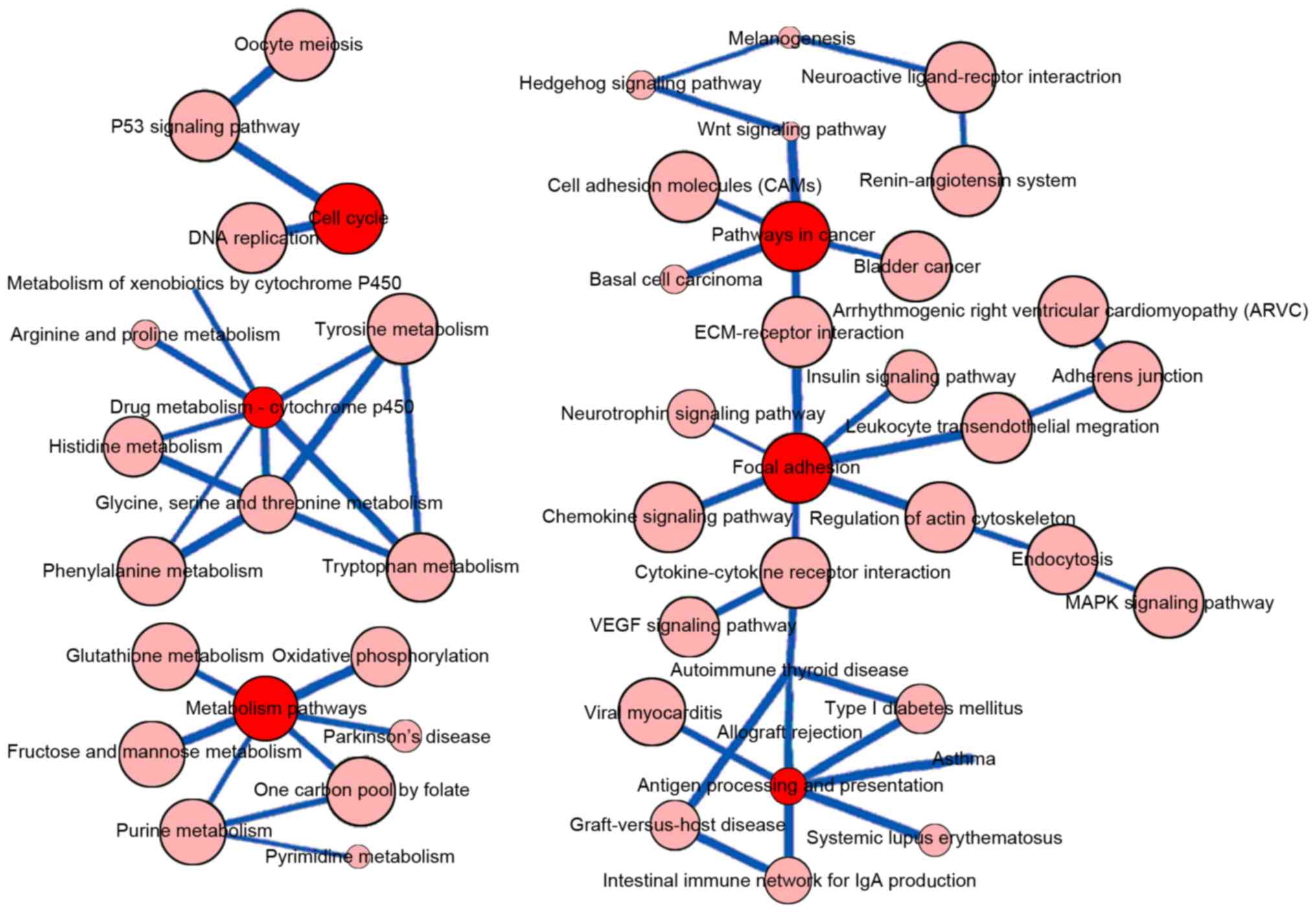
It was determined that the ‘cell cycle’, ‘drug
metabolism-cytochrome P450’, ‘metabolic pathways’, ‘pathways in
cancer’, ‘focal adhesion’ and ‘antigen processing and presentation’
KEGG pathways were central in the pathway-pathway crosstalk network
(Fig. 1).
Using the criteria defined for activated and
repressed pathways, 2 pathways (‘cell cycle’ and ‘P53 signaling
pathway’) were repressed, and 10 pathways were activated (Table II). The common DEGs between the
pathways are listed in Table III.
GADD45B was associated with three enriched pathways,
including the activated ‘MAPK signaling pathway’ and the repressed
‘cell cycle’ and ‘P53 signaling pathway’ terms (Fig. 2).
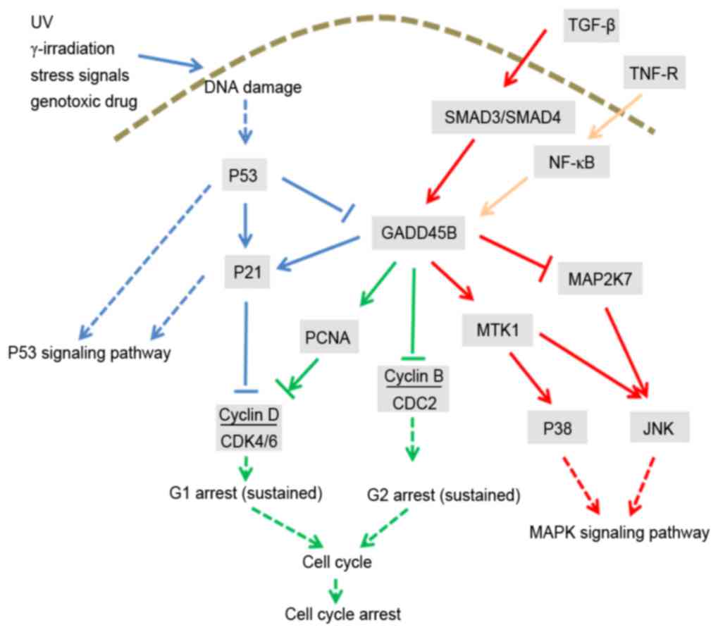 | Figure 2.Schematic diagram of the
pathway-pathway crosstalk via GADD45B. In this figure, the blue
arrows represent the interaction with the P53 signaling pathway,
the green arrows represent the interaction with the cell cycle
pathway and the red arrows represent the interaction with the MAPK
signaling pathway. Yellow arrows represent the interactions between
GADD45B and genes that are not associated with ‘P53 signaling
pathway’, ‘MAPK signaling pathway’ or ‘cell cycle’. Solid lines
represent direct interactions; dashed lines represent indirect
effects. GADD45B, growth arrest and DNA damage inducible β; UV,
ultraviolet radiation; TGF-β, transforming growth factor-β; TNF-R,
tumor necrosis factor receptor; NF-κB, nuclear factor-κB; CDK,
cyclin-dependent kinase; CDC2, cyclin-dependent kinase 1; PCNA,
proliferating cell nuclear antigen; MAPK, mitogen-activated protein
kinase; MTK1, MAPK kinase kinase 4; MAP2K7, MAPK kinase 7; JNK,
c-Jun N-terminal kinase. |
| Table II.Status of the Kyoto Encyclopedia of
Genes and Genomes pathways associated with the differentially
expressed genes in lung cancer. |
Table II.
Status of the Kyoto Encyclopedia of
Genes and Genomes pathways associated with the differentially
expressed genes in lung cancer.
|
| DEGs |
|
|---|
|
|
|
|
|---|
| Pathway | Upregulated | Downregulated | Both | Q-value |
|---|
| Cell cycle | 2 | 11 | 13 | −0.6923 |
| P53 signaling
pathway | 2 | 8 | 10 | −0.6000 |
| Cytokine-cytokine
receptor interaction | 13 | 3 | 16 | 0.6250 |
| Tight junction | 9 | 2 | 11 | 0.6364 |
| Neuroactive
ligand-receptor interaction | 15 | 3 | 18 | 0.6667 |
| Chemokine signaling
pathway | 11 | 2 | 13 | 0.6923 |
| Complement and
coagulation cascades | 14 | 2 | 16 | 0.7500 |
| Axon guidance | 9 | 1 | 10 | 0.8000 |
| Vascular smooth
muscle contraction | 12 | 1 | 13 | 0.8462 |
| MAPK signaling
pathway | 13 | 1 | 14 | 0.8571 |
| Regulation of actin
cytoskeleton | 11 | 0 | 11 | 1.0000 |
| Endocytosis | 10 | 0 | 10 | 1.0000 |
| Table III.Differentially expressed genes
associated with multiple enriched Kyoto Encyclopedia of Genes and
Genomes pathways. |
Table III.
Differentially expressed genes
associated with multiple enriched Kyoto Encyclopedia of Genes and
Genomes pathways.
| Pathway 1 | Pathway 2 | Gene IDs |
|---|
| Cytokine-cytokine
receptor interaction | Chemokine signaling
pathway | 6359, 1524, 2921,
2920, 6387 |
| Neuroactive
ligand-receptor interaction | Vascular smooth
muscle contraction | 1906, 10203, 1909,
185 |
| MAPK signaling
pathway | Regulation of actin
cytoskeleton | 2264, 6237,
2263 |
| MAPK signaling
pathway | Endocytosis | 2264, 7048,
2263 |
| Regulation of actin
cytoskeleton | Endocytosis | 2264, 2263,
8395 |
| Tight junction | Regulation of actin
cytoskeleton | 10398, 6237,
4628 |
| Cytokine-cytokine
receptor interaction | Neuroactive
ligand-receptor interaction | 3953, 2690 |
| Cytokine-cytokine
receptor interaction | Chemokine signaling
pathway | 10563, 9547 |
| Tight junction | Vascular smooth
muscle contraction | 10398, 4629 |
| Tight junction | MAPK signaling
pathway | 6237, 10000 |
| Vascular smooth
muscle contraction | Regulation of actin
cytoskeleton | 10398, 4638 |
| Cell cycle | P53 signaling
pathway | 4616 |
| Cell cycle | MAPK signaling
pathway | 4616 |
| Chemokine signaling
pathway | Axon guidance | 6387 |
| Chemokine signaling
pathway | Vascular smooth
muscle contraction | 115 |
| Chemokine signaling
pathway | MAPK signaling
pathway | 10000 |
| Chemokine signaling
pathway | Regulation of actin
cytoskeleton | 5295 |
| Chemokine signaling
pathway | Endocytosis | 2869 |
| Cytokine-cytokine
receptor interaction | Axon guidance | 6387 |
| Cytokine-cytokine
receptor interaction | MAPK signaling
pathway | 7048 |
| Cytokine-cytokine
receptor interaction | Endocytosis | 7048 |
| Neuroactive
ligand-receptor interaction | Complement and
coagulation cascades | 728 |
| Neuroactive
ligand-receptor interaction | Endocytosis | 154 |
| P53 signaling
pathway | MAPK signaling
pathway | 4616 |
| Tight junction | Chemokine signaling
pathway | 10000 |
| Vascular smooth
muscle contraction | MAPK signaling
pathway | 5319 |
| Vascular smooth
muscle contraction | MAPK signaling
pathway | 5321 |
Measurement of cell viability and
western blot analysis
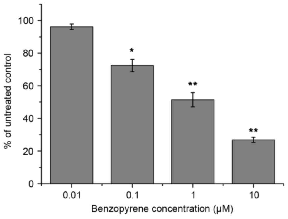
Benzopyrene treatment reduced the viability of lung
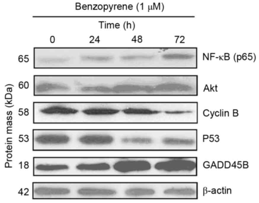
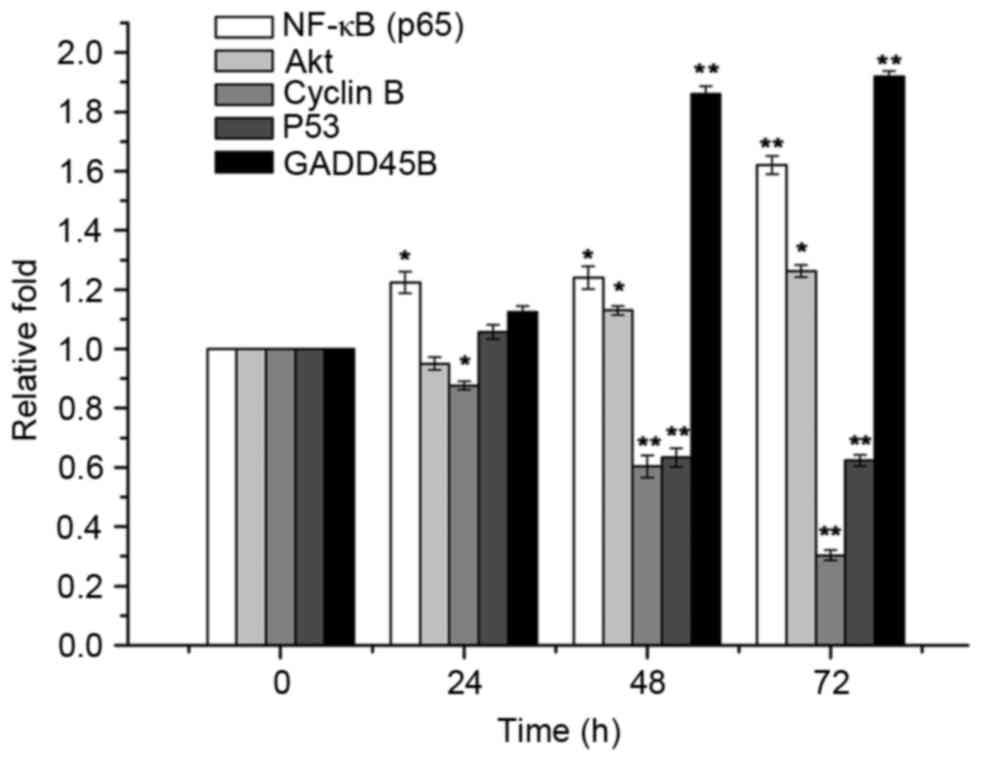
cells at all concentrations from 72 h (Fig. 3). GADD45B, P53, cyclin, Akt and NF-κB
protein levels were detected by Western blotting (Fig. 4). Western blotting demonstrated that
the expression of NF-κB, Akt and GADD45B increased over time in
lung cells treated with benzopyrene, whereas the expression of
cyclin B and P53 was decreased (Fig.
5).
Discussion
In the present study, significantly enriched
pathways were identified with impact analysis, and a
pathway-pathway crosstalk network was constructed. GADD45B was
identified as a connection between a number of the enriched
pathways, and experimental validation of the expression of this
gene and associated pathways was performed.
In comparison to classical pathway analysis, impact
analysis also considers the over-representation of DEGs in a given
pathway and the abnormal perturbation of that pathway, as measured
by the expression changes across the pathway topology (26). Previously, the roles of the P53
signaling pathway, cell adhesion, focal adhesion and the cell cycle
in the pathogenesis of lung cancer have been confirmed and studied
extensively (27,28). In contrast, the complement and
coagulation cascades in this disease have been less reported.
Corrales et al (29), reported
the activation of the complement system by detecting the
anaphylatoxin C5a, a potent immune mediator generated subsequent to
complement activation in lung cancer cell lines. Plasminogen
activator inhibitor (PAI) variants PAI-1 A15T and
PAI-2 S413C influence the prognosis of patients with lung
cancer (30), and PAI-1 has
been demonstrated to inhibit the activation of the coagulation
system (31). Levels of the
erythrocyte complement receptor 1 were significantly lower in
patients with small cell lung cancer (32). A previous study revealed that
activated coagulation factor X inhibited the migration of lung
cancer cells and may serve a key role in cell migration (33). Thus, it was indicated that the
complement and coagulation cascades may also serve an important
role in the pathogenesis of lung cancer.
The pathway-pathway cross-talk network indicated the
central roles of ‘cell cycle’, ‘drug metabolism-cytochrome P450’,
‘metabolic pathways’, ‘pathways in cancer’, ‘focal adhesion’ and
‘antigen processing and presentation’; metabolism-associated
pathways may therefore be particularly important in the
pathogenesis of lung cancer. Tumor cells sustain high rates of
glycolysis even in aerobic conditions to maintain their rapid
growth; alterations in in primary metabolites (34) and glycolysis-associated enzymes,
including hexokinase II and glyceraldehyde-3-phosphate
dehydrogenase (35,36), have been identified in lung cancer.
Additionally, the alteration of metabolic pathways may affect the
efficacy of anti-tumor drugs, e.g. the glutathione metabolic
pathway, which is involved in the detoxification or inactivation of
platinum drugs (37). The cytochromes
P450 are a group of enzymes that catalyze the oxidative
biotransformation of the majority of drugs and other lipophilic
xenobiotics (38).
In the present study, GADD45B, an upregulated
gene, was determined to be a common link between three KEGG
pathways, including the upregulated ‘MAPK signaling pathway’ and
the repressed ‘cell cycle’ and ‘P53 signaling pathway’. It was
therefore considered to potentially have an important role in the
pathogenesis of lung cancer. GADD45B, encoding MyD118, is
part of a highly conserved Gadd45 gene family with GADD45A,
encoding GADD45, and GADD45G, encoding CR6. Gadd45 proteins
have been implicated for their involvement in tumorigenesis
(39) and age-associated pathologies
(40); however, MyD118, GADD45 and
CR6 are considered to have similar but not identical functions via
different apoptotic and growth suppressive pathways (41). The MAPK signaling pathway has been
confirmed to have an important role in the pathogenesis of lung
cancer (42). In the present study,
this signaling pathway was activated in patients with lung cancer.
Gupta et al (43) demonstrated
that GADD45B promotes cell survival via activation of the
GADD45a-P38-NF-κB pathway and inhibition of the MAPK kinase 4-c-Jun
N-terminal kinases (JNK) pathway. P38 and JNK are members of the
MAPK family (44). TGF-β can activate
Smad by phosphorylation (45). One
previous study demonstrates that following the induction by TGF-β,
Smad transcription factors activate P38 via GADD45B (46). Another study demonstrates that SMAD3
and SMAD4 can activate GADD45B following the induction by TGF-β
(47). GADD45B and GADD45G are
cyclin-dependent kinase 2/cyclin B1 kinase inhibitors, and thus
function in in G2/M cell cycle arrest (48). This is in accord with the observation
in the present study that the cell cycle pathway was repressed.
It was also identified in the present study that the
P53 signaling pathway was repressed. Previously, Mi et al
(49) observed the upregulation of
GADD45B and the downregulation of P53 in human prostate cancer
cells exposed to silvestrol. This signaling pathway has been
reported to correlate with the radioresponse of NSCLC (13). GADD45A is a target gene of P53
(50). Lambert et al (51) reported the upregulation of GADD45B
mRNA in Saos-2-His273 cells exposed to proline rich membrane anchor
1, a P53-reactivating agent.
Benzopyrene is a carcinogen particularly associated
with lung cancer (52); therefore,
the normal lung cells were treated with benzopyrene in the present
study to investigate the process of lung carcinogenesis. The
harmful effects of benzopyrene against cell viability were observed
by an MTT assay at various concentrations. Western blotting
demonstrated that the expression of NF-κB, Akt and GADD45B were
increased over time in lung cells treated with benzopyrene, whereas
the expression of cyclin B and P53 were decreased. This
corresponded with the data that the AKT-MAPK pathway was activated
via NF-κB, while the P53 pathway and cell cycle pathway were
repressed; thus, GADD45B may contribute to lung carcinogenesis via
activating the MAPK signaling pathway and repressing the P53
signaling and cell cycle pathways.
Although a series of comprehensive bioinformatics
analyses and validation experiments were performed, there were two
major limitations in the present study: i) Only one lung cell line
was used in the current study, which may not eliminate the
heterogeneity of lung cancer; ii) knockdown of GADD45B was not
performed, and the expressions of other proteins after GADD45B
knockdown were not considered, which may weaken the regulatory
associations between them.
In addition to ‘ECM-receptor interaction’, ‘P53
signaling pathway’, ‘cell adhesion molecules (CAMs)’, ‘focal
adhesion’ and ‘cell cycle’, it was also identified that the
‘complement and coagulation cascades’ pathway may be associated
with the pathogenesis of lung cancer; therefore, it was speculated
that GADD45B may contribute to lung carcinogenesis via activating
the MAPK signaling pathway, and repressing the P53 signaling and
cell cycle pathways. Thus, the role of this gene in lung cancer
should be studied further; GADD45B siRNA knockdown experiments will
assist the further validation of this speculation.
Acknowledgements
Not applicable.
Funding
No funding was received.
Availability of data and material
The datasets used and/or analyzed during the current
study are available from the corresponding author on reasonable
request.
Authors' contributions
XJ and JL were involved in the conception and design
of the research and drafting the manuscript. ZZ participated in the
acquisition of data. RX performed the analysis and interpretation
of data. YG was involved in the statistical analysis, XL
participated in the design of the study and performed the
statistical analysis. All authors read and approved the final
manuscript.
Ethics approval and consent to
participate
Not applicable.
Patient consent for publication
Not applicable.
Competing interests
The authors declare that they have no competing
interests.
References
|
1
|
McGuire S: World cancer report 2014.
Geneva, switzerland: World health organization, international
agency for research on cancer, WHO Press, 2015. Adv Nutr.
7:418–419. 2016. View Article : Google Scholar : PubMed/NCBI
|
|
2
|
Travis WD, Travis LB and Devesa SS: Lung
cancer. Cancer. 75 Suppl 1:S191–S202. 1995. View Article : Google Scholar
|
|
3
|
Ahrendt SA, Decker PA, Alawi EA, Zhu Yr
YR, Sanchez-Cespedes M, Yang SC, Haasler GB, Kajdacsy-Balla A,
Demeure MJ and Sidransky D: Cigarette smoking is strongly
associated with mutation of the K-ras gene in patients with primary
adenocarcinoma of the lung. Cancer. 92:1525–1530. 2001. View Article : Google Scholar : PubMed/NCBI
|
|
4
|
Naoki K, Chen TH, Richards WG, Sugarbaker
DJ and Meyerson M: Missense mutations of the BRAF gene in human
lung adenocarcinoma. Cancer Res. 62:7001–7003. 2002.PubMed/NCBI
|
|
5
|
Weir BA, Woo MS, Getz G, Perner S, Ding L,
Beroukhim R, Lin WM, Province MA, Kraja A, Johnson LA, et al:
Characterizing the cancer genome in lung adenocarcinoma. Nature.
450:893–898. 2007. View Article : Google Scholar : PubMed/NCBI
|
|
6
|
Engels EA, Wu X, Gu J, Dong Q, Liu J and
Spitz MR: Systematic evaluation of genetic variants in the
inflammation pathway and risk of lung cancer. Cancer Res.
67:6520–6527. 2007. View Article : Google Scholar : PubMed/NCBI
|
|
7
|
Wenzlaff AS, Cote ML, Bock CH, Land SJ,
Santer SK, Schwartz DR and Schwartz AG: CYP1A1 and CYP1B1
polymorphisms and risk of lung cancer among never smokers: A
population-based study. Carcinogenesis. 26:2207–2212. 2005.
View Article : Google Scholar : PubMed/NCBI
|
|
8
|
Lin Z, Zhang X, Tuo J, Guo Y, Green B,
Chan CC, Tan W, Huang Y, Ling W, Kadlubar FF, et al: A variant of
the Cockayne syndrome B gene ERCC6 confers risk of lung cancer. Hum
Mutat. 29:113–122. 2008. View Article : Google Scholar : PubMed/NCBI
|
|
9
|
Amos CI, Wu X, Broderick P, Gorlov IP, Gu
J, Eisen T, Dong Q, Zhang Q, Gu X, Vijayakrishnan J, et al:
Genome-wide association scan of tag SNPs identifies a
susceptibility locus for lung cancer at 15q25.1. Nat Genet.
40:616–622. 2008. View
Article : Google Scholar : PubMed/NCBI
|
|
10
|
Westhoff B, Colaluca IN, D'Ario G,
Donzelli M, Tosoni D, Volorio S, Pelosi G, Spaggiari L, Mazzarol G,
Viale G, et al: Alterations of the Notch pathway in lung cancer.
Proc Natl Acad Sci USA. 106:pp. 22293–22298. 2009; View Article : Google Scholar : PubMed/NCBI
|
|
11
|
Leidner RS, Fu P, Clifford B, Hamdan A,
Jin C, Eisenberg R, Boggon TJ, Skokan M, Franklin WA, Cappuzzo F,
et al: Genetic abnormalities of the EGFR pathway in African
American Patients with non-small-cell lung cancer. J Clin Oncol.
27:5620–5626. 2009. View Article : Google Scholar : PubMed/NCBI
|
|
12
|
Gustafson AM, Soldi R, Anderlind C,
Scholand MB, Qian J, Zhang X, Cooper K, Walker D, McWilliams A, Liu
G, et al: Airway PI3K pathway activation is an early and reversible
event in lung cancer development. Sci Transl Med. 2:26ra252010.
View Article : Google Scholar : PubMed/NCBI
|
|
13
|
Jung IL, Kang HJ, Kim KC and Kim IG:
PTEN/pAkt/p53 signaling pathway correlates with the radioresponse
of non-small cell lung cancer. Int J Mol Med. 25:517–523.
2010.PubMed/NCBI
|
|
14
|
Ding L, Getz G, Wheeler DA, Mardis ER,
McLellan MD, Cibulskis K, Sougnez C, Greulich H, Muzny DM, Morgan
MB, et al: Somatic mutations affect key pathways in lung
adenocarcinoma. Nature. 455:1069–1075. 2008. View Article : Google Scholar : PubMed/NCBI
|
|
15
|
Bremnes RM, Veve R, Gabrielson E, Hirsch
FR, Baron A, Bemis L, Gemmill RM, Drabkin HA and Franklin WA:
High-throughput tissue microarray analysis used to evaluate biology
and prognostic significance of the E-cadherin pathway in
non-small-cell lung cancer. J Clin Oncol. 20:2417–2428. 2002.
View Article : Google Scholar : PubMed/NCBI
|
|
16
|
Yanaihara N, Caplen N, Bowman E, Seike M,
Kumamoto K, Yi M, Stephens RM, Okamoto A, Yokota J, Tanaka T, et
al: Unique microRNA molecular profiles in lung cancer diagnosis and
prognosis. Cancer Cell. 9:189–198. 2006. View Article : Google Scholar : PubMed/NCBI
|
|
17
|
Liu XG, Zhu WY, Huang YY, Ma LN, Zhou SQ,
Wang YK, Zeng F, Zhou JH and Zhang YK: High expression of serum
miR-21 and tumor miR-200c associated with poor prognosis in
patients with lung cancer. Med Oncol. 29:618–626. 2012. View Article : Google Scholar : PubMed/NCBI
|
|
18
|
Landi MT, Dracheva T, Rotunno M, Figueroa
JD, Liu H, Dasgupta A, Mann FE, Fukuoka J, Hames M, Bergen AW, et
al: Gene expression signature of cigarette smoking and its role in
lung adenocarcinoma development and survival. PLoS One.
3:e16512008. View Article : Google Scholar : PubMed/NCBI
|
|
19
|
Ogata H, Goto S, Sato K, Fujibuchi W, Bono
H and Kanehisa M: KEGG: Kyoto encyclopedia of genes and genomes.
Nucleic Acids Res. 27:29–34. 1999. View Article : Google Scholar : PubMed/NCBI
|
|
20
|
Joshi-Tope G, Gillespie M, Vastrik I,
D'Eustachio P, Schmidt E, de Bono B, Jassal B, Gopinath GR, Wu GR,
Matthews L, et al: Reactome: A knowledgebase of biological
pathways. Nucleic Acids Res. 33:D428–D432. 2005. View Article : Google Scholar : PubMed/NCBI
|
|
21
|
Gautier L, Cope L, Bolstad BM and Irizarry
RA: Affy-analysis of Affymetrix GeneChip data at the probe level.
Bioinformatics. 20:307–315. 2004. View Article : Google Scholar : PubMed/NCBI
|
|
22
|
Górecki T and Smaga Ł: A comparison of
tests for the one-way ANOVA problem for functional data.
Computational Statistics. 30:987–1010. 2015. View Article : Google Scholar
|
|
23
|
Benjamini Y and Hochberg Y: Controlling
the false discovery rate: A practical and powerful approach to
multiple testing. J Royal Statistical Society. Series B
(Methodological). 1–300. 1995.
|
|
24
|
Draghici S, Khatri P, Tarca AL, Amin K,
Done A, Voichita C, Georgescu C and Romero R: A systems biology
approach for pathway level analysis. Genome Res. 17:1537–1545.
2007. View Article : Google Scholar : PubMed/NCBI
|
|
25
|
Francesconi M, Remondini D, Neretti N,
Sedivy JM, Cooper LN, Verondini E, Milanesi L and Castellani G:
Reconstructing networks of pathways via significance analysis of
their intersections. BMC Bioinformatics. 9 Suppl 4:S92008.
View Article : Google Scholar : PubMed/NCBI
|
|
26
|
Tarca AL, Draghici S, Khatri P, Hassan SS,
Mittal P, Kim JS, Kim CJ, Kusanovic JP and Romero R: A novel
signaling pathway impact analysis. Bioinformatics. 25:75–82. 2009.
View Article : Google Scholar : PubMed/NCBI
|
|
27
|
Zhong G, Chen X, Fang X, Wang D, Xie M and
Chen Q: Fra-1 is upregulated in lung cancer tissues and inhibits
the apoptosis of lung cancer cells by the P53 signaling pathway.
Oncol Rep. 35:447–453. 2016. View Article : Google Scholar : PubMed/NCBI
|
|
28
|
Havel LS, Kline ER, Salgueiro AM and
Marcus AI: Vimentin regulates lung cancer cell adhesion through a
VAV2-Rac1 pathway to control focal adhesion kinase activity.
Oncogene. 34:1979–1990. 2015. View Article : Google Scholar : PubMed/NCBI
|
|
29
|
Corrales L, Ajona D, Rafail S, Lasarte JJ,
Riezu-Boj JI, Lambris JD, Rouzaut A, Pajares MJ, Montuenga LM and
Pio R: Anaphylatoxin C5a creates a favorable microenvironment for
lung cancer progression. J Immunol. 189:4674–4683. 2012. View Article : Google Scholar : PubMed/NCBI
|
|
30
|
Pappot H, Pedersen AN, Brünner N and
Christensen IJ: The complex between urokinase (uPA) and its type-1
inhibitor (PAI-1) in pulmonary adenocarcinoma: Relation to
prognosis. Lung Cancer. 51:193–200. 2006. View Article : Google Scholar : PubMed/NCBI
|
|
31
|
McVey JH: Tissue factor pathway.
Baillieres Best Pract Res Clin Haematol. 12:361–372. 1999.
View Article : Google Scholar : PubMed/NCBI
|
|
32
|
Currie MS, Vala M, Pisetsky DS, Greenberg
CS, Crawford J and Cohen HJ: Correlation between erythrocyte CR1
reduction and other blood proteinase markers in patients with
malignant and inflammatory disorders. Blood. 75:1699–1704.
1990.PubMed/NCBI
|
|
33
|
Borensztajn K, Peppelenbosch MP and Spek
CA: Coagulation Factor Xa inhibits cancer cell migration via
LIMK1-mediated cofilin inactivation. Thromb Res. 125:e323–e328.
2010. View Article : Google Scholar : PubMed/NCBI
|
|
34
|
Fan TW, Lane AN, Higashi RM, Farag MA, Gao
H, Bousamra M and Miller DM: Altered regulation of metabolic
pathways in human lung cancer discerned by (13)C stable
isotope-resolved metabolomics (SIRM). Mol Cancer. 8:412009.
View Article : Google Scholar : PubMed/NCBI
|
|
35
|
Koukourakis MI, Giatromanolaki A and
Sivridis E; Tumour and Angiogenesis Research Group, : Lactate
dehydrogenase isoenzymes 1 and 5: Differential expression by
neoplastic and stromal cells in non-small cell lung cancer and
other epithelial malignant tumors. Tumor Biol. 24:199–202. 2003.
View Article : Google Scholar
|
|
36
|
Koukourakis MI, Giatromanolaki A, Sivridis
E, Gatter KC and Harris AL; Tumor and Angiogenesis Research Group,
: Pyruvate dehydrogenase and pyruvate dehydrogenase kinase
expression in non small cell lung cancer and tumor-associated
stroma. Neoplasia. 7:1–6. 2005. View Article : Google Scholar : PubMed/NCBI
|
|
37
|
Yang P, Ebbert JO, Sun Z and Weinshilboum
RM: Role of the glutathione metabolic pathway in lung cancer
treatment and prognosis: A review. J Clin Oncol. 24:1761–1769.
2006. View Article : Google Scholar : PubMed/NCBI
|
|
38
|
Zanger UM, Turpeinen M, Klein K and Schwab
M: Functional pharmacogenetics/genomics of human cytochromes P450
involved in drug biotransformation. Anal Bioanal Chem.
392:1093–1108. 2008. View Article : Google Scholar : PubMed/NCBI
|
|
39
|
Tamura RE, de Vasconcellos JF, Sarkar D,
Libermann TA, Fisher PB and Zerbini LF: GADD45 proteins: Central
players in tumorigenesis. Curr Mol Med. 12:634–651. 2012.
View Article : Google Scholar : PubMed/NCBI
|
|
40
|
Moskalev AA, Smit-McBride Z, Shaposhnikov
MV, Plyusnina EN, Zhavoronkov A, Budovsky A, Tacutu R and Fraifeld
VE: Gadd45 proteins: Relevance to aging, longevity and age-related
pathologies. Ageing Res Rev. 11:51–66. 2012. View Article : Google Scholar : PubMed/NCBI
|
|
41
|
Azam N, Vairapandi M, Zhang W, Hoffman B
and Liebermann DA: Interaction of CR6 (GADD45gamma) with
proliferating cell nuclear antigen impedes negative growth control.
J Biol Chem. 276:2766–2774. 2001. View Article : Google Scholar : PubMed/NCBI
|
|
42
|
Hosokawa S, Toyooka S, Fujiwara Y, Tokumo
M, Soh J, Takigawa N, Hotta K, Yoshino T, Date H, Tanimoto M and
Kiura K: Comprehensive analysis of EGFR signaling pathways in
Japanese patients with non-small cell lung cancer. Lung Cancer.
66:107–113. 2009. View Article : Google Scholar : PubMed/NCBI
|
|
43
|
Gupta M, Gupta SK, Hoffman B and
Liebermann DA: Gadd45a and Gadd45b protect hematopoietic cells from
UV-induced apoptosis via distinct signaling pathways, including p38
activation and JNK inhibition. J Biol Chem. 281:17552–17558. 2006.
View Article : Google Scholar : PubMed/NCBI
|
|
44
|
Takekawa M, Tatebayashi K, Itoh F, Adachi
M, Imai K and Saito H: Smad-dependent GADD45beta expression
mediates delayed activation of p38 MAP kinase by TGF-beta. EMBO J.
21:6473–6482. 2002. View Article : Google Scholar : PubMed/NCBI
|
|
45
|
Macias MJ, Martin-Malpartida P and
Massagué J: Structural determinants of Smad function in TGF-β
signaling. Trends Biochem Sci. 40:296–308. 2015. View Article : Google Scholar : PubMed/NCBI
|
|
46
|
Howley BV, Hussey GS, Link LA and Howe PH:
Translational regulation of Inhibin βA by TGFβ via the RNA-binding
protein hnRNP E1 enhances the invasiveness of
epithelial-to-mesenchymal transitioned cells. Oncogene.
35:1725–1735. 2016. View Article : Google Scholar : PubMed/NCBI
|
|
47
|
Major MB and Jones DA: Identification of a
gadd45beta 3′enhancer that mediates SMAD3- and SMAD4-dependent
transcriptional induction by transforming growth factor beta. J
Biol Chem. 279:5278–5287. 2004. View Article : Google Scholar : PubMed/NCBI
|
|
48
|
Vairapandi M, Balliet AG, Hoffman B and
Liebermann DA: GADD45b and GADD45g are cdc2/cyclinB1 kinase
inhibitors with a role in S and G2/M cell cycle checkpoints induced
by genotoxic stress. J Cell Physiol. 192:327–338. 2002. View Article : Google Scholar : PubMed/NCBI
|
|
49
|
Mi Q, Kim S, Hwang BY, Su BN, Chai H,
Arbieva ZH, Kinghorn AD and Swanson SM: Silvestrol regulates G2/M
checkpoint genes independent of p53 activity. Anticancer Res.
26:3349–3356. 2006.PubMed/NCBI
|
|
50
|
Zhan Q: Gadd45a, a p53-and BRCA1-regulated
stress protein, in cellular response to DNA damage. Mutat Res.
569:133–143. 2005. View Article : Google Scholar : PubMed/NCBI
|
|
51
|
Lambert JM, Moshfegh A, Hainaut P, Wiman
KG and Bykov VJ: Mutant p53 reactivation by PRIMA-1MET induces
multiple signaling pathways converging on apoptosis. Oncogene.
29:1329–1338. 2010. View Article : Google Scholar : PubMed/NCBI
|
|
52
|
Denissenko MF, Pao A, Tang M and Pfeifer
GP: Preferential formation of benzo[a]pyrene adducts at lung cancer
mutational hotspots in p53. Science. 274:430–432. 1996. View Article : Google Scholar : PubMed/NCBI
|