Introduction
Prostate cancer has been reported as the second
leading cause of cancer-associated male mortality in the United
States (1,2). The majority of patients with early stage
prostate cancer can be treated by radical prostatectomy and
androgen deprivation therapy (3). It
has been indicated that after 1–3 years of treatment, the majority
of prostate cancer cases ultimately progress to hormone refractory
prostate cancer, which is classified as an incurable disease
(4). However, the underlying
mechanism associated with the carcinogenesis and progression of
prostate cancer requires further investigation. Therefore, it the
development of a novel, target-specific effective treatment
strategy for patients with prostate cancer exhibiting
androgen-resistance is urgently required.
Long non-coding RNAs (lncRNAs) are 200 nucleotides
in length (5) and serve crucial roles
in the progression of malignant tumors (6–9).
Plasmacytoma variant translocation 1 (PVT1), an oncogenic lncRNA,
is located in the chromosomal region 8q24 adjacent to MYC (10). Overexpression of lncRNA PVT1 has been
reported in various types of tumor (11–16),
including prostate cancer (17).
However, the biological function and underlying mechanism of lncRNA
PVT1 in prostate cancer requires further investigation. In the
present study, the effect of downregulation of lncRNA PVT1
expression on proliferation and migration was investigated, in
addition to its potential underlying mechanism in prostate
cancer.
Materials and methods
Cell culture
Human prostate cancer cell lines, PC-3 and DU145,
were obtained from American Type Culture Collection (Manassas, VA,
USA) and cultivated in Roswell Park Memorial Institute 1640 medium
(Gibco; Thermo Fisher Scientific, Inc., Waltham, MA, USA)
supplemented with 10% fetal bovine serum (HyClone; GE Healthcare
Bio-Sciences, Logan, UT, USA). The cells were maintained at 37°C in
5% CO2.
RNA extraction and reverse
transcription-quantitative polymerase chain reaction (RT-qPCR).
Total RNA was extracted from PC-3 and DU145 cells, using TRIzol™
reagent (Invitrogen; Thermo Fisher Scientific, Inc.) and reverse
transcribed into cDNA using the PrimeScript RT reagent kit (Takara
Bio, Inc., Otsu, Japan) from 1 µg of total RNA in a 20 µl reaction
volume, according to the manufacturers' protocols. The qPCR
reaction mixture was assessed using the ABI 7500 Fast Real Time PCR
system (Applied Biosystems; Thermo Fisher Scientific, Inc.)
utilizing SYBR Premix Ex Taq™ (Takara Bio, Inc.). The GAPDH
fragment was used as an internal control gene for standardizing the
expression of target genes. The primers were as follows: lncRNA
PVT1 forward, 5′-TGAGAACTGTCCTTACGTGACC-3′ and reverse,
5′-AGAGCACCAAGACTGGCTCT-3′; GAPDH forward,
5′-AGCCACATCGCTCAGACAC-3′ amd reverse, 5′-GCCCAATACGACCAAATCC-3′.
Each experiment was repeated three times. The reaction conditions
were 95°C for 10 min, followed by 95°C for 10 sec for 40 cycles and
60°C for 60 sec. The relative mRNA expression level of each sample
compared with GAPDH expression was derived using the
2−ΔΔCq method (18,19).
RNA interference
PC-3 and DU-145 cells (1×10⁵ cells) were
cultured to 60% confluency in 35-mm culture dishes. Subsequently,
the cells were transfected with small interfering (si)-lncRNA
PVT1-1# (10 nM) sense, 5′-GCUUGGAGGCUGAGGAGUUTT-3′ and antisense,
5′-AACUCCUCAGCCUCCAAGCTT3′; si-lncRNA PVT1-2# (10 nM) sense,
5′-CCCAACAGGAGGACAGCUUTT-3′ and antisense,
5′-AAGCUGUCCUCCUGUUGGGTT-3′; si-lncRNA PVT1-3# (10 nM) sense,
5′-CCUGUUACACCUGGGAUUUTT-3′ and antisense,
5′-AAAUCCCAGGUGUAACAGGTT-3′; and si-negative control (NC; 10 nM)
sense, 5′-UUCUCCGAACGUGUCACGUTT-3′ and antisense,
5′-ACGUGACACGUUCGGAGAATT-3′, using Lipofectamine® 2000
(Invitrogen; Thermo Fisher Scientific, Inc.), according to the
manufacturer's protocol. After 24 h, cells were harvested for RNA
isolation. The knockdown efficiency was assessed using RT-qPCR. All
siRNA senses were supplied from Shanghai Jima Company (Shanghai,
China). The most efficient siRNA fragment was subsequently packaged
into recombinant lentivirus vectors to construct stably transfected
cell lines exhibiting lncRNA PVT1 downregulation, which was
performed and produced by the Shanghai Jima Company. ShPVT1 was the
same sequence as siPVT1 and packaged into recombinant lentivirus
vectors by the Shanghai Jima Company.
Cell proliferation assay
A CCK-8 cell proliferation assay (Dojindo Molecular
Technologies, Inc., Kumamoto, Japan) was used to assess
proliferation, according to the manufacturer's protocol. PC3 and
DU145 cells were seeded in 96-well plates at 100 cells per well.
Cell were cultured in Dulbecco's modified Eagle's medium,
supplemented with 12% fetal bovine serum (both Gibco; Thermo Fisher
Scientific, Inc.) at 37°C with 5% CO2 for 5 days.
Absorbance of each well was subsequently measured at 450 nm using a
spectrophotometer (xMark; Bio-Rad Laboratories, Inc., Hercules, CA,
USA). All assays were performed at least in triplicate.
Wound healing assay
PC-3 and DU145 cells (1×10⁴) were seeded
with Dulbecco's modified Eagle's medium supplemented with 12% fetal
bovine serum (both Gibco; Thermo Fisher Scientific, Inc.) into
6-well plates at 80% confluency and incubated overnight at 4°C.
Wounds were created with a sterile 10-µl pipette tip. Subsequently,
detached cells were washed twice and removed with
phosphate-buffered saline (PBS). Migration and movement of cells
was assessed by measuring the same section of the wound at 0 and 24
h. This test was repeated three times under the same
conditions.
Colony formation assay
The PC-3 and DU145 cells stably transfected with
shRNA lncRNA PVT1 and NC were seeded in 6-well plates at a density
of 500 cells per well. The medium was replaced with fresh culture
medium every 4 days. After 14 days, plates were washed twice with
PBS, fixed with 4% paraformaldehyde for 30 min at 4°C, and stained
with Wright-Giemsa (Ruigen Co., Beijing, China) for 30 min at room
temperature. Subsequent to washing with PBS, colonies with >50
cells were counted under an Olympus confocal laser scanning
microscope (Olympus Corporation, Tokyo, Japan).
In vivo xenograft studies
A total of 8 male nude mice (4–6 weeks old; weight,
10–12 g) were purchased from the Model Animal Research Center of
Southern Medical University (Guangzhou, China). All animal
procedures and experiments were approved by the Southern Medical
University Animal Research Ethics Committee (Guangzhou, China). To
establish a prostate cancer xenograft model, 1×10⁷ PC3
cells in 200 µl PBS were injected subcutaneously into the left and
right flanks of mice. The mice were maintained in a pathogen-free
environment (Pa 0.65 cm H2O at 26–28°C, with light for
10 h), with access to sterile food, and tumor length and width were
measured every 3 and 4 days after the 13th day. After 33 days,
tumor weight and volume were calculated with the formula: Tumor
volume=(tumor width)2 × (tumor length) ×0.5.
Intracellular signaling array
The lysates of DU145 cells (1×10⁷ cells)
were prepared for detecting modifications of cellular proteins
using the PathScan® Intracellular Signaling Array kit
(cat. no. 7323; Cell Signaling Technology, Inc., Danvers, MA, USA),
according to the manufacturer's instructions. Each sample was
repeated in triplicate.
Western blot analysis
Total protein was obtained from DU145 and PC3 cells
using protease buffer and inhibitors containing
phenylmethanesufonyl fluoride (100 nM; Biyuntian Co. Shanghai,
China). Protein concentration was measured by bicinchoninic acid
assay. Protein lysates (50 µg) were separated by 10% SDS-PAGE and
transferred onto polyvinylidene difluoride membranes. Membranes
were blocked using 5% skimmed milk in PBST (0.1% triton in PBS) for
2 h at room temperature. Next, membranes were cultured with
polyclonal anti-p-P38 (dilution, 1:1,000; cat. no. BS6381; Bioworld
Technology, Inc., St. Louis Park, MN, USA) and monoclonal
anti-GAPDH (dilution, 1:2,000; cat. no. 5174; Cell Signaling
Technology, Inc.) primary antibodies overnight at4°C. Subsequent to
being incubated with a horseradish peroxidase-conjugated secondary
antibody (cat. no. PV-6000; OriGene Technologies, Inc., Beijing,
China) at room temperature for 30 min, signals were developed using
enhanced chemiluminescence detection kit (Thermo Fisher Scientific,
Inc.), according to the manufacturer's protocol.
Statistical analysis
All analyses were performed using SPSS 22.0 software
(IBM Corp., Armonk, NY, USA). All data are expressed as mean ±
standard deviation. Differences between two groups were determined
using two-tailed Student's t-test. P<0.05 was considered to
indicate a statistically significant difference.
Results
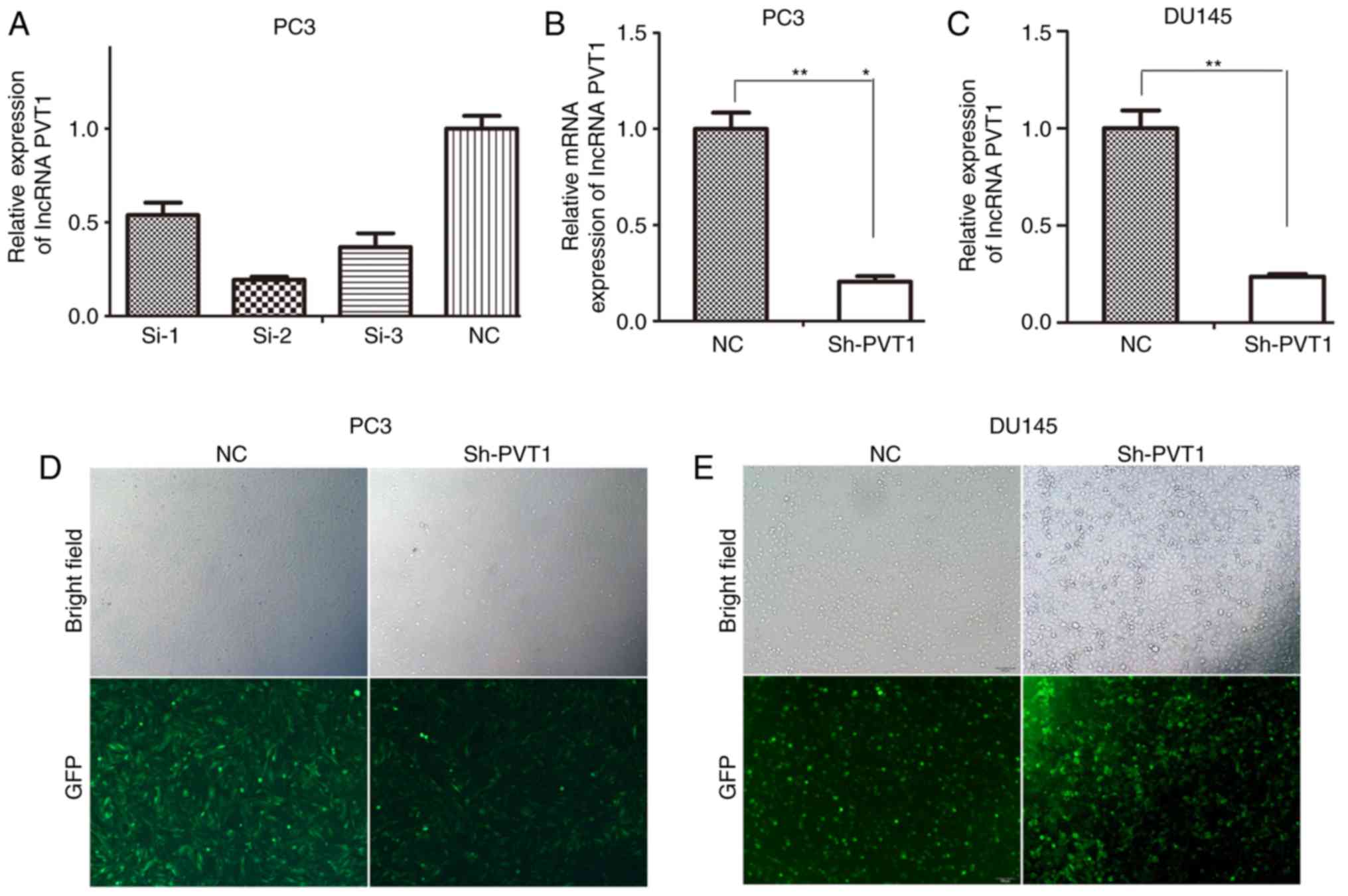
Identification of the most effective
siRNA
To identify the most effective siRNA for lncRNA
PVT1-knockdown, PC3 cells were transfected with three lncRNA PVT1
siRNAs and one NC si-RNA. si-RNA-2# was confirmed to be the most
effective siRNA for lncRNA PVT1-knockdown compared with other
siRNAs (Fig. 1A). si-RNA-2# was
subsequently recombined into a lentivirus vector. To obtain stable
cell lines exhibiting lncRNA PVT1-knockdown, the lentivirus vectors
were transfected into PC3 cells (Fig.
1D) and DU145 cells (Fig. 1E).
Subsequently, RT-qPCR results indicated significant knockdown of
lncRNA PVT1 expression levels in PC3 and DU145 cells (Fig. 1B and C).
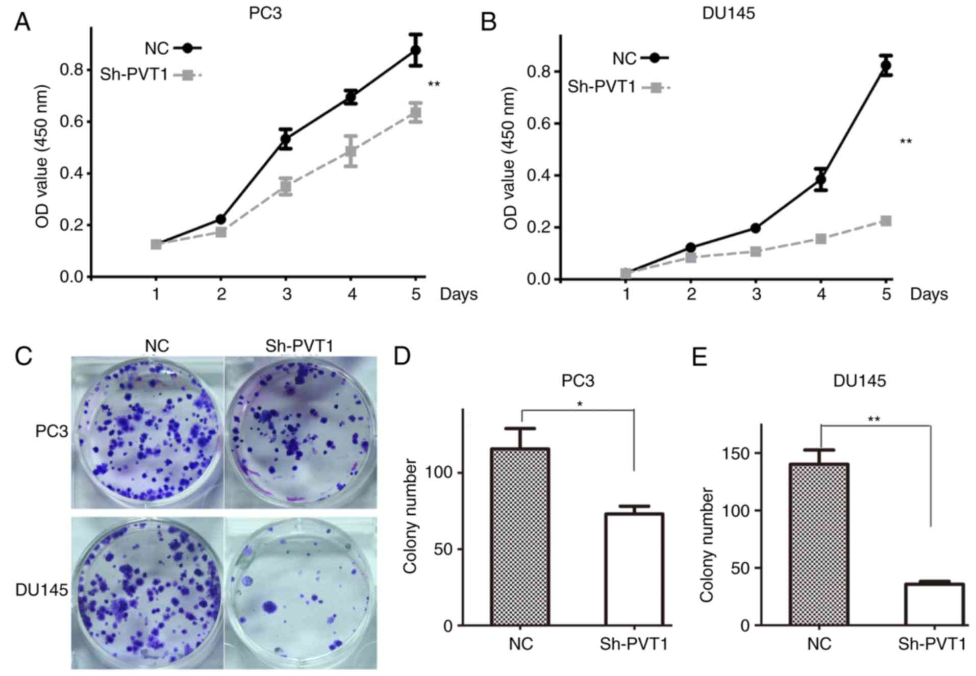
Downregulation of lncRNA PVT1
expression inhibits the proliferation, mobility and colony
formation abilities of prostate cancer cells
To investigate the effect of lncRNA PVT1 on the
proliferation of prostate cancer cells, a CCK-8 assay was performed
in PC3 and DU145 cells. The results indicated that lncRNA PVT1
downregulation significantly inhibited the proliferation of PC3
(Fig. 2A) and DU145 (Fig. 2B) cells, compared with the NC
group.
The effect of lncRNA PVT1 was also examined on the
colony formation ability of prostate cancer cells. The colony
number was significantly decreased in shPVT1 PC3 and DU145 cells
compared with NC cells (Fig. 2C and
D). These results indicated that downregulation of lncRNA PVT1
expression inhibited the colony formation ability of prostate
cancer cells.
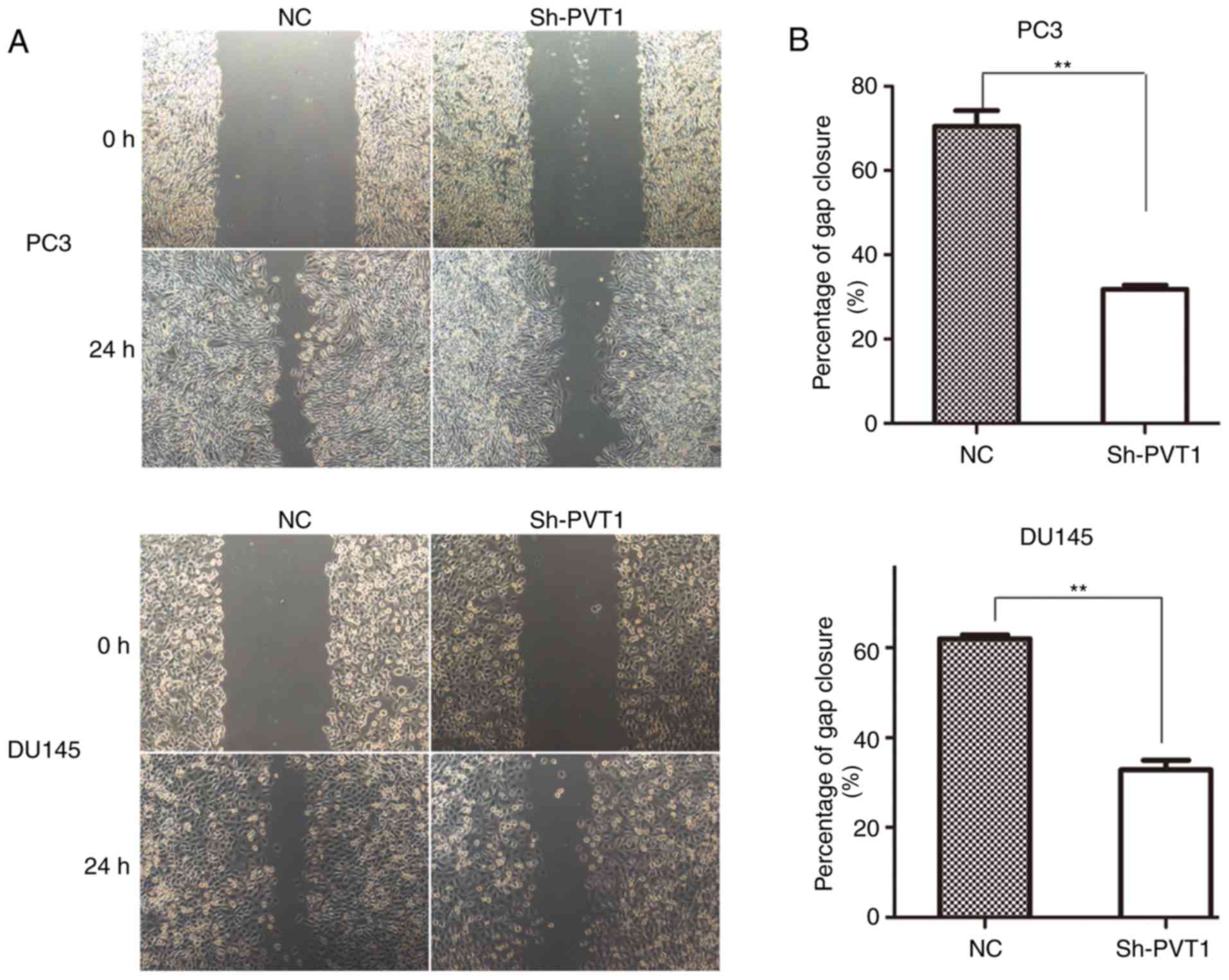
A wound-healing assay was conducted to assess the
effect of lncRNA PVT1 expression on the mobility of prostate cancer
cells. Downregulation of lncRNA PVT1 expression significantly
inhibited the migration of PC3 cells (Fig. 3A) and DU145 (Fig. 3B) cells compared with the NC
group.
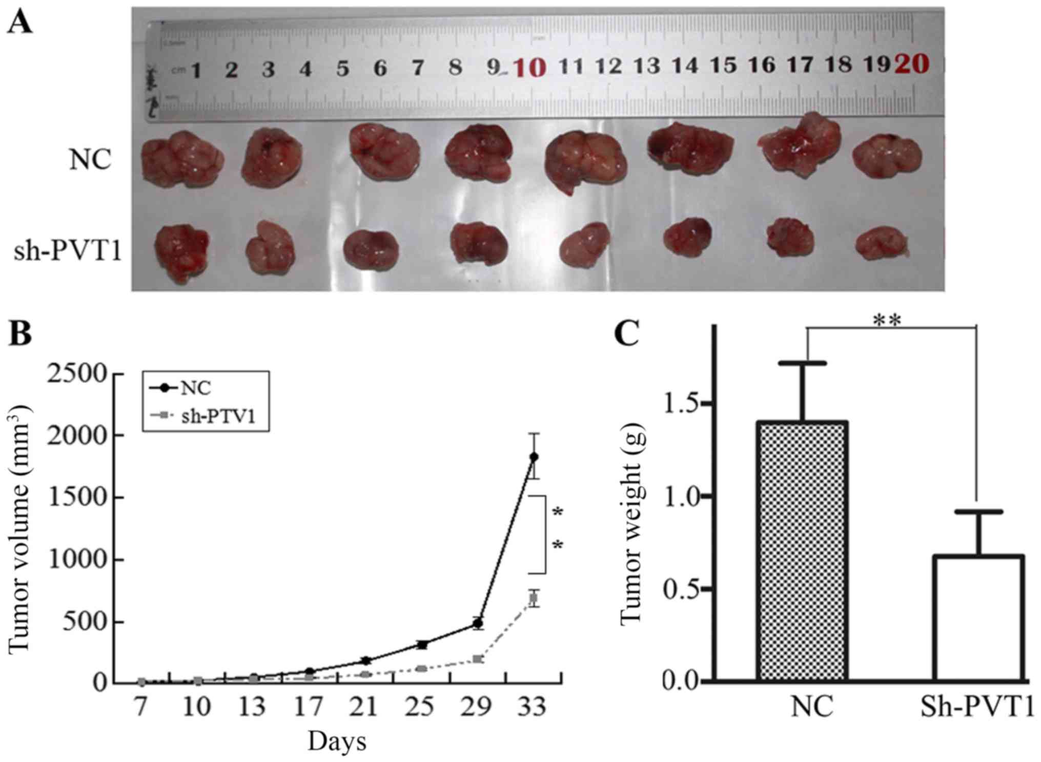
LncRNA PVT1 downregulation inhibited
tumorigenesis in a tumor xenograft model
To confirm the effect of lncRNA PVT1 on
tumorigenesis in prostate cancer cells, a xenograft tumor formation
assay was performed by injecting PC3 cells into mice. After 33
days, all mice were sacrificed, and the size and weight of
subcutaneous tumors were calculated (Fig.
4A). Compared with the NC group, the growth rate of xenograft
tumors was decreased in the shPVT1 group (Fig. 4B). The mean tumor weight of the shPVT1
group was significantly reduced compared with that of the NC group
(Fig. 4C). The results of the present
study indicated that lncRNA PVT1 downregulation inhibited
tumorigenesis in prostate cancer cells.
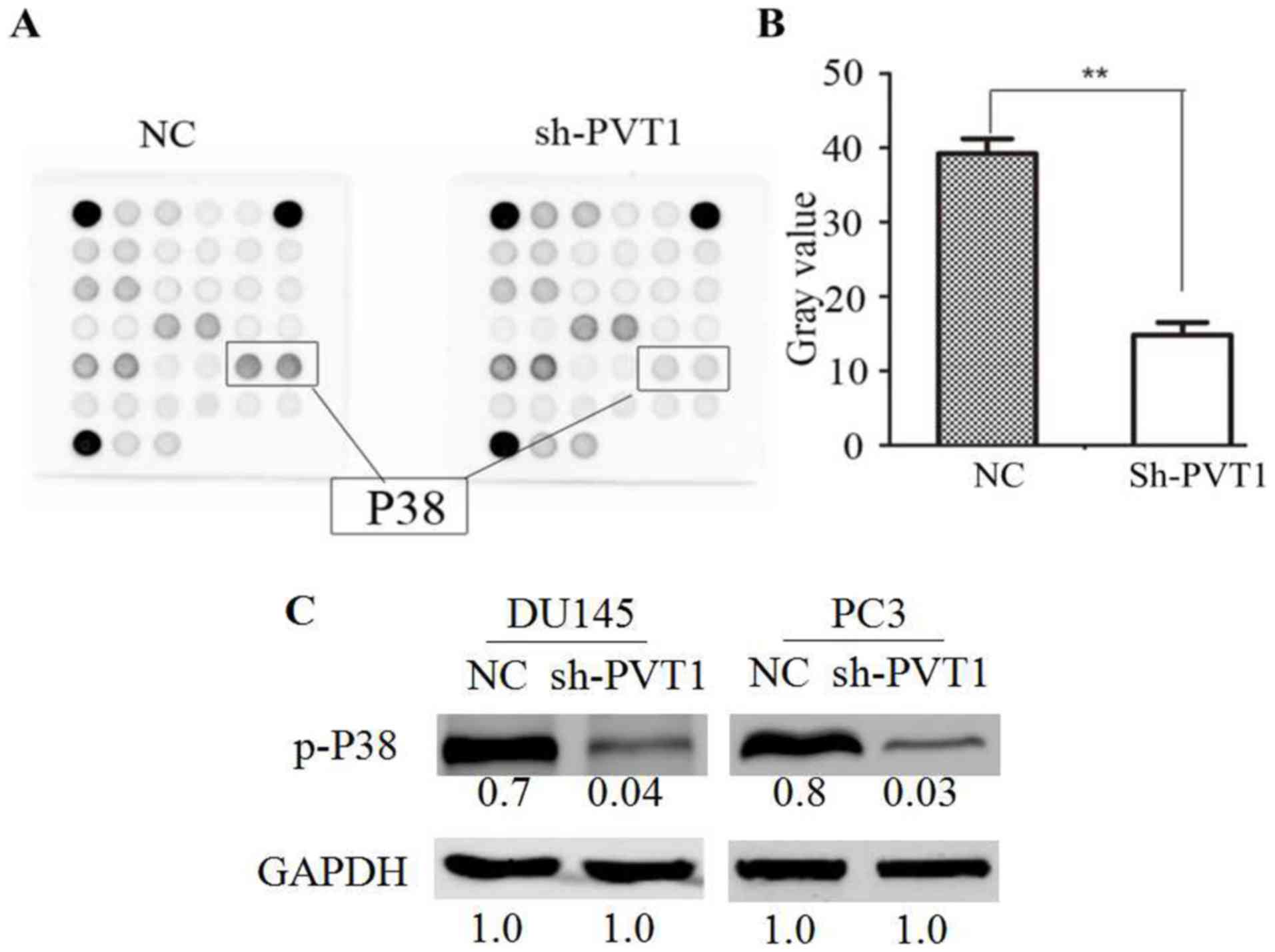
Downregulation of lncRNA PVT1
expression inhibits p38 activation
To explore the underlying molecular mechanisms of
lncRNA PVT1 downregulation in prostate cancer cells, the
PathScan® Intracellular Signaling Array kit
(Chemiluminescent Readout) was utilized in DU145 cells. The
phosphorylation level of p38 (Thr180/Tyr182) was demonstrated to be
significantly reduced in the shPVT1 group compared with the NC
group (Fig. 5A and B). Western blot
analysis indicated that the phosphorylation levels of p38 cells
were significantly reduced in the DU145 and PC3 cells with
downregulated PVT1 expression compared with the NC group (Fig. 5C). Therefore, the data of the present
study indicated that lncRNA PVT1 downregulation suppressed p38
phosphorylation.
Discussion
In previous studies it has been reported that lncRNA
PVT1 expression is upregulated in various types of cancer,
including non-small cell lung cancer (NSCLC) (20,21),
gastric cancer (22,23), renal cell carcinoma (24), cervical cancer (25), bladder cancer (26), hepatocellular carcinoma (HCC)
(27), ovarian cancer (28), acute promyelocytic leukemia (29) and colorectal carcinoma (30). It has been indicated that lncRNA PVT1
is overexpressed in prostate cancer cells and may be a potential
biomarker for prostate cancer (31).
In previous studies, lncRNA PVT1 expression has been identified as
a potential oncogene in multifarious types of cancer. For example,
in HCC, high lncRNA PVT1 expression levels have been reported to be
associated with a poor clinical prognosis (32). In addition, lncRNA PVT1 has been
reported to promote HCC proliferation, the cell cycle, and the
acquisition of stem cell-like properties (33). In another study, ectopic lncRNA PVT1
expression was indicated to significantly accelerate proliferation
in NSCLC by downregulating p15 and p21 expression (34). In cervical cancer, it has been
demonstrated that lncRNA PVT1 expression promoted cervical cell
proliferation and migration depending on miR-200b silencing
(25). Guan et al (35) reported that lncRNA PVT1-knockdown
inhibited proliferation and enhanced apoptosis in breast and
ovarian cancer cell lines. Downregulation of lncRNA PVT1 expression
has been demonstrated to decrease proliferation and colony
formation abilities in vitro and to suppress xenograft tumor
proliferation in vivo by regulating p15 and p16 expression
(36). The aforementioned findings
indicate that lncRNA PVT1 could be a potential therapeutic target
for these types of cancer (37).
However, the functional role of lncRNA PVT1 in
prostate cancer cells requires further investigation. To examine
the detailed underlying mechanism of lncRNA PVT1, lncRNA PVT1
expression was knocked down in PC3 and DU145 cell lines by
lentivirus-mediated shRNA. The effect of lncRNA PVT1 on
proliferation and migration of prostate cancer cells was evaluated
by CCK-8, colony formation, and wound healing assays. The results
of the present study indicated that lncRNA PVT1-knockdown inhibited
prostate cancer cell proliferation and mobility in vitro.
Furthermore, to determine the impact of lncRNA PVT1 on in
vivo proliferation, a PC3 cell line stably transfected with
lncRNA PVT1 was used to establish tumorigenicity in a tumor
xenograft model. The results of the present study indicated that
the tumorigenic capacity and weight in the shPVT1 group were
decreased compared with the control group. Furthermore, knockdown
of lncRNA PVT1 expression decreased the phosphorylation of p38,
which has been identified as a mitosis-associated molecule,
involved in the modulation of proliferation and migration of cancer
cells (38,39). Therefore, lncRNA PVT1 expression may
regulate prostate cancer proliferation through phosphorylation of
p38. The results of the present study demonstrated that lncRNA PVT1
downregulation inhibited proliferation and migration of prostate
cancer cells, which are associated with p38 expression. Therefore,
lncRNA PVT1 may represent a novel diagnostic marker and a
therapeutic target for prostate cancer. However, further
investigation is required.
Acknowledgements
The authors would like to thank the Department of
Pathology (Southern Medical University) for technical support.
Funding
The present study was supported by the National
Natural Science Foundation of China (grant nos. 81773277 and
81270597; Beijing, China), Science and Technology Projects of
Guangdong (grant no. c1430611500534; Guandong, China) and Science
and Technology Projects of Shenzhen (grant no. 201404153000668;
Shenzhen, China).
Availability of data and materials
All data generated or analyzed during this study are
included in this published article.
Authors' contributions
XMM conceived and designed the experiments. BW, HYW,
DJL and XMZ performed the experiments. LRZ, BL and SBZ acquired and
analyzed the data. XMM and BW contributed to drafting and revising
the manuscript.
Ethics approval and consent to
participate
All animal procedures and experiments were approved
by the Southern Medical University Animal Research Ethics Committee
(Guangzhou, China).
Patient consent for publication
Not applicable.
Competing interests
The authors declare that they have no conflicts of
interest.
References
|
1
|
Lilja H, Ulmert D and Vickers AJ:
Prostate-specific antigen and prostate cancer: Prediction,
detection and monitoring. Nat Rev Cancer. 8:268–278. 2008.
View Article : Google Scholar : PubMed/NCBI
|
|
2
|
Crawford ED: Epidemiology of prostate
cancer. Urology. 62 6 Suppl 1:S3–S12. 2003. View Article : Google Scholar
|
|
3
|
Zhu Y, Yang XQ, Han CT, Dai B, Zhang HL,
Shi GH, Wang CF and Ye DW: Pathological features of localized
prostate cancer in China: A contemporary analysis of radical
prostatectomy specimens. PLoS One. 10:e01210762015. View Article : Google Scholar : PubMed/NCBI
|
|
4
|
Hellerstedt BA and Pienta KJ: The current
state of hormonal therapy for prostate cancer. CA Cancer J Clin.
52:154–179. 2002. View Article : Google Scholar : PubMed/NCBI
|
|
5
|
Mercer TR, Dinger ME and Mattick JS: Long
non-coding RNAs: Insights into functions. Nat Rev Genet.
10:155–159. 2009. View
Article : Google Scholar : PubMed/NCBI
|
|
6
|
Tsai M-C, Spitale RC and Chang HY: Long
intergenic noncoding RNAs: New links in cancer progression. Cancer
Res. 71:3–7. 2011. View Article : Google Scholar : PubMed/NCBI
|
|
7
|
Geisler S and Coller J: RNA in unexpected
places: Long non-coding RNA functions in diverse cellular contexts.
Nat Rev Mol Cell Biol. 14:699–712. 2013. View Article : Google Scholar : PubMed/NCBI
|
|
8
|
Zeng C, Xu Y, Xu L, Yu X, Cheng J, Yang L,
Chen S and Li Y: Inhibition of long non-coding RNA NEAT1 impairs
myeloid differentiation in acute promyelocytic leukemia cells. BMC
Cancer. 14:6932014. View Article : Google Scholar : PubMed/NCBI
|
|
9
|
Gibb EA, Brown CJ and Lam WL: The
functional role of long non-coding RNA in human carcinomas. Mol
Cancer. 10:38. 2011. View Article : Google Scholar : PubMed/NCBI
|
|
10
|
Luo X, Yang W, Ye DQ, Cui H, Zhang Y,
Hirankarn N, Qian X, Tang Y, Lau YL, de Vries N, et al: A
functional variant in microRNA-146a promoter modulates its
expression and confers disease risk for systemic lupus
erythematosus. PLoS Genet. 7:e10021282011. View Article : Google Scholar : PubMed/NCBI
|
|
11
|
Ding J, Li D, Gong M, Wang J, Huang X, Wu
T and Wang C: Expression and clinical significance of the long
non-coding RNA PVT1 in human gastric cancer. Onco Targets Ther.
7:1625–1630. 2014. View Article : Google Scholar : PubMed/NCBI
|
|
12
|
Ding C, Yang Z, Lv Z, Du C, Xiao H, Peng
C, Cheng S, Xie H, Zhou L, Wu J and Zheng S: Long non-coding RNA
PVT1 is associated with tumor progression and predicts recurrence
in hepatocellular carcinoma patients. Oncol Lett. 9:955–963. 2015.
View Article : Google Scholar : PubMed/NCBI
|
|
13
|
Barsotti AM, Beckerman R, Laptenko O,
Huppi K, Caplen NJ and Prives C: p53-dependent Induction of PVT1
and miR-1204. J Biol Chem. 287:2509–2519. 2012. View Article : Google Scholar : PubMed/NCBI
|
|
14
|
Chapman MH, Tidswell R, Dooley JS,
Sandanayake NS, Cerec V, Deheragoda M, Lee AJ, Swanton C, Andreola
F and Pereira SP: Whole genome RNA expression profiling of
endoscopic biliary brushings provides data suitable for biomarker
discovery in cholangiocarcinoma. J Hepatol. 56:877–885. 2012.
View Article : Google Scholar : PubMed/NCBI
|
|
15
|
Li Q, Dai Y, Wang F and Hou S:
Differentially expressed long non-coding RNAs and the prognostic
potential in colorectal cancer. Neoplasma. 63:977–983. 2016.
View Article : Google Scholar : PubMed/NCBI
|
|
16
|
Zhang XW, Bu P, Liu L, Zhang Xz and Li J:
Overexpression of long non-coding RNA PVT1 in gastric cancer cells
promotes the development of multidrug resistance. Biochem Biophys
Res Commun. 462:227–232. 2015. View Article : Google Scholar : PubMed/NCBI
|
|
17
|
Ilboudo A, Chouhan J, McNeil BK, Osborne
JR and Ogunwobi OO: PVT1 Exon 9: A potential biomarker of
aggressive prostate cancer? Int J Environ Res Public Health.
13:ijerph13010012. 2016.
|
|
18
|
Livak KJ and Schmittgen TD: Analysis of
relative gene expression data using real-time quantitative PCR and
the 2(-Delta Delta C(T)) method. Methods. 25:402–408. 2001.
View Article : Google Scholar : PubMed/NCBI
|
|
19
|
Lei B, Zhou X, Lv D, Wan B, Wu H, Zhong L,
Shu F and Mao X: Apoptotic and nonapoptotic function of caspase 7
in spermatogenesis. Asian J Androl. 719:47–51. 2007.
|
|
20
|
Yang YR, Zang SZ, Zhong CL, Li YX, Zhao SS
and Feng XJ: Increased expression of the lncRNA PVT1 promotes
tumorigenesis in non-small cell lung cancer. Int J Clin Exp Pathol.
7:6929–6935. 2014.PubMed/NCBI
|
|
21
|
Wan L, Sun M, Liu GJ, Wei CC, Zhang EB,
Kong R, Xu TP, Huang MD and Wang ZX: Long noncoding RNA PVT1
Promotes promotes non-small cell lung cancer cell proliferation
through epigenetically regulating LATS2 expression. Mol Cancer
Ther. 15:1082–1094. 2016. View Article : Google Scholar : PubMed/NCBI
|
|
22
|
Yuan CL, Li H, Zhu L, Liu Z, Zhou J and
Shu Y: Aberrant expression of long noncoding RNA PVT1 and its
diagnostic and prognostic significance in patients with gastric
cancer. Neoplasma. 63:442–449. 2016. View Article : Google Scholar : PubMed/NCBI
|
|
23
|
Cao WJ, Wu HL, He BS, Zhang YS and Zhang
ZY: Analysis of long non-coding RNA expression profiles in gastric
cancer. World J Gastroenterol. 19:3658–3664. 2013. View Article : Google Scholar : PubMed/NCBI
|
|
24
|
Wu Y, Wang YQ, Weng WW, Zhang QY, Yang XQ,
Gan HL, Yang YS, Zhang PP, Sun MH, Xu MD and Wang CF: A
serum-circulating long noncoding RNA signature can discriminate
between patients with clear cell renal cell carcinoma and healthy
controls. Oncogenesis. 5:e1922016. View Article : Google Scholar : PubMed/NCBI
|
|
25
|
Zhang S, Zhang G and Liu J: Long noncoding
RNA PVT1 promotes cervical cancer progression through
epigenetically silencing miR-200b. APMIS. 124:649–658. 2016.
View Article : Google Scholar : PubMed/NCBI
|
|
26
|
Zhuang C, Li J, Liu Y, Chen M, Yuan J, Fu
X, Zhan Y, Liu L, Lin J, Zhou Q, et al: Tetracycline-inducible
shRNA targeting long non-coding RNA PVT1 inhibits cell growth and
induces apoptosis in bladder cancer cells. Oncotarget.
6:41194–41203. 2015. View Article : Google Scholar : PubMed/NCBI
|
|
27
|
Yan H, Yang Y, Zhang L, Tang G, Wang Y,
Xue G, Zhou W and Sun S: Characterization of the genotype and
integration patterns of hepatitis B virus in early- and late-onset
hepatocellular carcinoma. Hepatology. 61:1821–1831. 2015.
View Article : Google Scholar : PubMed/NCBI
|
|
28
|
Liu E, Liu Z, Zhou Y, Mi R and Wang D:
Overexpression of long non-coding RNA PVT1 in ovarian cancer cells
promotes cisplatin resistance by regulating apoptotic pathways. Int
J Clin Exp Med. 8:20565–20572. 2015.PubMed/NCBI
|
|
29
|
Zeng C, Yu X, Lai J, Yang L, Chen S and Li
Y: Overexpression of the long non-coding RNA PVT1 is correlated
with leukemic cell proliferation in acute promyelocytic leukemia. J
Hematol Oncol. 8:1262015. View Article : Google Scholar : PubMed/NCBI
|
|
30
|
Takahashi Y, Sawada G, Kurashige J, Uchi
R, Matsumura T, Ueo H, Takano Y, Eguchi H, Sudo T, Sugimachi K, et
al: Amplification of PVT-1 is involved in poor prognosis via
apoptosis inhibition in colorectal cancers. Br J Cancer.
110:164–171. 2014. View Article : Google Scholar : PubMed/NCBI
|
|
31
|
Bawa P, Zackaria S, Verma M, Gupta S,
Srivatsan R, Chaudhary B and Srinivasan S: Integrative analysis of
normal long intergenic non-coding rnas in prostate cancer. PLoS
One. 10:e01221432015. View Article : Google Scholar : PubMed/NCBI
|
|
32
|
Yu J, Han J, Zhang J, Li G, Liu H, Cui X,
Xu Y, Li T, Liu J and Wang C: The long noncoding RNAs PVT1 and
uc002mbe.2 in sera provide a new supplementary method for
hepatocellular carcinoma diagnosis. Medicine (Baltimore).
95:e44362016. View Article : Google Scholar : PubMed/NCBI
|
|
33
|
Wang F, Yuan JH, Wang SB, Yang F, Yuan SX,
Ye C, Yang N, Zhou WP, Li WL, Li W and Sun SH: Oncofetal long
noncoding RNA PVT1 promotes proliferation and stem cell-like
property of hepatocellular carcinoma cells by stabilizing NOP2.
Hepatology. 60:1278–1290. 2014. View Article : Google Scholar : PubMed/NCBI
|
|
34
|
Cui D, Yu CH, Liu M, Xia QQ, Zhang YF and
Jiang WL: Long non-coding RNA PVT1 as a novel biomarker for
diagnosis and prognosis of non-small cell lung cancer. Tumour Biol.
37:4127–4134. 2016. View Article : Google Scholar : PubMed/NCBI
|
|
35
|
Guan Y, Kuo WL, Stilwell JL, Takano H,
Lapuk AV, Fridlyand J, Mao JH, Yu M, Miller MA, Santos JL, et al:
Amplification of PVT1 contributes to the pathophysiology of ovarian
and breast cancer. Clin Cancer Res. 13:5745–5755. 2007. View Article : Google Scholar : PubMed/NCBI
|
|
36
|
Kong R, Zhang EB, Yin DD, You LH, Xu TP,
Chen WM, Xia R, Wan L, Sun M, Wang ZX, et al: Long noncoding RNA
PVT1 indicates a poor prognosis of gastric cancer and promotes cell
proliferation through epigenetically regulating p15 and p16. Mol
Cancer. 14:822015. View Article : Google Scholar : PubMed/NCBI
|
|
37
|
Tseng YY, Moriarity BS, Gong W, Akiyama R,
Tiwari A, Kawakami H, Ronning P, Reuland B, Guenther K, Beadnell
TC, et al: PVT1 dependence in cancer with MYC copy-number increase.
Nature. 512:82–86. 2014. View Article : Google Scholar : PubMed/NCBI
|
|
38
|
Xia P, Zhang R and Ge G: C/EBPβ mediates
tnf-α-induced cancer cell migration by inducing mmp expression
dependent on p38 MAPK. J Cell Biochem. 116:2766–2777. 2015.
View Article : Google Scholar : PubMed/NCBI
|
|
39
|
Cheng G, Gao F, Sun X, Bi H and Zhu Y:
Paris saponin VII suppresses osteosarcoma cell migration and
invasion by inhibiting MMP-2/9 production via the p38 MAPK
signaling pathway. Mol Med Rep. 14:3199–3205. 2016. View Article : Google Scholar : PubMed/NCBI
|