Introduction
Cervical cancer (CC) remains one of the most
frequent gynecological malignancies among females worldwide, which
leads to the highest morbidity and mortality in young women,
particularly in developing country (1). Considered as the major etiologic
contributor to the pathogenesis of CC, HPVs have been associated
with more than 99% of cervical carcinomas (2). It is clear that persistent infection of
high-risk human papillomavirus (HR-HPV), especially HPV18 and HPV16
are the most important etiologic agent in cervical carcinogenesis
(3,4).
The viral oncoproteins HPV18 and HPV16 E7 and E6 can inactivate pRB
and p53, thereby influencing their regulation and subsequently
contributing to cell cycle checkpoint escape and cervical
carcinogenesis (5,6). However, due to the CC complex mechanism,
the regulatory mechanism and the biological functions underlying
HPV pathogenesis need to be further investigated.
Glucose-6-phosphate dehydrogenase (G6PD) catalyses
the first rate-limiting step in the pentose phosphate pathway (PPP)
(7). G6PD produces nicotinamide
adenine dinucleotide phosphate (NADPH) that affect antioxidant
defense and biosynthesis in the cells and is especially important
in red blood cells functionally (8).
G6PD is taken into account as one oncogene on account of its high
expression in a great range of tumors, including breast cancers
(9), melanoma (10), colorectal cancer (11) and lung cancer (12). A previous study has demonstrated that
the expression of G6PD was high and there is a positive correlation
with cervical patients infected with HPV18 and HPV16 of 30 to 40
years female (13). The high
expression of G6PD may affect the progression and development of
HR-HPV16/18-infected CC. Its underlying molecular mechanisms and
the biological functions for its oncogenic roles in HR-HPV16/18
infected CC are still unknown.
MicroRNAs (miRNAs/miRs) are a class of highly
conserved, non-coding and endogenous RNAs (ranging in 18–23
nucleotides length) (14,15), which can modulate the physiological
process or pathogenesis through partial complementary binding to
the 3′-UTR of mRNAs (16). miR-206
has been demonstrated to be involved in different physiological and
pathological processes (17).
Dysfunctions of miR-206 occurred in a group of tumors, such as
hepatocellular carcinoma (18), head
and neck squamous cell carcinoma (19) and medulloblastoma (17), which can regulate tumor progression
that is invovled in cell differentiation, proliferation,
metastasis, and apoptosis. However, the specific functional
molecular mechanisms of miR-206 in CC are still elusive, and the
potential of miR-206 as a therapeutic target of CC remains to be
evaluated.
We demonstrated that miR-206 was frequently
downregulated while G6PD was upregulated in HR-HPV(+) CC.
Overexpression of miR-206 or low expressed G6PD suppressed cell
proliferation, and miR-206 low expressed or G6PD overexpressed
predicted poor prognosis. Furthermore, we identified G6PD as a
direct target of miR-206. We also measured the overall survival
(OS) according to the expression of miR-206 and G6PD. The newly
identified miR-206/G6PD axis partially elucidates the molecular
mechanism of proliferation and is a novel potential therapeutic
target for CC treatment.
Materials and methods
Patient selection and human
tissues
A total of 56 CC patients (including 42
HPV16/18-positive CCs, 14 HPV-negative CCs) who were treated at the
Yantaishan Hospital (Yantai, China)between March, 2014 and August,
2016 participated in this study. Written informed consent was
provided by patients. The study was approved by the Ethics
Committee of Yantaishan Hospital. All the tissues were
independently and histologically diagnosed, and CC was classified
based on the International Federation of Gynecology and Obstetrics
(FIGO) staging system (20). All the
specimens were stored at −80°C. PCR amplification was used to
detect cervical HPV infection with the presence of HPV DNA
(7).
Cell culture and treatments
HPV16-positive SiHa (HPV16+SiHa), and
HPV18-positive HeLa (HPV18+HeLa) were purchased from the
American Type Culture Collection (Manassas, VA, USA). The cells
were cultured in DMEM (Gibco; Thermo Fisher Scientific, Inc.,
Waltham, MA, USA) supplemented with 10% FCS (Invitrogen; Thermo
Fisher Scientific, Inc.) at 37°C and 5% CO2.
miR-206 mimics, G6PD siRNA and the negative controls
(NC) were obtained from GenePharma, Co., Ltd. (Shanghai, China).
HeLa and SiHa cells were transfected with G6PD siRNA or miR-206
mimics as well as the NC using Lipofectamine 2000 Reagent
(Invitrogen; Thermo Fisher Scientific, Inc.). Cells were used for
proliferation after transfection. All transfection was conducted
three times.
CCK-8 assay
CCK-8 (Dojindo Molecular Technologies, Inc.,
Kumamoto, Japan) was performed to detect cell proliferation.
Cervical cells transfected with miR-206 mimics or G6PD siRNA were
seeded into 96-well plates. Then, 10 µl CCK-8 reagent was added to
the wells. The absorbance of each well at 24, 48, 72 and 96 h was
detected at 450 nm.
Western blot analysis
Western blot analysis was used to detect the G6PD
protein expression in HeLa and SiHa cells transfected with miR-206
mimics. Proteins were isolated from cervical cells with different
transfections using RIPA lysis buffer (Thermo Fisher Scientific,
Inc.). The protein was then transferred onto a PVDF membrane
(Bio-Rad Laboratories, Inc., Hercules, CA, USA) and sealed with
Tris-buffered saline Tween-20 (TBST). Then, the membranes were
blocked by 5% bovine serum albumin and incubated with specific
primary antibody rabbit polyclonal anti-G6PD antibody (1:1,000;
cat. no. ab993; Abcam, Cambridge, MA, USA) or GAPDH (1:3,000; cat.
no. ab226408). After that, the membrane was incubated in the
secondary antibody goat polyclonal anti-rabbit IgG H&L
secondary antibody (1:2,000; cat. no. ab150077; Abcam). The ECL
detection system was used to detect the protein level (BestBio,
Shanghai, China).
RNA isolation and RT-qPCR
Total RNA of cervical cells and tissues were
extracted using TRIzol Reagent. RT-qPCR for miR-206/G6PD was
performed with SYBR Premix Ex Taq™ (Takara Biotechnology Co., Ltd.,
Dalian, China). Relative gene expression was determined using the
2−ΔΔCq method (21). U6
and GAPDH acted as the internal control for the expressions of
miR-206 and SPARC. G6PD and GAPDH primers were produced by
Invitrogen; Thermo Fisher Scientific, Inc. The transcription primer
and PCR primer of miR-206 and U6 were purchased from Guangzhou
RiboBio Co., Ltd. (Guangzhou, China). Primer sequences were as
follows: miR-206 forward, 5′-CCAAAGCGGAGTCTCGCAT-3′ and reverse,
5′-GCCTAGCATCTTGCTTAGCTC-3′; U6 forward,
5′-GCTTCGGCAGCACATATACTAAAAT-3′ and reverse,
5′-CGCTTCACGAATTTGCGTGTCAT-3′; G6PD forward,
5′-TGCCTTCCATCAGTCGGATACAC-3′ and reverse,
5′-TGGTGGGGTAGATCTTCTTCTTGG-3′; and GAPDH forward,
5′-CCCTTCATTGACCTCAACTACATG-3′ and reverse,
5′-TGGGATTTCCATTGATGACAAGC-3′.
Luciferase assay
The bioinformatics analysis software TargetScan
(http://www.targetscan.org/) and miRanda
(http://www.microrna.org/microrna/home.do) were chosen
for predicting the targets of miR-206. For luciferase reporter, the
wild-type (WT) and mutant type (Mut) 3′-UTR of G6PD were cloned
into pcDNA3.1 vector (Ambion; Thermo Fisher Scientific, Inc.) and
verified by sequencing. For the luciferase assay, the cells were
co-transfected with miR-206 mimics and WT or Mut 3′-UTR of G6PD
luciferase reporter plasmid. Then we used Dual-Luciferase Reporter
Assay System (Promega Corporation, Madison, WI, USA) for measuring
the reporter activities.
Statistical analysis
Statistical analyses were presented as the mean ±
standard deviation using SPSS19.0 software (SPSS, Inc., Chicago,
IL, USA). Differences between groups were evaluated by Student's
t-test or Tukey's post hoc test after ANOVA in SPSS. Correlation
between mRNA and miRNA were estimated using the Spearman's
correlation method. In addition, the Kaplan-Meier method with
log-rank test was used for analyzing survival. P<0.05 was
considered to indicate a statistically significant difference.
Results
miR-206 is downregulated, while G6PD
is upregulated in HR-HPV(+) CC
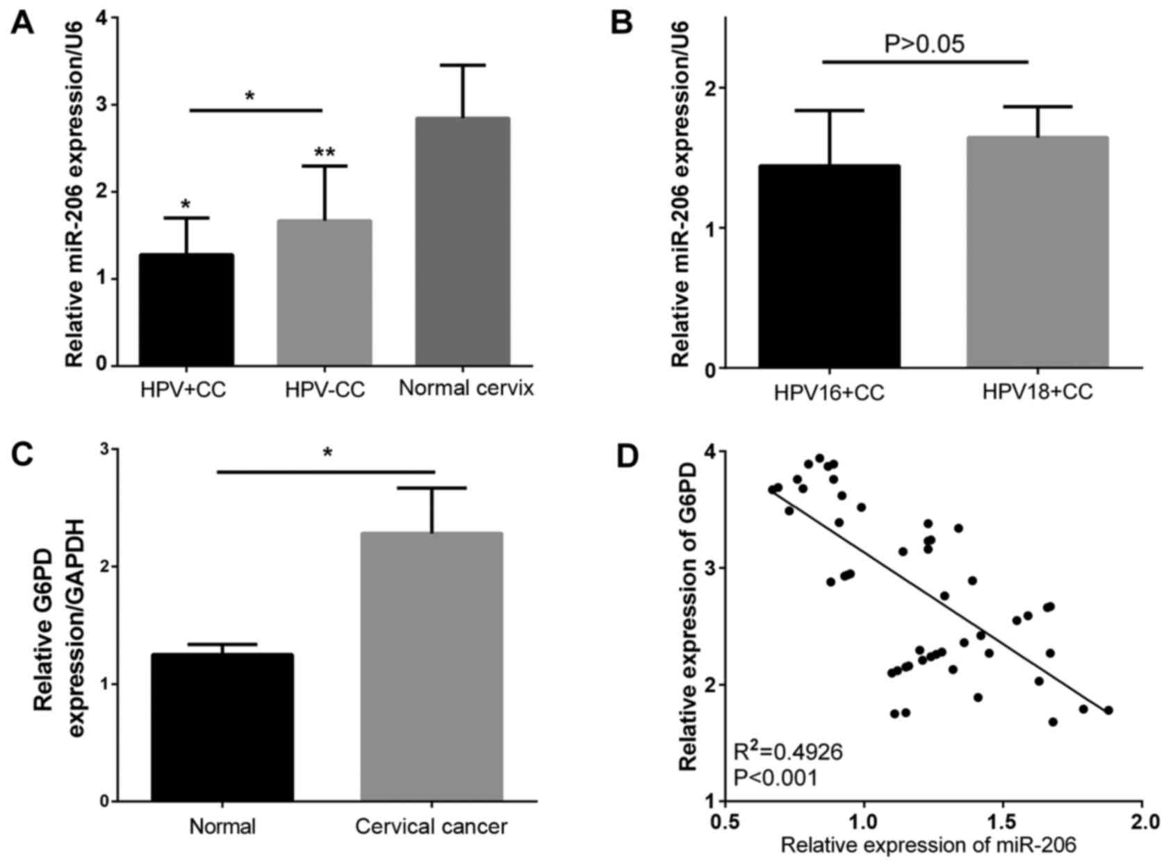
To investigate whether the miR-206 expression was
altered in HR-HPV(+) cervical tissues, RT-qPCR was performed in CC
and normal cervical tissues, obtained from 46 HPV16/18-positive
patients and 10 HPV-negative patients. miR-206 expression was
significantly lower in CC tissues (Fig.
1A), and miR-206 expression was lower in HPV16/18-positive CC
compared to HPV-negative tissues (Fig.
1A). However, the results showed no significant difference
between HPV18(+) CC tissues (n=20) and HPV16(+) CC tissues (n=26)
(Fig. 1B). Generally, G6PD level was
significantly higher in CC patients compared to normal control
patients (Fig. 1C). An inverse
correlation between miR-206 expression and the G6PD level in these
clinical specimens (R2=0.4926, P<0.0001) (Fig. 1D). Thus, miR-206 and G6PD may have
possible roles in modulating the progression of HR-HPV(+) CC.
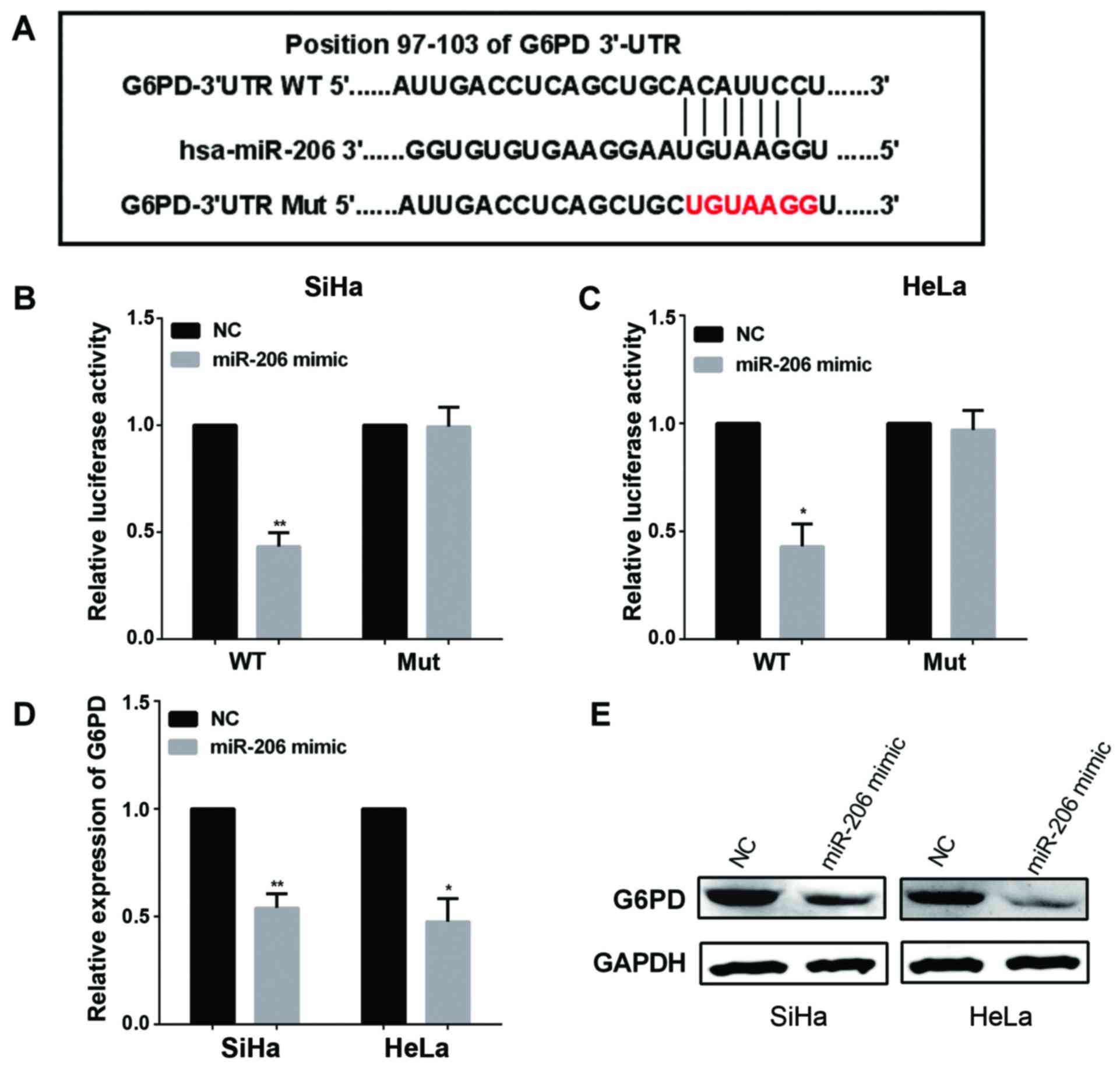
miR-206 directly targets the 3′-UTR of
G6PD
We predicted that G6PD was a downstream target of
miR-206 by bioinformatics analysis software TargetScan and miRanda,
the binding site of G6PD was at its 3′-UTR located at 97 to 103 as
shown in Fig. 2A. To further confirm
whether the 3′-UTR of G6PD can be directly targeted by miR-206, we
performed luciferase reporter assay. Following the protocol, G6PD
3′-UTR-WT and G6PD 3′-UTR-Mut were cloned into plasmids with either
miR-206 mimic or NC, then following by the measurement of
luciferase reporter assays. Luciferase activity decreased when
miR-206 mimics were co-transfected with the G6PD 3′-UTR-WT plasmid
(P<0.01), but there was no change with the G6PD 3′-UTR-Mut
plasmid (P>0.05) in SiHa and HeLa cells (Fig. 2B and C). These results suggested
miR-206 can downregulate the expression of G6PD by binding to its
predicted regions of 3′-UTR. Furthermore, when overexpressed
miR-206 by transfected miR-206 mimic, the expression of G6PD was
decreased in SiHa and HeLa cell (Fig.
2D). In Fig. 2E, overexpression
of miR-206 reduced the protein level of G6PD in the SiHa and HeLa
cells. Together, these results demonstrated miR-206 negatively
regulated endogenous G6PD expression.
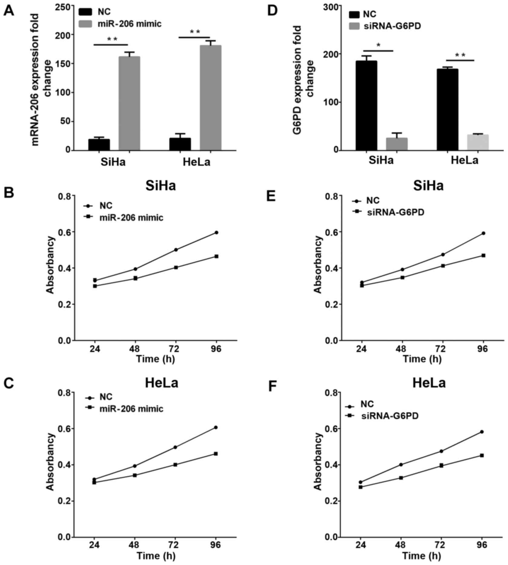
miR-206 overexpressed or G6PD low
expressed suppressed cell proliferation
Due to the downregulation of miR-206 and its inverse
correlation with G6PD, we hypothesized that miR-206 is a tumor
suppressor of CC, and affected CC cell proliferation. For the sake
of testing the impact of miR-206 on proliferation, we utilized
miR-206 mimic to overexpress miR-206 in CC SiHa and HeLa cells and
then the expression levels in cells were determined by RT-qPCR
(Fig. 3A). Then, we measured cell
proliferative ability and found that for overexpressed miR-206 the
proliferation ability was decreased both in SiHa and HeLa (Fig. 3B and C). To examine the effect of G6PD
on the proliferation of CC, we used siRNA-G6PD to interfere with
G6PD expression and the results (P<0.01) were measured by
RT-qPCR, as shown in Fig. 3D, and
then we calculated the capabilities of cell proliferation. Under
these conditions, the results indicated cell proliferative ability
was inhibited (Fig. 3E and F).
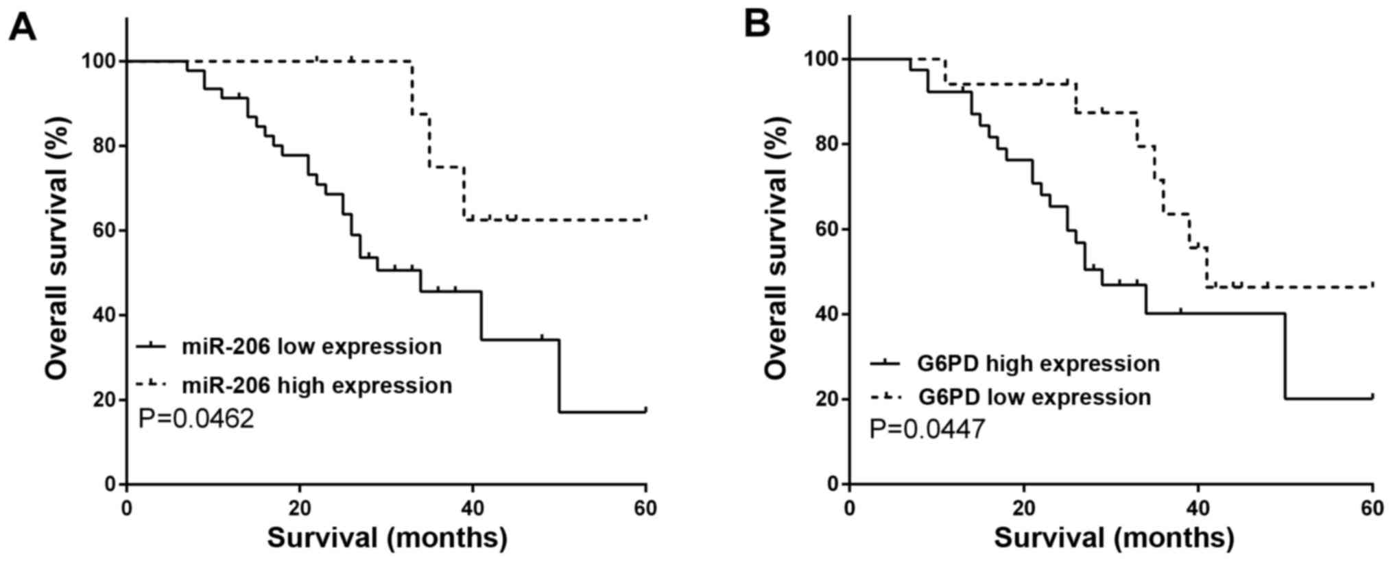
miR-206 low expressed or G6PD
overexpressed predicted poor prognosis
We divided 56 gastric cancer patients into the
miR-206 high expression group (n=10) and miR-206 low expression
group (n=46) according to miR-206 expression level. In addition,
the 56 patients were separated into
HPV16−/18− negative group (n=14) and
HPV16+/18+ (n=42) according to HPV status.
The 56 patients were separated on the basis of FIGO stage,
differentiation, tumor diameter, respectively, and the detailed
grouping is shown in Table I.
 | Table I.HPV status and miR-206 levels in women
diagnosed as cervical cancer (n=56). |
Table I.
HPV status and miR-206 levels in women
diagnosed as cervical cancer (n=56).
|
| HPV status
(HR-HPV16/18) | miR-206
expression |
|---|
|
|
|
|
|---|
| Variable |
HPV16−/18− negative,
n (%) |
HPV16+/18+ positive,
n (%) | miR-206 low level (≤
median), n (%) | miR-206 high level
(> median), n (%) |
|---|
| FIGO stage |
|
|
|
|
| I | 2 (3.6) | 2 (3.6) | 24 (42.8) | 2 (3.6) |
| II | 4 (7.1) | 8 (14.3) | 14 (25) | 2 (3.6) |
| III | 2 (3.6) | 12 (21.4) | 6 (10.7) | 2 (3.6) |
| IV | 6 (10.7) | 20 (35.7) | 2 (3.6) | 4 (7.1) |
|
Differentiation |
|
|
|
|
|
Well | 4 (7.1) | 8 (14.3) | 30 (53.6) | 2 (3.6) |
|
Moderate | 2 (3.6) | 20 (35.7) | 13 (23.2) | 2 (3.6) |
|
Poor | 8 (14.3) | 12 (21.4) | 3 (5.4) | 6
(10.7) |
| Tumor diameter
(cm) |
|
|
|
|
| ≤4 | 10 (17.8) | 19 (33.9) | 32 (57.1) | 8
(14.3) |
|
>4 | 4 (7.1) | 23 (41.2) | 14 (25) | 2 (3.6) |
To further evaluate whether miR-206 levels were
associated with CC prognosis, we performed Kaplan-Meier analysis to
evaluate five year OS in CC. OS was significantly poorer in
patients with low tissue miR-206 expression than those with high
miR-206 expression (log-rank, P=0.0462; Fig. 4A). In addition, we measured OS
according to G6PD expression, and the opposite results were
obtained, whereby the OS was lower with G6PD overexpression
compared with low expression (log-rank, P=0.0447; Fig. 4B).
Discussion
CC was once considered to be one of the most serious
cancers in women worldwide, and almost 90% of CC deaths occurred in
developing countries of the world (22). Although cancer treatments have been
improved in recent years, the outcomes of patients with CC remain
unsatisfactory (23). Thus,
identifying new targets for the development of effective
therapeutics for CC is urgent. Dysregulation of miRNAs may lead to
uncontrolled and progressive cancer growth and has been thoroughly
reported in almost all types of human malignancies (24,25),
including CC (26). In this study, we
found miR-206 was significantly downregulated in CC tissues, and
was reduced in HPV16+/18+ CC. However, there
was no significant difference between HPV16(+) CC tissues and
HPV18(+) CC tissues. Moreover, G6PD was identified as a direct
target of miR-206 and the inverse relationship between them was
also observed. We demonstrated that miR-206/G6PD may act as a novel
potential therapeutic target and treatment for CC, and a low
expression of miR-206 may contribute to tumor progression and cell
proliferation in CC patients.
Accumulating evidence has shown that miRNAs can
function as a crucial point in gene expressions, and then influence
tumor development and progression (27). Mounting evidences have demonstrated
that miR-206 is downregulated in breast cancer (28), gastric cancer (29) and various types of human tumors. The
low expression of miR-206 may be linked with physiological and
pathological processes of tumors, as those researchers proposed. A
previous study has reported that miR-206 was downregulated and
inhibited cell proliferation, invasion and migration in CC
(30), but the underlying molecular
mechanisms are still elusive. Our findings were consistent with all
the findings, as we have demonstrated that the overexpression of
miR-206 could inhibit proliferation by directly targeting G6PD in
SiHa and HeLa. In addition, we identified that miR-206
downregulation and/or G6PD upregulation predicted poor
prognosis.
This study revealed the relative expression of G6PD
was higher in HPV16+/18+ CC tissues. G6PD can
be found widely expressed in tumors and could function as an
important member in regulating cell invasiveness, survival and
oxidative stress (10,31). The bioinformatics analysis software
was used for predicting the targets of miR-206. And then G6PD was
looked for as the potential gene effectors which may participate in
the function of miR-206. We confirmed that G6PD was a direct target
of miR-206, and it was confirmed that a higher G6PD expression
played a significant role in tumor proliferation and predicted poor
prognosis. G6PD may therefore be independent prognostic factors for
OS of patients suffering from CC.
In conclusion, we have indicated that miR-206 acts
as a tumor suppressor in CC by inhibiting cancer proliferation.
Furthermore, we demonstrated that miR-206 has an inverse
correlation with G6PD and directly targets it. This newly
identified miR-206 may provide new insight into the progression of
CC and offer a promising therapeutic target for the treatment of
CC. Nevertheless, further investigation to examine the function
miR-206/G6PD axis in tumorigenesis and progression of CC is
needed.
Acknowledgements
Not applicable.
Funding
No funding was received.
Availability of data and materials
The datasets used and/or analyzed during the present
study are available from the corresponding author on reasonable
request.
Authors' contributions
JC and YP contributed to the study design, data
acquisition and analysis and drafted the manuscript; JW and YL
contributed to the conception of the study. HW and HL contributed
significantly to the data analysis and study preparation. All
authors have read and approved the final study.
Ethics approval and consent to
participate
Written informed consent was obtained from all the
patients. The study was approved by the Ethics Committee of
Yantaishan Hospital (Yantai, China).
Patient consent for publication
Not applicable.
Competing interests
The authors declare that they have no competing
interests.
References
|
1
|
Siegel RL, Miller KD and Jemal A: Cancer
statistics, 2016. CA Cancer J Clin. 66:7–30. 2016. View Article : Google Scholar : PubMed/NCBI
|
|
2
|
Munagala R, Kausar H, Munjal C and Gupta
RC: Withaferin A induces p53-dependent apoptosis by repression of
HPV oncogenes and upregulation of tumor suppressor proteins in
human cervical cancer cells. Carcinogenesis. 32:1697–1705. 2011.
View Article : Google Scholar : PubMed/NCBI
|
|
3
|
Zur Hausen H: Papillomaviruses and cancer:
From basic studies to clinical application. Nat Rev Cancer.
2:342–350. 2002. View
Article : Google Scholar : PubMed/NCBI
|
|
4
|
Moody CA and Laimins LA: Human
papillomavirus oncoproteins: Pathways to transformation. Nat Rev
Cancer. 10:550–560. 2010. View
Article : Google Scholar : PubMed/NCBI
|
|
5
|
Woodman CB, Collins SI and Young LS: The
natural history of cervical HPV infection: Unresolved issues. Nat
Rev Cancer. 7:11–22. 2007. View
Article : Google Scholar : PubMed/NCBI
|
|
6
|
Wang X, Wang HK, McCoy JP, Banerjee NS,
Rader JS, Broker TR, Meyers C, Chow LT and Zheng ZM: Oncogenic HPV
infection interrupts the expression of tumor-suppressive miR-34a
through viral oncoprotein E6. RNA. 15:637–647. 2009. View Article : Google Scholar : PubMed/NCBI
|
|
7
|
Hu T, Li YS, Chen B, Chang YF, Liu GC,
Hong Y, Chen HL and Xiyang YB: Elevated glucose-6-phosphate
dehydrogenase expression in the cervical cancer cases is associated
with the cancerigenic event of high-risk human papillomaviruses.
Exp Biol Med (Maywood). 240:1287–1297. 2015. View Article : Google Scholar : PubMed/NCBI
|
|
8
|
Salimi A, Paeezi M, Yousefsani BS, Shadnia
S, Hassanian-Moghaddam H, Zamani N and Pourahmad J: Inhibition of
glucose-6-phosphate dehydrogenase protects hepatocytes from
aluminum phosphide-induced toxicity. Pestic Biochem Physiol.
143:141–146. 2017. View Article : Google Scholar : PubMed/NCBI
|
|
9
|
Polat MF, Taysi S, Gul M, Cikman O, Yilmaz
I, Bakan E and Erdogan F: Oxidant/antioxidant status in blood of
patients with malignant breast tumour and benign breast disease.
Cell Biochem Funct. 20:327–331. 2002. View
Article : Google Scholar : PubMed/NCBI
|
|
10
|
Hu T, Zhang C, Tang Q, Su Y, Li B, Chen L,
Zhang Z, Cai T and Zhu Y: Variant G6PD levels promote tumor cell
proliferation or apoptosis via the STAT3/5 pathway in the human
melanoma xenograft mouse model. BMC Cancer. 13:2512013. View Article : Google Scholar : PubMed/NCBI
|
|
11
|
Pes GM, Bassotti G and Dore MP: Colorectal
cancer mortality in relation to glucose − 6 - phosphate
dehydrogenase deficiency and consanguinity in Sardinia: A spatial
correlation analysis. Asian Pac J Cancer Prev. 18:2403–2407.
2017.PubMed/NCBI
|
|
12
|
Giatromanolaki A, Sivridis E, Arelaki S
and Koukourakis MI: Expression of enzymes related to glucose
metabolism in non-small cell lung cancer and prognosis. Exp Lung
Res. 43:167–174. 2017. View Article : Google Scholar : PubMed/NCBI
|
|
13
|
Hu T, Chang YF, Xiao Z, Mao R, Tong J,
Chen B, Liu GC, Hong Y, Chen HL, Kong SY, et al: miR-1 inhibits
progression of high-risk papillomavirus-associated human cervical
cancer by targeting G6PD. Oncotarget. 7:86103–86116. 2016.
View Article : Google Scholar : PubMed/NCBI
|
|
14
|
Ambros V and Lee RC: Identification of
microRNAs and other tiny noncoding RNAs by cDNA cloning. Methods
Mol Biol. 265:131–158. 2004.PubMed/NCBI
|
|
15
|
Gimpel C, Avni FE, Bergmann C, Cetiner M,
Habbig S, Haffner D, König J, Konrad M, Liebau MC, Pape L, et al:
Perinatal diagnosis, management, and follow-up of cystic renal
diseases: A clinical practice recommendation with systematic
literature reviews. JAMA Pediatr. 172:74–86. 2018. View Article : Google Scholar : PubMed/NCBI
|
|
16
|
Calin GA and Croce CM: MicroRNA signatures
in human cancers. Nat Rev Cancer. 6:857–866. 2006. View Article : Google Scholar : PubMed/NCBI
|
|
17
|
Pan X, Wang Z, Wan B and Zheng Z:
MicroRNA-206 inhibits the viability and migration of
medulloblastoma cells by targeting LIM and SH3 protein 1. Exp Ther
Med. 14:3894–3900. 2017. View Article : Google Scholar : PubMed/NCBI
|
|
18
|
Pang C, Huang G, Luo K, Dong Y, He F, Du
G, Xiao M and Cai W: miR-206 inhibits the growth of hepatocellular
carcinoma cells via targeting CDK9. Cancer Med. 6:2398–2409. 2017.
View Article : Google Scholar : PubMed/NCBI
|
|
19
|
Liu F, Zhao X, Qian Y, Zhang J, Zhang Y
and Yin R: MiR-206 inhibits head and neck squamous cell carcinoma
cell progression by targeting HDAC6 via PTEN/AKT/mTOR pathway.
Biomed Pharmacother. 96:229–237. 2017. View Article : Google Scholar : PubMed/NCBI
|
|
20
|
FIGO Committee on Gynecologic Oncology, .
FIGO staging for carcinoma of the vulva, cervix, and corpus uteri.
Int J Gynaecol Obstet. 125:97–98. 2014. View Article : Google Scholar : PubMed/NCBI
|
|
21
|
Livak KJ and Schmittgen TD: Analysis of
relative gene expression data using real-time quantitative PCR and
the 2(-Delta Delta C(T)) Method. Methods. 25:402–408. 2001.
View Article : Google Scholar : PubMed/NCBI
|
|
22
|
Torre LA, Bray F, Siegel RL, Ferlay J,
Lortet-Tieulent J and Jemal A: Global cancer statistics, 2012. CA
Cancer J Clin. 65:87–108. 2015. View Article : Google Scholar : PubMed/NCBI
|
|
23
|
Rob L, Halaska M and Robova H:
Nerve-sparing and individually tailored surgery for cervical
cancer. Lancet Oncol. 11:292–301. 2010. View Article : Google Scholar : PubMed/NCBI
|
|
24
|
Pandima Devi K, Rajavel T, Daglia M,
Nabavi SF, Bishayee A and Nabavi SM: Targeting miRNAs by
polyphenols: Novel therapeutic strategy for cancer. Semin Cancer
Biol. 46:146–157. 2017. View Article : Google Scholar : PubMed/NCBI
|
|
25
|
Muluhngwi P and Klinge CM: Identification
of miRNAs as biomarkers for acquired endocrine resistance in breast
cancer. Mol Cell Endocrinol. 456:76–86. 2017. View Article : Google Scholar : PubMed/NCBI
|
|
26
|
Chuanyin L, Xiaona W, Zhiling Y, Yu Z,
Shuyuan L, Jie Y, Chao H, Li S, Hongying Y and Yufeng Y: The
association between polymorphisms in microRNA genes and cervical
cancer in a Chinese Han population. Oncotarget. 8:87914–87927.
2017. View Article : Google Scholar : PubMed/NCBI
|
|
27
|
Manikandan J, Aarthi JJ, Kumar SD and
Pushparaj PN: Oncomirs: The potential role of non-coding microRNAs
in understanding cancer. Bioinformation. 2:330–334. 2008.
View Article : Google Scholar : PubMed/NCBI
|
|
28
|
Yin K, Yin W, Wang Y, Zhou L, Liu Y, Yang
G, Wang J and Lu J: MiR-206 suppresses epithelial mesenchymal
transition by targeting TGF-β signaling in estrogen receptor
positive breast cancer cells. Oncotarget. 7:24537–24548.
2016.PubMed/NCBI
|
|
29
|
Yan B, Zhu CD, Guo JT, Zhao LH and Zhao
JL: miR-206 regulates the growth of the teleost tilapia
(Oreochromis niloticus) through the modulation of IGF-1 gene
expression. J Exp Biol. 216:1265–1269. 2013. View Article : Google Scholar : PubMed/NCBI
|
|
30
|
Ling S, Ruiqin M, Guohong Z, Bing S and
Yanshan C: Decreased microRNA-206 and its function in cervical
cancer. Eur J Gynaecol Oncol. 36:716–721. 2015.PubMed/NCBI
|
|
31
|
Gao LP, Cheng ML, Chou HJ, Yang YH, Ho HY
and Chiu DT: Ineffective GSH regeneration enhances G6PD-knockdown
Hep G2 cell sensitivity to diamide-induced oxidative damage. Free
Radic Biol Med. 47:529–535. 2009. View Article : Google Scholar : PubMed/NCBI
|