Introduction
Colorectal cancer (CRC) is the second most common
cancer worldwide (1). It is widely
accepted that inactivating mutations in the adenomatous polyposis
coli (APC) gene are the initiating event in the traditional
pathway of CRC development, with further accumulation of oncogenic
mutations leading to cancer development and progression (2,3).
Morphologically normal intestinal crypts containing APC-deficient
cells are observed prior to the formation of adenomas (4). This suggests the initiating event in
adenoma formation is an APC mutation in a crypt stem cell
that subsequently divides to form all other crypt cells, thereby
passing on the APC mutation to daughter cells and eventually
leading to the formation of APC-deficient crypts (4). This supports the notion that crypt stem
cells are the cell-of-origin in CRC (5).
Normal intestinal stem cells are identified by
expression of a membrane receptor named leucine-rich
repeat-containing G-protein coupled receptor-5 (Lgr5) (6). This marker may also identify the
cell-of-origin in intestinal cancers (5). Expression of Lgr5 increases during
progression from normal intestinal cells to adenomas and cancers,
suggesting that expansion of crypt stem cells occurs upon tumour
formation (7–10). Interestingly, Lgr5 positive
(Lgr5+) cells in early adenoma development reside at the
luminal surface before migrating towards the basal region of crypts
during progression of disease (9,11).
Although the majority of intestinal adenomas are thought to
originate from Lgr5+ cells, Lgr5− cells also
may also develop into early adenomas (11,12).
The transcription factor referred to as sex
determining region Y-box 2 (SOX2) is essential for the induction of
pluripotency in adult cells (13).
SOX2 expression in CRC has been associated with features of
‘stemness’ and is as a marker of poor prognosis (14–17).
Although SOX2 expression in early CRC has not yet been
characterised, increased expression has been associated with
disease progression in head and neck cancer (18), gynaecological cancers (19) and squamous cell carcinoma of the lung
(20).
Both Lgr5 and SOX2 have been described as markers of
cancer stem-like cells (CSC), a population of cancer cells with
stem cell-like properties such as self-renewal, chemoresistance and
enhanced tumourigenicity (21).
Accumulating evidence suggests that CSC may also have enhanced
ability for immune evasion, thus increasing their survival
advantage (22). Immune escape has
been described as the final stage of the immunoediting process
whereby tumours gain the ability to avoid immune recognition,
leading to invasive cancer (23,24).
Infiltration of the tumour by lymphocytes is a well-established
prognostic factor in CRC (25,26). Cell
densities for the T lymphocyte subsets CD8+ cytotoxic T
cells and Foxp3+ T regulatory (Treg) cells have also
been shown to increase with progression of disease (27–29).
The expression of programmed death-ligand 1 (PD-L1)
can inhibit T cell activity and may provide a mechanism for
premalignant tumour cells to escape immune recognition (30). PD-L1 can be expressed by both immune
cells and tumour cells, and has recently become the target of
novel, immune-modulating therapies that antagonise PD-1 signalling
to improve tumour-specific immune responses (31,32). The
expression of PD-L1 has been found to increase with disease
progression in early oral cancer (33) and in respiratory papilloma (34). However, its expression in early CRC
has yet to be characterised.
The aim of this exploratory study was to assess
expression of the putative CSC markers Lgr5 and SOX2, and the
immune cell-related markers CD8, Foxp3 and PD-L1 through the stages
of rectal cancer development from low grade adenoma (LG) to
invasive cancer, and to investigate any correlation between CSC and
immune cell-related markers.
Materials and methods
Patient selection and ethic
approval
Patients who underwent Trans-anal Endoscopic
MicroSurgery (TEMS) for resection of benign polyps or early stage
carcinoma with no evidence of local spread during the period from
November 2006 to April 2014 were identified from our institutional
research database. At the time of diagnosis, each case was assessed
by a pathologist for the degree of dysplasia and the presence or
absence of invasive cancer. This information was used to allocate
cases into one of four groups for the purposes of this study: LG,
low grade adenoma with high grade areas (LGwHG), high-grade adenoma
(HG), and invasive cancer. The study was approved by the St John of
God Healthcare (SJ-737) and the University of Western Australia
Human Research Ethics Committees (RA/4/1/7278). All patients gave
written informed consent for the use of their biological material
and health information for research purposes.
Immunohistochemistry (IHC)
4 µm sections were cut from selected samples and
mounted on positively charged slides for IHC. Staining was
performed for the markers Lgr5, SOX2, CD8, Foxp3 and PD-L1.
Sections were dewaxed and rehydrated in a graded xylene and ethanol
series before performing antigen retrieval in a DakoCytomation
laboratory pressure cooker (Dako, Copenhagen, Denmark) at 121°C for
6 min. Sections for Lgr5 and SOX2 were submersed in 10 mM sodium
citrate retrieval buffer (pH 6.0) and sections for CD8, Foxp3 and
PD-L1 in CINtec epitope retrieval buffer (pH 9.0; CINtec Histology
kit; Roche Australia, Castle Hill, New South Wales, Australia).
Endogenous peroxidase activity and non-specific IgG binding was
blocked using Peroxidazed 1 and Background Sniper solutions,
respectively (Biocare Medical LLC., Concord, CA, USA). Lgr5 (1:200;
MC-1235; MBL International, Woburn, MA, USA) and CD8 (1:100;
C8/144B; Dako) primary antibodies were incubated for 30 min at room
temperature. Primary antibodies for SOX2 (1:50; EPR3131; Abcam,
Cambridge, MA, USA), Foxp3 (1:100; 236A/E7; Abcam) and PD-L1
(1:100; E1L3N; Cell Signalling Technology, Inc., Danvers, MA, USA)
were incubated for one hour at room temperature. The REAL™ EnVision
HRP/DAB staining system (Dako) was used to complete immunostaining
for Lgr5, SOX2 and PD-L1. SOX2 and PD-L1 were also incubated with
EnVision FLEX Linker prior to secondary antibody application. The
MACH-2 Mouse HRP-polymer and Betazoid DAB (Biocare Medical, LLC.)
was used for CD8 immunostaining. The MACH-3 Mouse HRP detection
system (Biocare Medical, LLC.) was used for Foxp3 immunostaining.
All sections were briefly counterstained in Mayer's haematoxylin
(Hurst Scientific, Perth, Western Australia, Australia) before
dehydration and mounting.
Marker expression analysis
Sections were scanned on a high-resolution scanner
(Aperio Scanscope XT; Leica Biosystems, North Ryde, New South
Wales, Australia) and manually annotated to ensure the tissue
analysed matched the histology type to which samples had been
assigned. Image analysis software was then used to quantify marker
expression as previously described (Aperio Imagescope version 11)
(35,36). Expression of PD-L1 by immune and
tumour cells was included in the analysis. Data were reported as
cell density (cells/mm2) for SOX2, CD8 and Foxp3 and as
percentage of positively staining area for Lgr5 and PD-L1.
Statistical analysis
Correlations between marker expression as continuous
variables were analysed using linear regression. Comparisons
between expression in each group was assessed using the
Kruskal-Wallis test. Categorical variables were created for each
marker using median expression values as the cut-off to distinguish
low and high expression. As group was a non-binary, ordered outcome
variable the associations between groups and categorical expression
levels were assessed using multinomial logistic regression with
P-values generated using the Wald Chi-squared statistic. P<0.05
was considered to indicate a statistically significant difference.
SAS version 9.4 (SAS Institute, Inc., Cary, NC, USA) and Graphpad
prism version 7 (GraphPad Software, Inc., La Jolla, CA, USA) were
used for statistical analysis.
Results
Tumour classification
Patient samples were classified into one of four
groups based on the level of dysplasia and the invasion status as
determined by a pathologist at the time of diagnosis. There were 20
LG, 18 LGwHG, 19 HG and 22 cases of adenocarcinoma (AdCa). Of the
57 cases with preinvasive disease (LG, LGwHG or HG), all were
considered to contain villous, tubular or tubulovillous
architecture. All tumours were localised to the rectum.
Immunohistochemical staining
Lgr5 was predominantly expressed in the cytoplasm of
tumour cells with a granulated appearance. For SOX2 and Foxp3,
nuclear expression was primarily observed in tumour cells and in
lymphocytes, respectively. Membranous expression on lymphocytes was
seen for CD8 and PD-L1 with some stromal and membranous tumour cell
expression also seen for PD-L1 (Fig.
1). The median expression level for each marker was used to
stratify patients into low and high expression groups (Table I).
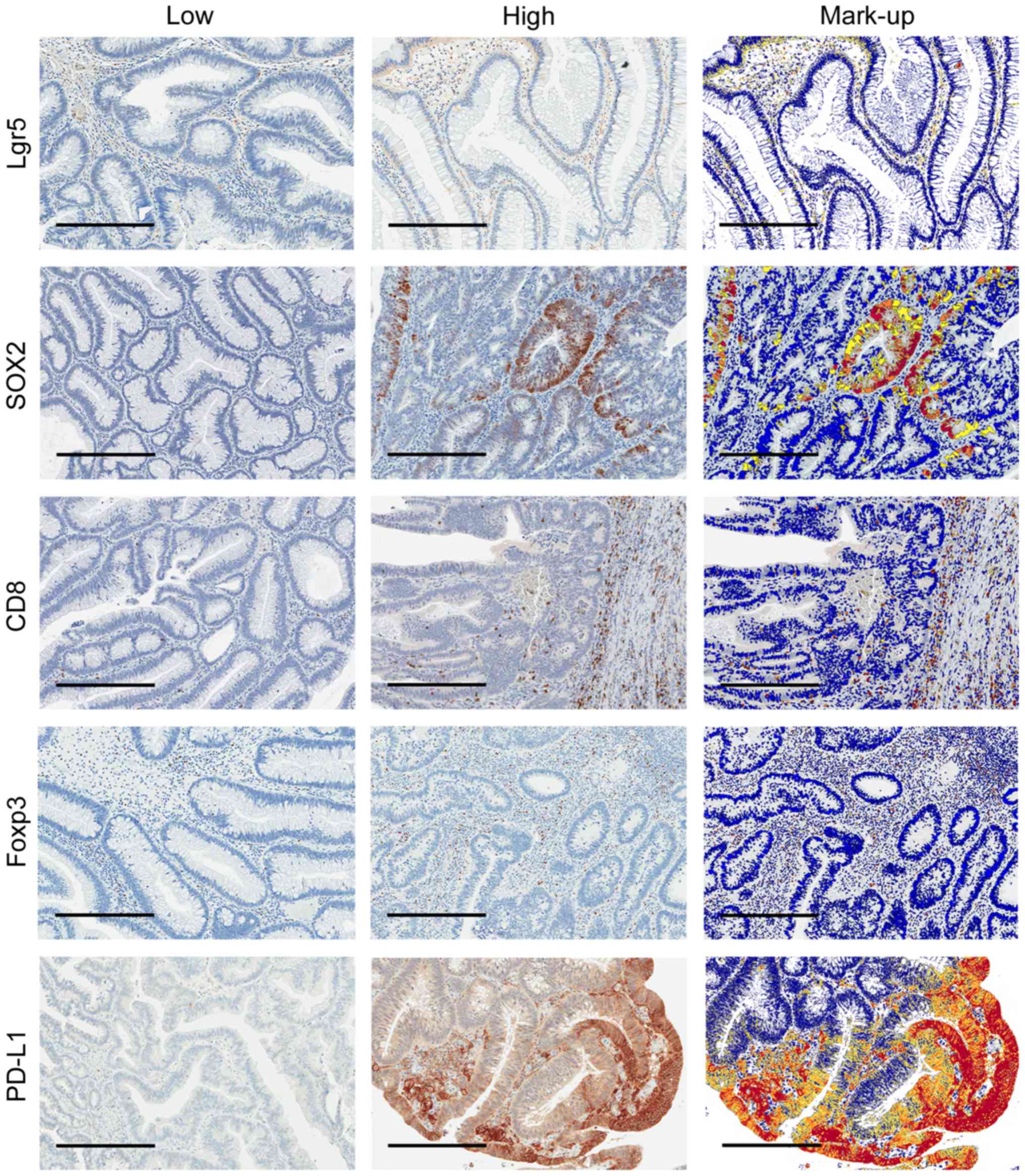 | Figure 1.Low and high immunostaining of Lgr5,
SOX2, CD8, Foxp3 and PD-L1 with corresponding digital mark-up of
high immunostaining images. Representative images (70X) of each
marker where blue represents negative staining, yellow weak, orange
moderate and red strong staining in digital mark-up images. Scale
bars=300 µm. Lgr5, Leucine-rich repeat-containing G-protein coupled
receptor-5; SOX2, sex determining region Y-box 2; CD8, Cluster of
differentiation 8; Foxp3, Forkhead box P3; PD-L1, Programmed
death-ligand 1. |
 | Table I.Median expression and number of cases
classified as high for each marker. |
Table I.
Median expression and number of cases
classified as high for each marker.
| Marker | Median | LG, n (%) | LGwHG, n (%) | HG, n (%) | AdCa, n (%) |
|---|
| Lgr5 | 9.84% | 10 (50.0) | 10 (55.6) | 8
(42.1) | 12 (54.5) |
| SOX2 | 50.5
cells/mm2 | 8
(40.0) | 10 (55.6) | 9
(47.4) | 12 (54.5) |
| CD8 | 293.5
cells/mm2 | 5
(25.0) | 9
(50.0) | 12 (63.2) | 13 (59.1) |
| Foxp3 | 194.8
cells/mm2 | 2
(10.0) | 7
(38.9) | 13 (68.4) | 18 (81.8) |
| PDL1 | 8.01% | 2
(10.0) | 8
(44.4) | 14 (73.7) | 16 (72.7) |
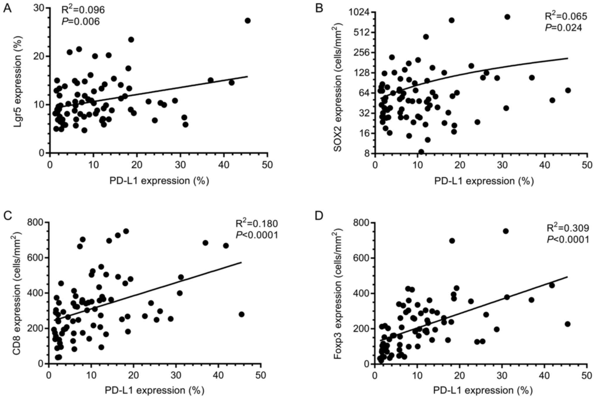
Correlations between marker
expression
Linear regression was used to assess correlations in
marker expression. PD-L1 expression significantly correlated to all
other markers studied (Fig. 2). CD8
and Foxp3 expression also demonstrated a highly significant
positive correlation (R2=0.27; P<0.0001). Expression
of Lgr5 and SOX2 demonstrated no significant correlation
(R2=0.0004; P=0.87) suggesting that different
sub-populations of tumour cells are identified by these
markers.
Marker expression during disease
progression
Kruskal-Wallis tests showed that there was
significant difference between the expression of CD8, Foxp3 and
PD-L1 with disease progression using expression as a continuous
variable (Fig. 3A-C). This was
confirmed in multinomial regression analysis using expression as a
categorical variable (Table II).
 | Figure 3.Expression of each marker with
progression of disease. Dots represent individual cases within each
group for (A) CD8, (B) Foxp3, (C) PD-L1, (D) Lgr5 and (E) SOX2.
Bars represent median values. Immune-related marker expression
trends upwards with progression of disease. Although not
significant, the median expression of SOX2 in the AdCa groups is
considerably higher than that observed in other groups. Expression
between groups was compared using the Kruskal-Wallis test.
*P<0.05, ***P<0.001. Lgr5, Leucine-rich repeat-containing
G-protein coupled receptor 5; SOX2, sex determining region Y-box 2;
CD8, Cluster of differentiation 8; Foxp3, Forkhead box P3; PD-L1,
Programmed death-ligand 1. |
| Table II.Expression of IHC markers as
categorical variables (high vs. low) throughout the stages of
rectal tumorigenesis. |
Table II.
Expression of IHC markers as
categorical variables (high vs. low) throughout the stages of
rectal tumorigenesis.
|
| Lgr5 | SOX2 | CD8 | Foxp3 | PD-L1 |
|---|
|
|
|
|
|
|
|
|---|
| Variable | OR | 95% CI | P-value | OR | 95% CI | P-value | OR | 95% CI | P-value | OR | 95% CI | P-value | OR | 95% CI | P-value |
|---|
| LG | 1.00 |
| 0.840 | 1.00 |
| 0.75 | 1.00 |
| 0.089 | 1.00 |
| <0.001 | 1.00 |
| 0.001 |
| LGwHG | 1.25 | 0.35–4.49 | 0.732 | 1.88 | 0.52–6.81 | 0.340 | 3.00 | 0.76–11.8 | 0.116 | 5.73 | 1.01–32.7 | 0.049 | 7.20 | 1.27–40.7 | 0.026 |
| HG | 0.73 | 0.21–2.57 | 0.622 | 1.35 | 0.38–4.80 | 0.643 | 5.14 | 1.30–20.4 | 0.020 | 19.5 | 3.38–112.4 | <0.001 | 25.2 | 4.24–149.8 | <0.001 |
| AdCa | 1.20 | 0.36–4.04 | 0.768 | 1.80 | 0.53–6.14 | 0.348 | 4.33 | 1.16–16.3 | 0.030 | 40.5 | 6.57–249.6 | <0.001 | 24.0 | 4.23–136.2 | <0.001 |
The expression of Foxp3 and PD-L1 both significantly
increased with progression of disease (overall P=0.0003 and
P=0.0010 respectively). Compared to LG adenoma, the greatest
increase in expression for Foxp3 was observed in AdCa [odds ratio
(OR)=40.5; 95% confidence interval (CI), 6.57–249.6; P<0.0001;
Table II]. For PD-L1 expression, the
largest increase was observed in HG adenoma when compared to LG
adenoma (OR=25.2; 95% CI, 4.24–49.8; P=0.0004; Table II).
Although not significant overall using multinomial
regression (P=0.089), increased expression of CD8 was nevertheless
seen in HG adenoma (OR=5.14; 95% CI, 1.30–20.4; P=0.02) and AdCa
(OR=4.33; 95% CI, 1.16–16.3; P=0.03).
No significant differences between groups were seen
for Lgr5 or SOX2 expression (P=0.84 and P=0.75, respectively).
However, in cases classified as LGwHG, higher SOX2 expression was
observed the high grade areas (Fig.
4).
 | Figure 4.Expression of SOX2 in areas of high
grade dysplasia. Representative immunostaining for (B, D, F and H)
SOX2 and matched (A, C, E and G) H&E stains in areas of high
grade dysplasia for a case diagnosed as low grade adenoma
containing high grade features. Low power images (magnification,
×50) of region 1 and 2 (A-D, respectively) with boxes indicating
higher power images (magnification, ×120)of each region (E-H,
respectively) are shown. SOX2 expression is present in high grade
areas of low grade adenomas, suggesting a function for SOX2 in
tumour cell invasion. Scale bars=500 µm for A-D and 200 µm for E-H.
Lgr5, Leucine-rich repeat-containing G-protein coupled receptor 5;
SOX2, Sex determining region Y-box 2; CD8, Cluster of
differentiation 8; Foxp3, Forkhead box P3; PD-L1, Programmed
death-ligand 1. |
Discussion
In this exploratory study we assessed expression of
the putative CSC markers Lgr5 and SOX2 and the immune cell-related
markers CD8, Foxp3 and PD-L1 at different stages of rectal
tumourigenesis. Expression of each of the immune cell-related
markers increased significantly during progression from LG adenoma
to AdCa, but no changes were observed for the CSC markers.
The increased expression of CD8 and Foxp3 in HG
adenomas and AdCa groups found here is consistent with previous
reports (27–29) and is suggestive of a tumour-specific
immune response attempting to control the progression of disease
(expansion of tumour-specific CD8+ T cells with
concurrent expansion of Foxp3+ regulatory T cells).
However, a concurrent increase in PD-L1 expression was also
observed, particularly in HG adenoma and AdCa, which may represent
an immune escape mechanism. Secretion of interferon-γ by T cells
can induce expression of PD-L1 (37),
so the increased T cell infiltrates in the later stages of disease
progression observed here may induce PD-L1 expression in tumour
cells, potentially permitting tumour cell invasion and the
development of AdCa.
The antibody and protocol used here for PD-L1
immunostaining was carefully optimised in-house and has been
directly compared to clinically validated protocols (38). However, it is important to note that
the analysis method used in this study could not distinguish
between tumour and stromal expression of PD-L1, even though it is
less subjective than traditional scoring methods. Although
assessment of PD-L1 expression to date has generally been limited
to tumour cells, there is evidence that immune-cell expression is
also clinically relevant in CRC (39–41).
Tumour cells may constitutively express PD-L1, or the expression
may be induced via T cell responses (39). The ability to distinguish PD-L1
expression in terms of contribution from the T cell infiltrate and
from tumour cells may provide important information with respect to
tumour-specific immune responses. It is also interesting to note
that recent findings suggested the PD-L1 gene can be
transcriptionally activated by SOX2, thus providing a potential
intrinsic mechanism for the induction of PD-L1 expression in tumour
cells (42). This is consistent with
the present result showing that PD-L1 and SOX2 expression are
significantly correlated.
Increased expression of the putative CSC markers
CD44, CD133 and Musashi-1 have been correlated with the progression
of premalignant disease in gastric cancer (43). In contrast, no significant
relationship between CSC marker expression and the progression of
rectal cancer was observed in the present study. Other studies have
reported increased expression of Lgr5 mRNA using in situ
hybridisation (ISH) in LG adenoma and AdCa compared to normal
tissue (7,8). Since IHC was used in the present study
to assess protein expression, it is possible that poor antibody
specificity may explain the discrepant results. Nonetheless, the
distribution of Lgr5 expression observed here was consistent with
previous reports using IHC (9,10) and ISH
(7,8),
with the expression localised primarily to the luminal surfaces in
LG adenoma, and heterogeneous expression throughout the crypts at
later stages of tumourigenesis.
One explanation for the observed lack of change in
SOX2 expression during rectal tumourigenesis may be due to the
small sample size. Also, SOX2 expression may have greater relevance
for proximal colon cancers with higher expression in right-sided,
primary colonic tumours associated with metastatic spread and
poorer outcomes (14). Since the
samples used in this study were all resected using the TEMS
procedure they were all rectal. However, an increase in the median
expression of SOX2 was observed in the AdCa group (Fig. 3E). Increased SOX2 expression was also
seen in LGwHG where malignant progression may be occurring, as
signified by areas of higher grade dysplasia (Fig. 4). This result suggests that SOX2 could
play a role in progression of dysplasia by promoting
de-differentiation or by promoting cellular migration and invasion.
The mechanism underlying SOX2 up-regulation in tumour cells could
therefore be relevant as a possible target to inhibit disease
progression. Recent evidence has shown that stem-like
characteristics of Lgr5+ CSC subsets can be intrinsic to
these cells, rather than being induced by stromal signals (44). It is not known whether the stem-like
characteristics of SOX2 expressing tumour cells is induced via
signals from the stromal microenvironment or if these cells also
undergo intrinsic changes independent of external signals. These
findings could provide novel insights into the mechanisms of
increasing dysplasia and invasion, as well as how tumour cells
escape immune recognition in the early stages of
tumourigenesis.
No correlation was observed between expression of
Lgr5 and SOX2 in this study. This indicates that perhaps these two
markers are identifying two different subsets of tumour cells or
possibly different CSC subsets. There was also no significant
increase in Lgr5 or SOX2 expression with disease progression. As
CSC only account for a small minority of tumour cells it is
possible that the methods used here could not detect subtle changes
in positive CSC marker expression. It is also possible that the
functional status of CSC is more clinically relevant than their
density. It is important to note that Lgr5 and SOX2 are only
thought to identify cancer cells with stem-like potential. Markers
of functional CSC have yet to be identified and this warrants
further investigation.
Here, we have demonstrated increased expression of
the T cell markers CD8 and Foxp3 concurrently with PD-L1 expression
during rectal tumourigenesis. To our knowledge, this is the first
study to demonstrate elevated expression of PD-L1 in HG adenoma and
early AdCa, when compared to LG adenoma in rectal lesions. This may
represent a mechanism by which tumour cells avoid T cell-mediated
immunity. Another novel finding in this study was that higher SOX2
expression was observed in high grade areas, which could be
associated with de-differentiation and cellular migration. However,
further investigation is required to determine the exact role of
SOX2 expression in rectal tumourigenesis.
Acknowledgements
The authors would like to thank Mr. Bob Mirzai and
Dr Kathy Fuller at the School of Pathology and Laboratory Medicine,
University of Western Australia for assistance with sectioning, Ms.
Cheryl Penter of St John of God Subiaco Hospital for assistance
with data collection, and Professor Paul Rigby and Ms. Alysia
Hubbard of the Centre for Microscopy, Characterisation and Analysis
at the University of Western Australia for assistance with digital
slide scanning.
Funding
Dr Timothy J. Miller was funded by a University
Postgraduate Award and a John Clauscen Murray Postgraduate
Scholarship. Purchasing of the image analysis software was funded
by the Tonkinson Colorectal Cancer Research Fund.
Availability of data and materials
The datasets used and/or analyzed during the current
study are available from the corresponding author on reasonable
request, subject to review by the institutional ethics
committee.
Authors' contributions
TJM was involved in the study design, pathological
review, performed the laboratory experiments, data analysis and the
manuscript review. MJM was involved in the study design and
manuscript review. CH assisted with the pathological review and was
involved in the study design and manuscript review. BI was involved
in the study design and manuscript review. CFP was involved in the
study design and manuscript review.
Ethics approval and consent to
participate
The study was approved by the St John of God
Healthcare (SJ-737) and the University of Western Australia Human
Research Ethics Committees (RA/4/1/7278). All patients provided
written informed consent for the use of their biological material
and health information for research purposes.
Patient consent for publication
All patients provided written informed consent to
the publication of research results as part of the consent
procedure.
Competing interests
The authors declare that they have no competing
interests.
References
|
1
|
American Cancer Society, . Global cancer
facts & figures. 2nd. American Cancer Society; Atlanta, GA:
2012
|
|
2
|
Vogelstein B, Fearon ER, Hamilton SR, Kern
SE, Preisinger AC, Leppert M, Nakamura Y, White R, Smits AM and Bos
JL: Genetic alterations during colorectal-tumor development. N Engl
J Med. 319:525–532. 1988. View Article : Google Scholar : PubMed/NCBI
|
|
3
|
Fearon ER and Vogelstein B: A genetic
model for colorectal tumorigenesis. Cell. 61:759–767. 1990.
View Article : Google Scholar : PubMed/NCBI
|
|
4
|
Fischer JM, Schepers AG, Clevers H,
Shibata D and Liskay RM: Occult progression by Apc-deficient
intestinal crypts as a target for chemoprevention. Carcinogenesis.
35:237–246. 2014. View Article : Google Scholar : PubMed/NCBI
|
|
5
|
Barker N, Ridgway RA, van Es JH, van de
Wetering M, Begthel H, van den Born M, Danenberg E, Clarke AR,
Sansom OJ and Clevers H: Crypt stem cells as the cells-of-origin of
intestinal cancer. Nature. 457:608–611. 2009. View Article : Google Scholar : PubMed/NCBI
|
|
6
|
Barker N, van Es JH, Kuipers J, Kujala P,
van den Born M, Cozijnsen M, Haegebarth A, Korving J, Begthel H,
Peters PJ and Clevers H: Identification of stem cells in small
intestine and colon by marker gene Lgr5. Nature. 449:1003–1007.
2007. View Article : Google Scholar : PubMed/NCBI
|
|
7
|
Baker AM, Graham TA, Elia G, Wright NA and
Rodriguez-Justo M: Characterization of LGR5 stem cells in
colorectal adenomas and carcinomas. Sci Rep. 5:86542015. View Article : Google Scholar : PubMed/NCBI
|
|
8
|
Jang BG, Kim HS, Kim KJ, Rhee YY, Kim WH
and Kang GH: Distribution of intestinal stem cell markers in
colorectal precancerous lesions. Histopathology. 68:567–577. 2016.
View Article : Google Scholar : PubMed/NCBI
|
|
9
|
Takeda K, Kinoshita I, Shimizu Y, Matsuno
Y, Shichinohe T and Dosaka-Akita H: Expression of LGR5, an
intestinal stem cell marker, during each stage of colorectal
tumorigenesis. Anticancer Res. 31:263–270. 2011.PubMed/NCBI
|
|
10
|
Becker L, Huang Q and Mashimo H:
Immunostaining of Lgr5, an intestinal stem cell marker, in normal
and premalignant human gastrointestinal tissue.
ScientificWorldJournal. 8:1168–1176. 2008. View Article : Google Scholar : PubMed/NCBI
|
|
11
|
Choi JW, Kim JK, Choi M, Kim YR and Yun
SH: In vivo imaging of Lgr5-positive cell populations using
confocal laser endomicroscopy during early colon tumorigenesis.
Endoscopy. 46:1110–1115. 2014. View Article : Google Scholar : PubMed/NCBI
|
|
12
|
Davis H, Irshad S, Bansal M, Rafferty H,
Boitsova T, Bardella C, Jaeger E, Lewis A, Freeman-Mills L, Giner
FC, et al: Aberrant epithelial GREM1 expression initiates colonic
tumorigenesis from cells outside the stem cell niche. Nat Med.
21:62–70. 2015. View
Article : Google Scholar : PubMed/NCBI
|
|
13
|
Takahashi K and Yamanaka S: Induction of
pluripotent stem cells from mouse embryonic and adult fibroblast
cultures by defined factors. Cell. 126:663–676. 2006. View Article : Google Scholar : PubMed/NCBI
|
|
14
|
Neumann J, Bahr F, Horst D, Kriegl L,
Engel J, Luque RM, Gerhard M, Kirchner T and Jung A: SOX2
expression correlates with lymph-node metastases and distant spread
in right-sided colon cancer. Bmc Cancer. 11:5182011. View Article : Google Scholar : PubMed/NCBI
|
|
15
|
Lundberg IV, Burström Löfgren A, Edin S,
Eklöf V, Öberg Å, Stenling R, Palmqvist R and Wikberg ML: SOX2
expression is regulated by BRAF and contributes to poor patient
prognosis in colorectal cancer. PLoS One. 9:e1019572014. View Article : Google Scholar : PubMed/NCBI
|
|
16
|
Lundberg IV, Edin S, Eklöf V, Öberg Å,
Palmqvist R and Wikberg ML: SOX2 expression is associated with a
cancer stem cell state and down-regulation of CDX2 in colorectal
cancer. BMC Cancer. 16:4712016. View Article : Google Scholar : PubMed/NCBI
|
|
17
|
Ong CW, Chong PY, McArt DG, Chan JY, Tan
HT, Kumar AP, Chung MC, Clément MV, Soong R, Van Schaeybroeck S, et
al: The prognostic value of the stem-like group in colorectal
cancer using a panel of immunohistochemistry markers. Oncotarget.
6:12763–12773. 2015. View Article : Google Scholar : PubMed/NCBI
|
|
18
|
Mazibrada J, Longo L, Vatrano S, Cappia S,
Giorcelli J, Pentenero M, Gandolfo S, Volante M, dell'Oste V, Lo
Cigno I, et al: Differential expression of HER2, STAT3, SOX2, IFI16
and cell cycle markers during HPV-related head and neck
carcinogenesis. New Microbiol. 37:129–143. 2014.PubMed/NCBI
|
|
19
|
Hellner K, Miranda F, Chedom Fotso D,
Herrero-Gonzalez S, Hayden DM, Tearle R, Artibani M,
KaramiNejadRanjbar M, Williams R, Gaitskell K, et al: Premalignant
SOX2 overexpression in the fallopian tubes of ovarian cancer
patients: Discovery and validation studies. EBioMedicine.
10:137–149. 2016. View Article : Google Scholar : PubMed/NCBI
|
|
20
|
Yuan P, Kadara H, Behrens C, Tang X, Woods
D, Solis LM, Huang J, Spinola M, Dong W, Yin G, et al: Sex
determining region Y-Box 2 (SOX2) is a potential cell-lineage gene
highly expressed in the pathogenesis of squamous cell carcinomas of
the lung. PLoS One. 5:e91122010. View Article : Google Scholar : PubMed/NCBI
|
|
21
|
Kreso A and Dick JE: Evolution of the
cancer stem cell model. Cell Stem Cell. 14:275–291. 2014.
View Article : Google Scholar : PubMed/NCBI
|
|
22
|
Bruttel VS and Wischhusen J: Cancer stem
cell immunology: Key to understanding tumorigenesis and tumor
immune escape? Front Immunol. 5:3602014. View Article : Google Scholar : PubMed/NCBI
|
|
23
|
Dunn GP, Bruce AT, Ikeda H, Old LJ and
Schreiber RD: Cancer immunoediting: From immunosurveillance to
tumor escape. Nat Immunol. 3:991–998. 2002. View Article : Google Scholar : PubMed/NCBI
|
|
24
|
Schreiber RD, Old LJ and Smyth MJ: Cancer
immunoediting: Integrating immunity's roles in cancer suppression
and promotion. Science. 331:1565–1570. 2011. View Article : Google Scholar : PubMed/NCBI
|
|
25
|
Pagès F, Berger A, Camus M, Sanchez-Cabo
F, Costes A, Molidor R, Mlecnik B, Kirilovsky A, Nilsson M, Damotte
D, et al: Effector memory T cells, early metastasis, and survival
in colorectal cancer. New Engl J Med. 353:2654–2666. 2005.
View Article : Google Scholar : PubMed/NCBI
|
|
26
|
Galon J, Costes A, Sanchez-Cabo F,
Kirilovsky A, Mlecnik B, Lagorce-Pagès C, Tosolini M, Camus M,
Berger A, Wind P, et al: Type, density, and location of immune
cells within human colorectal tumors predict clinical outcome.
Science. 313:1960–1964. 2006. View Article : Google Scholar : PubMed/NCBI
|
|
27
|
Maglietta A, Maglietta R, Staiano T,
Bertoni R, Ancona N, Marra G and Resta L: The immune landscapes of
polypoid and nonpolypoid precancerous colorectal lesions. PLoS One.
11:e01593732016. View Article : Google Scholar : PubMed/NCBI
|
|
28
|
Mariani F, Sena P, Pedroni M, Benatti P,
Manni P, Di Gregorio C, Manenti A, Palumbo C, de Leon MP and
Roncucci L: Th inducing POZ-Kruppel Factor (ThPOK) is a key
regulator of the immune response since the early steps of
colorectal carcinogenesis. PLoS One. 8:e544882013. View Article : Google Scholar : PubMed/NCBI
|
|
29
|
McLean MH, Murray GI, Stewart KN, Norrie
G, Mayer C, Hold GL, Thomson J, Fyfe N, Hope M, Mowat NA, et al:
The inflammatory microenvironment in colorectal neoplasia. PLoS
One. 6:e153662011. View Article : Google Scholar : PubMed/NCBI
|
|
30
|
Dong H, Zhu G, Tamada K and Chen L: B7-H1,
a third member of the B7 family, co-stimulates T-cell proliferation
and interleukin-10 secretion. Nat Med. 5:1365–1369. 1999.
View Article : Google Scholar : PubMed/NCBI
|
|
31
|
Le DT, Uram JN, Wang H, Bartlett BR,
Kemberling H, Eyring AD, Skora AD, Luber BS, Azad NS, Laheru D, et
al: PD-1 blockade in tumors with mismatch-repair deficiency. New
Engl J Med. 372:2509–2520. 2015. View Article : Google Scholar : PubMed/NCBI
|
|
32
|
Page DB, Postow MA, Callahan MK, Allison
JP and Wolchok JD: Immune modulation in cancer with antibodies.
Annu Rev Med. 65:185–202. 2014. View Article : Google Scholar : PubMed/NCBI
|
|
33
|
Yagyuu T, Hatakeyama K, Imada M, Kurihara
M, Matsusue Y, Yamamoto K, Obayashi C and Kirita T: Programmed
death ligand 1 (PD-L1) expression and tumor microenvironment:
Implications for patients with oral precancerous lesions. Oral
Oncol. 68:36–43. 2017. View Article : Google Scholar : PubMed/NCBI
|
|
34
|
Hatam LJ, DeVoti JA, Rosenthal DW, Lam F,
Abramson AL, Steinberg BM and Bonagura VR: Immune suppression in
premalignant respiratory papillomas: enriched functional CD4+Foxp3+
regulatory T cells and PD-1/PD-L1/L2 expression. Clin Cancer Res.
18:1925–1935. 2012. View Article : Google Scholar : PubMed/NCBI
|
|
35
|
Miller TJ, McCoy MJ, Hemmings C, Bulsara
MK, Iacopetta B and Platell CF: Objective analysis of cancer stem
cell marker expression using immunohistochemistry. Pathology.
49:24–29. 2017. View Article : Google Scholar : PubMed/NCBI
|
|
36
|
McCoy MJ, Hemmings C, Miller TJ, Austin
SJ, Bulsara MK, Zeps N, Nowak AK, Lake RA and Platell CF: Low
stromal Foxp3+ regulatory T-cell density is associated with
complete response to neoadjuvant chemoradiotherapy in rectal
cancer. Br J Cancer. 113:1677–1686. 2015. View Article : Google Scholar : PubMed/NCBI
|
|
37
|
Lee SJ, Jang BC, Lee SW, Yang YI, Suh SI,
Park YM, Oh S, Shin JG, Yao S, Chen L and Choi IH: Interferon
regulatory factor-1 is prerequisite to the constitutive expression
and IFN-gamma-induced upregulation of B7-H1 (CD274). FEBS Lett.
580:755–762. 2006. View Article : Google Scholar : PubMed/NCBI
|
|
38
|
Anyaegbu CC, Garrett K, Hemmings C,
Lee-Pullen TF and McCoy MJ: Immunohistochemical detection of PD-L1
for research studies: which antibody and what protocol? Pathology.
49:427–430. 2017. View Article : Google Scholar : PubMed/NCBI
|
|
39
|
Ribas A and Hu-Lieskovan S: What does
PD-L1 positive or negative mean? J Exp Med. 213:2835–2840. 2016.
View Article : Google Scholar : PubMed/NCBI
|
|
40
|
Li Y, Liang L, Dai W, Cai G, Xu Y, Li X,
Li Q and Cai S: Prognostic impact of programed cell death-1 (PD-1)
and PD-ligand 1 (PD-L1) expression in cancer cells and tumor
infiltrating lymphocytes in colorectal cancer. Mol Cancer.
15:552016. View Article : Google Scholar : PubMed/NCBI
|
|
41
|
Lee KS, Kwak Y, Ahn S, Shin E, Oh HK, Kim
DW, Kang SB, Choe G, Kim WH and Lee HS: Prognostic implication of
CD274 (PD-L1) protein expression in tumor-infiltrating immune cells
for microsatellite unstable and stable colorectal cancer. Cancer
Immunol Immunother. 66:927–939. 2017. View Article : Google Scholar : PubMed/NCBI
|
|
42
|
Zhong F, Cheng X, Sun S and Zhou J:
Transcriptional activation of PD-L1 by Sox2 contributes to the
proliferation of hepatocellular carcinoma cells. Oncol Rep.
37:3061–3067. 2017. View Article : Google Scholar : PubMed/NCBI
|
|
43
|
Wang T, Ong CW, Shi J, Srivastava S, Yan
B, Cheng CL, Yong WP, Chan SL, Yeoh KG, Iacopetta B and
Salto-Tellez M: Sequential expression of putative stem cell markers
in gastric carcinogenesis. Br J Cancer. 105:658–665. 2011.
View Article : Google Scholar : PubMed/NCBI
|
|
44
|
de Sousa e Melo F, Kurtova AV, Harnoss JM,
Kljavin N, Hoeck JD, Hung J, Anderson JE, Storm EE, Modrusan Z,
Koeppen H, et al: A distinct role for Lgr5+ stem cells
in primary and metastatic colon cancer. Nature. 543:676–680. 2017.
View Article : Google Scholar : PubMed/NCBI
|