Introduction
Thyroid cancer is rare, but it is the most frequent
endocrine malignancy. Thyroid cancer is divided into four types
including papillary, follicular, medullary, and anaplastic thyroid
cancer (1,2). Papillary thyroid carcinoma (PTC)
accounts for approximately more than 85% of all thyroid cancers.
Although PTC exhibits a satisfactory outcome after surgery or
radioiodine, however, approximately 15% of PTC patients who have
relapsed and frequently distant metastases present poor prognosis
(3,4).
Thus, to investigate underling molecular mechanisms involved in
carcinogenesis of PTC was urgent.
Long non coding RNAs (lncRNAs) have been identified
as crucial regulators in many biological processes, such as the
cell cycle, cell apoptosis, senescence, cell differentiation and
tumorigenesis (5,6). Abnormal expression of lncRNAs was
involved in PTC development and progression. For example, PVT1
(7), HOTAIR (8), and MEG3 (9) and so on, potentially function as
oncogenes or tumor suppressors of PTC. LINC01186 was reported to
acts as a tumor suppressor in lung cancer and inhibits migration
and invasion through Epithelial-Mesenchymal-Transition of lung
cancer (10). However, little was
known for LINC01186 in PTC development and progression.
In the study, we demonstrated that LINC01186 was
significantly downregulated in PTC tissues compared to adjacent
normal tissues. LINC01186 overexpression suppressed cell
proliferation and invasion ability. Moreover, we demonstrated that
LINC01186 overexpression suppressed thelarge tumor suppressor
kinase (LATS1)/ YY1 associated protein 1 (YAP1) signaling in PTC
cells. Thus, our data suggested that LINC01186 may serve as a
target for therapy in PTC.
Materials and methods
Tissue samples
A total of 65 cases PTC tissue sample and adjacent
normal tissue sample were obtained between April 2012 and January
2014 at Department of Thyroid Surgery, Affiliated Hospital of
Guizhou Medical University (Guizhou, China). All patients (22 male
and 43 female; age range, 23–76 years; mean age, 45 years) were
enrolled in the study did not receive any preoperative therapies
including radiotherapy, chemotherapy and levothyroxine, prior to
the thyroidectomy. All of tissue samples were immediately frozen in
liquid nitrogen and stored at −80°C following operation until
further RNA extraction. The study protocol was approved by the
Ethics Committee of Affiliated Hospital of Guizhou Medical
University and written informed consent was obtained from all of
patients.
Cell lines culture
Three human PTC cell lines (TPC-1, K1 and IHH-4) and
a human thyroid epithelial cell line Nthy-ori3-1 were purchased
from the Cell Bank of the Chinese Academy of Sciences (Shanghai,
China). Although K1 cells are contaminated with GLAG-66 (11), the resulting phenotypic and genotypic
differences between K1 and GLAG-66 cells were considered unlikely
to affect our study. The PTC cells were supplemented with
Dulbecco's modified Eagle's medium (DMEM) containing 10% fetal
bovine serum (FBS; both Gibco; Thermo Fisher Scientific Inc.,
Waltham, MA, USA). Cells were cultured at 37°C in humidified air
with 5% CO2.
Plasmid construction
The full-length LINC01186 sequences were chemically
synthesized and cloned into the pcDNA3.1 plasmid by Guangzhou
RiboBio Co., Ltd. (Guangzhou, China). The pcDNA3.1-LINC01186
plasmid was transfected into TPC-1 and IHH-4 cells using
Lipofectamine 2000 (Invitrogen; Thermo Fisher Scientific,
Inc.).
RNA extraction and reverse
transcription-quantitative PCR (RT-qPCR)
Total RNA was extracted using Trizol reagent
(Invitrogen; Thermo Fisher Scientific, Inc.) following the
manufacturer's instructions. LINC01186 cDNA was reverse-transcribed
using Prime Script TM RT-PCR kit and were detected using SYBR
Premix Ex TaqTM (both Takala, Dalian, China) under an ABI 7500 Fast
Sequence Detection System (Applied Biosystems, Foster City, CA,
USA). The GAPDH mRNA expression was used as internal control. The
primer sequences were as follows: LINC01186-forward,
5′-CTAAAACGGGAAAGATGCCTTC-3′ and reverse,
5′-TCAAGATTAGGGAACATACTGGC-3′; GAPDH forward,
5′-GGAGCGAGATCCCTCCAAAAT-3′ and reverse,
5′-GGCTGTTGTCATACTTCTCATGG-3′. Data was quantified using the
2−ΔΔCq method (12).
Cell proliferation assay
PTC cells (3,000 cells per well) were seeded in
96-well plates were and cultured for 24 h. Then, cells were
transfected with si-NC and si-LINC01186 and cultured at 37°C in
humidified air with 5% CO2. Cell proliferation ability
was measured by using the Cell Counting kit-8 (CCK-8; Dojindo,
Kumamoto, Japan) at 12, 24, 48, and 72 h. The absorbance was at 450
nm under a microplate spectrophotometer (Thermo Labsystems, Vantaa,
Finland).
Cell colony formation assay
PTC cells (3,000 cells per well) were seeded in
6-well plates were and cultured for 24 h. Then, cells were
transfected with si-NC and si-LINC01186 and cultured at 37°C in
humidified air with 5% CO2 for 7 days. Cell colonies
were fixed with 4% paraformaldehyde, stained with 0.1% crystal
violet. Cells colonies were calculated under a microscope (Olympus,
Tokyo, Japan).
Cell invasion assay
Cell invasion assays were performed using transwell
chambers with membrane pore size of 8.0 µm (Corning Inc., Union
City, CA, USA). Transfected cells (1×10⁵ cells) were
added into the upper chamber, while the lower chamber contained
DMEM medium with 10% FBS. After cell culture for 48 h, cells in the
lower chamber were fixed with 4% paraformaldehyde and stained with
0.1% crystal violet. Invasive cells were calculated under a
microscope (Olympus) in five random fields.
Western blot assay
Total proteins were extracted from transfecting
cells using RIPA lysis buffer (Pierce, Rockford, IL, USA). After
centrifugation at 14,000 g, 4°C for 15 min, the supernatants were
collected. The protein concentration was detected using a BCA
protein assay kit (Thermo Fisher Scientific Inc.). Equal protein
was separated at sodium dodecyl sulfate-polyacrylamide gel
electrophoresis (SDS-PAGE), and then transferred to PVDF membranes
(Millipore, Billerica MA, USA). Then, the membranes were blocked
with 5% non-fat milk and incubated with primary antibody: Anti-YAP1
(1:1,000), anti-LATS1 (1:1,000) and GAPDH (1:1,000 all Cell
Signaling Technology, Inc., Danvers, MA, USA). After incubation
with the appropriate horseradish peroxidase (HRP)-conjugated
secondary antibody for 2 h, the blots were detected using an
enhanced chemiluminescence detection system (Amersham, Little
Chalfont, UK). GAPDH were used as an internal control.
Statistical analysis
The SPSS 20.0 software system (IBM Corp., Armonk,
NY, USA) was used for statistical analyses. The results are showed
as the mean ± SD (standard deviation) from at least three
independent experiments. The chi-square test was applied to examine
the relationship between LINC01186 expression and
clinicopathological characteristics. Significant differences
between two groups were compared using two-tailed Student's
t-tests. Significant differences among multiple groups were
performed using one-way analysis of variance followed by a
Bonferroni post hoc test. P<0.05 was considered to indicate a
statistically significant difference.
Results
Expression of LINC01186 is
significantly downregulated in PTC tissues and cells
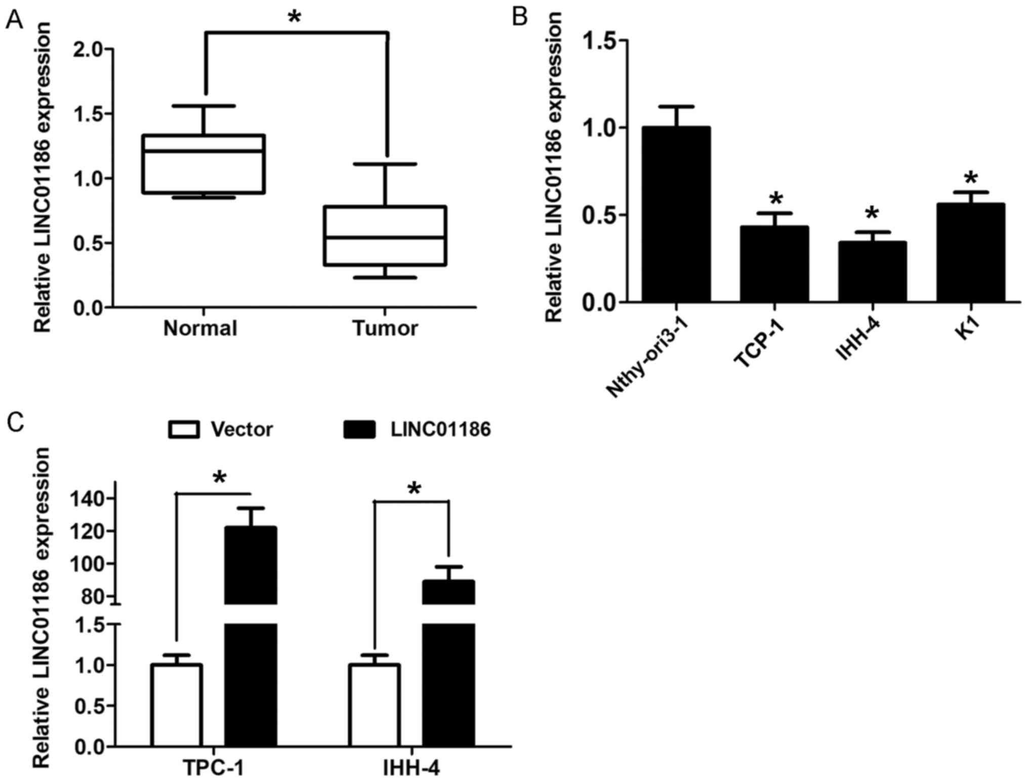
To explore the clinical role of LINC01186 in PTC, we
examined the expression of LINC01186 in PTC tissue samples and
adjacent normal tissue samples using RT-qPCR. The results showed
that LINC01186 expression levels were notably downregulated in PTC
tissues compared to adjacent normal tissues (P<0.05; Fig. 1A). The median expression of LINC01186
was used as a cut-off value to divide 65 cases into two groups
(lower or higher group). We found that lower LINC01186 expression
was associated with lymph node metastasis of PTC patients
(P<0.05; Table I). Moreover, a
decreased expression of LINC01186 was also observed in three
different PTC cell lines compared to human thyroid epithelial cell
line Nthy-ori3-1 (P<0.05; Fig.
1B). Collectively, all the results indicated that LINC01186 is
downregulated in PTC.
 | Table I.Association between LINC01186
expression and clinicopathological factors in PTC patients. |
Table I.
Association between LINC01186
expression and clinicopathological factors in PTC patients.
|
| LINC01186
expression |
|
|---|
|
|
|
|
|---|
| Clinicopathological
factors | Patients (n=65) | Lower (n=32) | Higher (n=33) | P-value |
|---|
| Age, years |
|
|
| 0.821 |
| ≤55 | 50 | 25 | 25 |
|
|
>55 | 15 | 7 | 8 |
|
| Sex |
|
|
| 0.261 |
| Male | 22 | 10 | 12 |
|
|
Female | 43 | 22 | 21 |
|
| Tumor size, cm |
|
|
| 0.511 |
| ≤2 | 31 | 13 | 18 |
|
|
>2 | 34 | 19 | 15 |
|
| Lymph node
metastasis |
|
|
| 0.009a |
|
Negative | 35 | 12 | 23 |
|
|
Positive | 30 | 20 | 10 |
|
| Tumor location |
|
|
| 0.494 |
|
Unilateral | 36 | 20 | 16 |
|
|
Bilateral | 18 | 7 | 11 |
|
|
Multifocality | 11 | 5 | 6 |
|
| TNM stage |
|
|
| 0.223 |
| I | 33 | 13 | 20 |
|
| II | 20 | 11 | 9 |
|
|
III–IV | 12 | 8 | 4 |
|
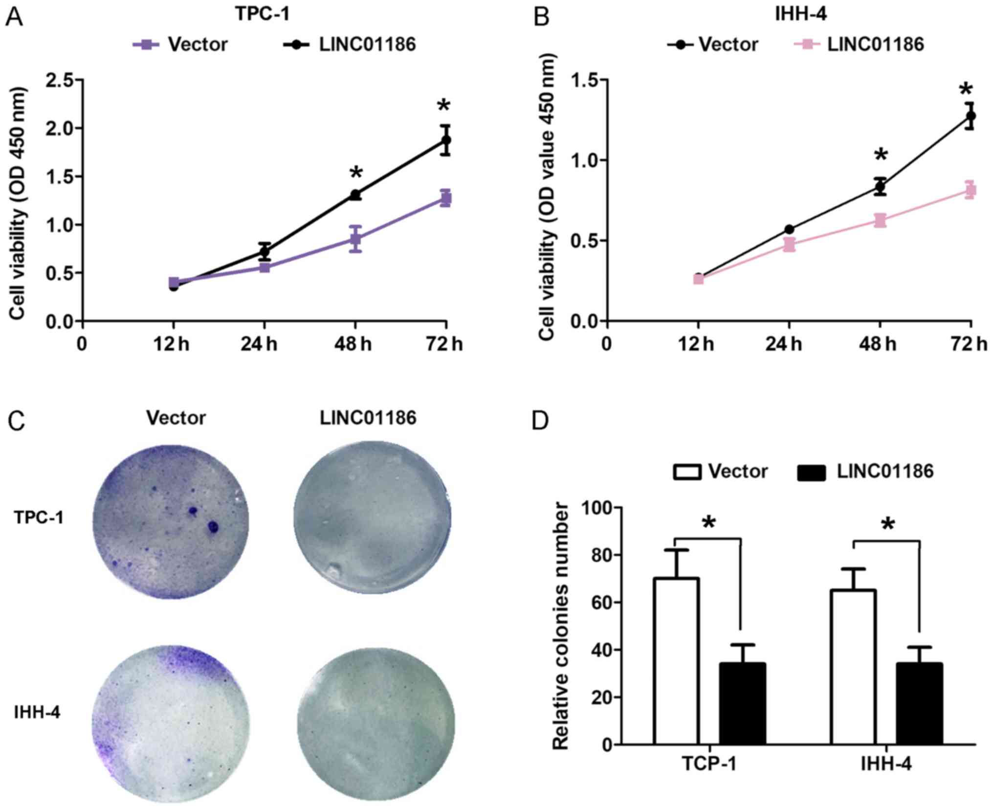
LINC01186 inhibits cell proliferation
and migration of PTC in vitro
To explore the biological role of LINC01186 in PTC
progression, we performed gain-function assays by transfection with
pcDNA3.1-LINC01186 plasmids into TPC-1 and IHH-4 cells. The
efficiency of LINC01186 overexpression by pcDNA3.1-LINC01186
plasmid was confirmed by RT-qPCR and shown in Fig. 1C (P<0.05). CCK8 cell proliferation
assays demonstrated that enhanced expression of LINC01186
suppressed the proliferation ability of TPC-1 and IHH-4 cells
compared with the control cells transfected with empty vector
(P<0.05; Fig. 2A and B). Cell
colony formed number was reduced when LINC01186 was overexpressed
after transfected with pcDNA3.1-LINC01186 plasmids into TPC-1 and
IHH-4 cells compared with the control cells transfected with empty
vector (P<0.05; Fig. 2C and D).
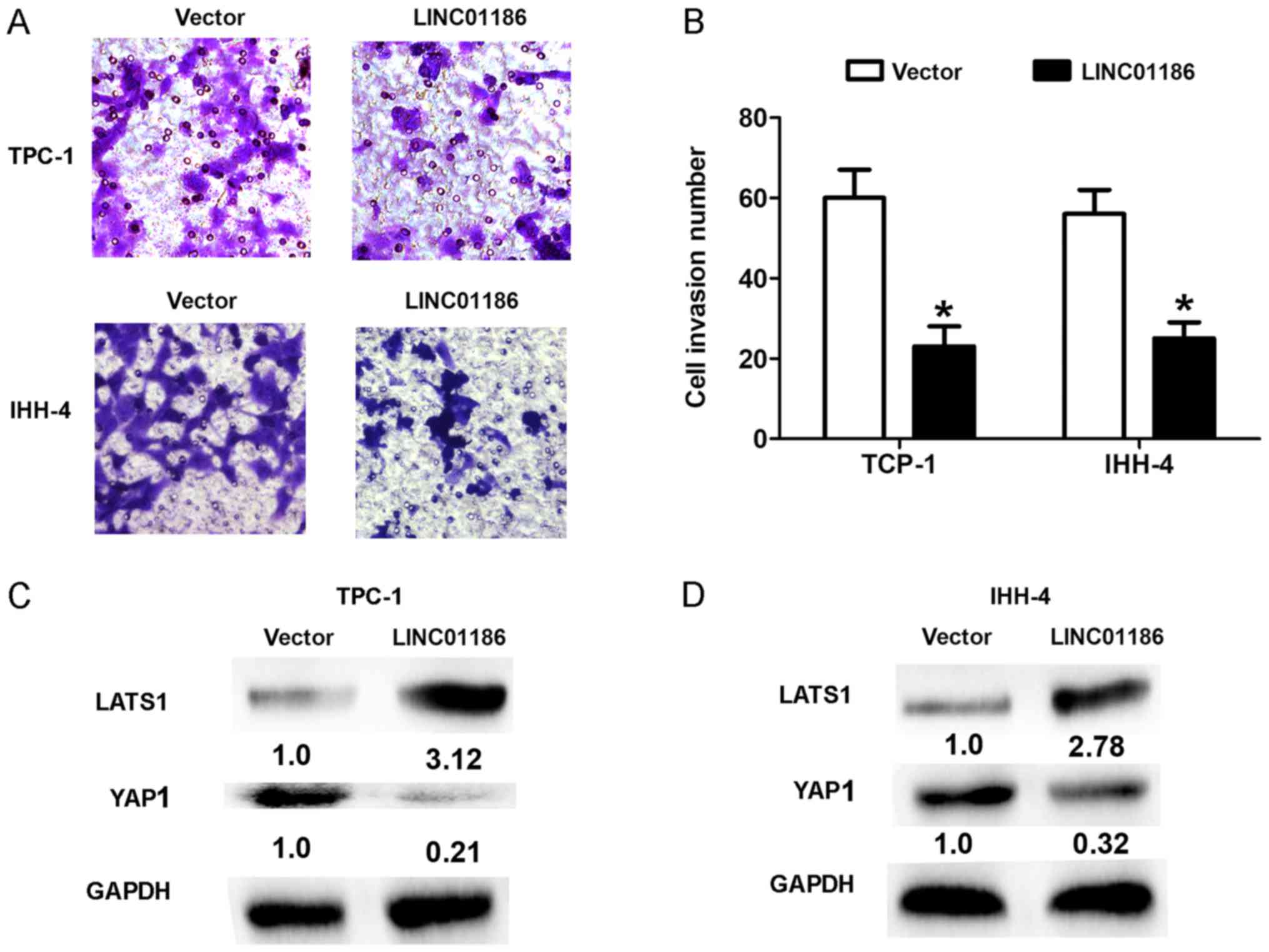
Moreover, we demonstrated that upregulated expression of LINC01186
suppressed the cell invasion ability of TPC-1 and IHH-4 cells
compared with the control cells transfected with empty vector
(P<0.05, Fig. 3A and B). Thus,
these results indicated that LINC01186 inhibited cell proliferation
and invasion of PTC in vitro.
LINC01186 inhibits LATS1/YAP signaling
pathway in PTC cells
LATS1/YAP signaling pathway executes important
function in PTC progression. High expression of Yes-activated
protein-1 in PTC correlates with poor prognosis (13). In thyroid cancer cell lines,
Yes-associated protein 1 promotes papillary thyroid cancer cell
proliferation by activating the ERK/MAPK signaling pathway
(14). In the study, we demonstrated
that upregulated expression of LINC01186 suppressed the expression
levels of YAP1 and upregualted the LATS1 in TPC-1 and IHH-4 cells
compared with the control cells transfected with empty vector
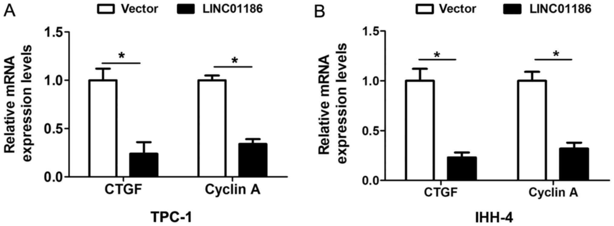
(P<0.05; Fig. 3C and D). Moreover,
we also detected the mRNA expression of YAP downstream targeted
gene CTGF and Cyclin A. The results showed that upregulated of
LINC01186 expression suppressed the mRNA expression levels of CTGF
and Cyclin A in TPC-1 and IHH-4 cells (Fig. 4A and B). Thus, these results indicated
that LINC01186 inhibited LATS1/YAP signaling pathway in PTC
cells.
Discussion
LncRNAs have been recently found to be dysregulated
and act as crucial regulators in the progression of thyroid cancer.
For instance, the lncRNA n340790 accelerates carcinogenesis of
thyroid cancer by regulating miR-1254 (15). Knockdown of lncRNA-PANDAR suppresses
the proliferation, cell cycle and promotes apoptosis in thyroid
cancer cells (16). Long non-coding
RNA ANRIL promotes the invasion and metastasis of thyroid cancer
cells through TGF-β/Smad signaling pathway (17). Overexpression of long intergenic
noncoding RNA LINC00312 inhibits the invasion and migration of
thyroid cancer cells by downregulating microRNA-197-3p (18). In the study, we demonstrated that long
noncoding RNA LINC01186 was significantly downregulated in PTC
tissues compared to adjacent normal tissues. Furthermore, in
vitro, CCK8 cell proliferation and cell colony formation showed
that LINC01186 overexpression suppressed cell proliferation
ability. Meanwhile, cell invasion was also been inhibited by
overexpression of LINC01186 in thyroid carcinoma cells. These
results indicated that LINC01186 suppressed cell proliferation and
invasion.
Many studies have demonstrated YAP, core downstream
target of Hippo signaling pathway, is altered in different
malignant tumors including thyroid carcinoma (19). In thyroid carcinoma, Ugolini et
al reported that YAP-1 is overexpressed in papillary and
anaplastic thyroid cancers and associated with poor prognosis of
thyroid cancer (13). Liu et
al showed that YAP expression positively correlated with TNM
stage and lymph node metastasis and knockdown of YAP inhibited cell
proliferation, migration and invasion in thyroid carcinoma cells
(20). Yes-associated protein 1
promotes papillary thyroid cancer cell proliferation by activating
the ERK/MAPK signaling pathway (14).
In the study, we demonstrated that LINC01186 overexpression
inhibited LATS1/YAP signaling pathway by downregulaing the YAP1
expression and increasing the LATS1 expression in PTC cells. Thus,
our data suggested that LINC01186 expression closely related to
LATS1/YAP in PTC. Of course, in the previous study, LINC01186 was
found to suppress cell EMT process in lung cancer (10). In the future, we hope to investigate
whether LINC01186 expression affects PTC cell EMT process by
LATS1/YAP signaling pathway.
In conclusion, we found that LINC01186 was
downregulated in PTC tissues and cells. LINC01186 acts as tumor
suppressor to inhibit cell proliferation and invasion of PTC.
Moreover, we demonstrated that LINC01186 overexpression inhibited
LATS1/YAP signaling pathway by downregulating the YAP expression.
Thus, our data suggested that LINC01186 may serve as a target for
therapy in PTC.
Acknowledgements
Not applicable.
Funding
No funding was received.
Availability of data and materials
The datasets used and/or analyzed in the present
study are available from the corresponding author upon reasonable
request.
Authors' contributions
NW and HD designed the study that led to the
submission. CZ, RG and YZ performed functional experiments to
investigate the roles of LINC01186 in PTC. NW and HD drafted the
manuscript.
Ethics approval and consent to
participate
The study protocol was approved by the Ethics
Committee of Affiliated Hospital of Guizhou Medical University and
written informed consent was obtained from all of patients.
Patient consent for publication
Not applicable.
Competing interest
The authors declare that they have no competing
interests.
References
|
1
|
Sipos JA and Mazzaferri EL: Thyroid cancer
epidemiology and prognostic variables. Clin Oncol (R Coll Radiol).
22:395–404. 2010. View Article : Google Scholar : PubMed/NCBI
|
|
2
|
Lee YS, Lim YS, Lee JC, Wang SG, Kim IJ
and Lee BJ: Clinical implication of the number of central lymph
node metastasis in papillary thyroid carcinoma: Preliminary report.
World J Surg. 34:2558–2563. 2010. View Article : Google Scholar : PubMed/NCBI
|
|
3
|
Siegel RL, Miller KD and Jemal A: Cancer
statistics, 2017. CA Cancer J Clin. 67:7–30. 2017. View Article : Google Scholar : PubMed/NCBI
|
|
4
|
Roh JL, Park JY and Park CI: Total
thyroidectomy plus neck dissection in differentiated papillary
thyroid carcinoma patients: Pattern of nodal metastasis, morbidity,
recurrence, and postoperative levels of serum parathyroid hormone.
Ann Surg. 245:604–610. 2007. View Article : Google Scholar : PubMed/NCBI
|
|
5
|
Cheetham SW, Gruhl F, Mattick JS and
Dinger ME: Long noncoding RNAs and the genetics of cancer. Br J
Cancer. 108:2419–2425. 2013. View Article : Google Scholar : PubMed/NCBI
|
|
6
|
Murugan AK, Munirajan AK and Alzahrani AS:
Long noncoding RNAs: Emerging players in thyroid cancer
pathogenesis. Endocr Relat Cancer. 25:R59–R82. 2018. View Article : Google Scholar : PubMed/NCBI
|
|
7
|
Zhou Q, Chen J, Feng J and Wang J: Long
noncoding RNA PVT1 modulates thyroid cancer cell proliferation by
recruiting EZH2 and regulating thyroid-stimulating hormone receptor
(TSHR). Tumour Biol. 37:3105–3113. 2016. View Article : Google Scholar : PubMed/NCBI
|
|
8
|
Zhu H, Lv Z, An C, Shi M, Pan W, Zhou L,
Yang W and Yang M: Onco-lncRNA HOTAIR and its functional genetic
variants in papillary thyroid carcinoma. Sci Rep. 6:319692016.
View Article : Google Scholar : PubMed/NCBI
|
|
9
|
Wang C, Yan G, Zhang Y, Jia X and Bu P:
Long non-coding RNA MEG3 suppresses migration and invasion of
thyroid carcinoma by targeting of Rac1. Neoplasma. 62:541–549.
2015. View Article : Google Scholar : PubMed/NCBI
|
|
10
|
Hao Y, Yang X, Zhang D, Luo J and Chen R:
Long noncoding RNA LINC01186, regulated by TGF-β/SMAD3, inhibits
migration and invasion through Epithelial-Mesenchymal-Transition in
lung cancer. Gene. 608:1–12. 2017. View Article : Google Scholar : PubMed/NCBI
|
|
11
|
Ribeiro FR, Meireles AM, Rocha AS and
Teixeira MR: Conventional and molecular cytogenetics of human
non-medullary thyroid carcinoma: Characterization of eight cell
line models and review of the literature on clinical samples. BMC
Cancer. 8:3712008. View Article : Google Scholar : PubMed/NCBI
|
|
12
|
Livak KJ and Schmittgen TD: Analysis of
relative gene expression data using real-time quantitative PCR and
the 2(-Delta Delta C(T)) method. Methods. 25:402–408. 2001.
View Article : Google Scholar : PubMed/NCBI
|
|
13
|
Ugolini C, Borrelli N, Niccoli C, Elisei
R, Viola D, Vitti P, Miccoli P and Basolo F: Role of YAP-1 in
thyroid tumor progression and outcome. Appl Immunohistochem Mol
Morphol. 25:581–585. 2017. View Article : Google Scholar : PubMed/NCBI
|
|
14
|
Liao T, Wen D, Ma B, Hu JQ, Qu N, Shi RL,
Liu L, Guan Q, Li DS and Ji QH: Yes-associated protein 1 promotes
papillary thyroid cancer cell proliferation by activating the
ERK/MAPK signaling pathway. Oncotarget. 8:11719–11728. 2017.
View Article : Google Scholar : PubMed/NCBI
|
|
15
|
Li Q, Shen W, Li X, Zhang L and Jin X: The
lncRNA n340790 accelerates carcinogenesis of thyroid cancer by
regulating miR-1254. Am J Transl Res. 9:2181–2194. 2017.PubMed/NCBI
|
|
16
|
Li Z, Gao B, Hao S, Tian W, Chen Y, Wang
L, Zhang X and Luo D: Knockdown of lncRNA-PANDAR suppresses the
proliferation, cell cycle and promotes apoptosis in thyroid cancer
cells. EXCLI J. 16:354–362. 2017.PubMed/NCBI
|
|
17
|
Zhao JJ, Hao S, Wang LL, Hu CY, Zhang S,
Guo LJ, Zhang G, Gao B, Jiang Y, Tian WG and Luo DL: Long
non-coding RNA ANRIL promotes the invasion and metastasis of
thyroid cancer cells through TGF-β/Smad signaling pathway.
Oncotarget. 7:57903–57918. 2016.PubMed/NCBI
|
|
18
|
Liu K, Huang W, Yan DQ, Luo Q and Min X:
Overexpression of long intergenic noncoding RNA LINC00312 inhibits
the invasion and migration of thyroid cancer cells by
down-regulating microRNA-197-3p. Biosci Rep. 37(pii):
BSR201701092017. View Article : Google Scholar : PubMed/NCBI
|
|
19
|
Celano M, Mignogna C, Rosignolo F,
Sponziello M, Iannone M, Lepore SM, Lombardo GE, Maggisano V,
Verrienti A, Bulotta S, et al: Expression of YAP1 in aggressive
thyroid cancer. Endocrine. 59:209–212. 2018. View Article : Google Scholar : PubMed/NCBI
|
|
20
|
Liu Z, Zeng W, Wang S, Zhao X, Guo Y, Yu
P, Yin X, Liu C and Huang T: A potential role for the Hippo pathway
protein, YAP, in controlling proliferation, cell cycle progression,
and autophagy in BCPAP and KI thyroid papillary carcinoma cells. Am
J Transl Res. 9:3212–3223. 2017.PubMed/NCBI
|