Introduction
Head and neck squamous cell carcinoma (HNSCC) is the
sixth most common type of cancer globally, with ~600,000 incident
cases diagnosed per year (1–3). It is well known that this carcinoma is
primarily a loco-regional disease. Improved local control of HNSCC
has been achieved over previous decades due to recent advances in
multidisciplinary treatments, including the surgical approach,
reconstruction techniques and adjuvant treatment modalities
(2,3).
However, HNSCC is characterized by high rates of recurrence or
metastasis following initial treatment, with rates of 40–60%
(4). In consequence, the quality of
life and life expectancy of patients with advanced HNSCC have not
markedly improved over previous decades (2).
Human papillomavirus (HPV) infection is one of the
major risk factors for HNSCC (5).
Oropharynx squamous cell carcinoma (OPSCC) is most markedly
associated with high-risk HPV infection (6–8). HNSCCs of
other sites, including the oral cavity (9–11) and
larynx (12–15), also have been demonstrated to harbor
the virus. HPV status in tumors may be determined by numerous
assays, including HPV E6/E7 RNA expression detected by reverse
transcription-quantitative polymerase chain reaction (RT-qPCR) and
HPV DNA detection by in situ hybridization or PCR.
Cyclin-dependent kinase inhibitor 2A (p16INK4a), a
tumor-suppressor protein that is overexpressed in cells, is a
surrogate immunohistochemical marker of oncogenic subtypes of HPV
infection in the oropharynx (6,16–18). However, the immunohistochemical
studies of p16INK4a in non-oropharyngeal HNSCC have
demonstrated variable results (13–15,19–23);
thus, the validity of p16INK4a protein as a marker of
HPV infection in non-OPSCC requires additional validation.
Furthermore, patients who are positive for p16INK4a have
improved outcomes compared with those of patients with OPSCC who
are negative for p16INK4a (6,17).
Compared with the significance of the data regarding HPV and OPSCC,
the significance of HPV in HNSCC sites outside the oropharynx has
not been established. Major gaps in the literature remain,
including very little data for Asia, particularly China.
Additionally, to the best of our knowledge, with the exception of a
small number of studies investigating laryngeal cancer in Eastern
China (24,25), there has been no analysis of
p16INK4a expression in Chinese patients in oral cavity
cancer. The aim of the present study was to evaluate the frequency
of p16INK4a protein positive expression in patients with
non-OPSCC in Southern China, and to assess its prognostic
value.
Patients and methods
Patients and specimens
Patients with non-oropharyngeal HNSCC were treated
at the Department of Head and Neck Surgery of Sun Yat-sen
University Cancer Center (SYSUCC; Guangzhou, China) between January
2001 and December 2008, and were selected randomly and
retrospectively by stratified sampling. The present study was
approved by the Ethics Review Board of SYSUCC and written informed
consent was provided by all patients. All activities were in accord
with the 1964 Declaration of Helsinki. The oral cavity, glottis and
supraglottic larynx cancer were defined as non-OPSCC. The inclusion
criteria were as follows: i) Patients had undergone complete
excision, with or without unilateral or bilateral neck dissection;
ii) the diagnosis was confirmed by pathology; iii) pathologic
samples and complete follow-up data were available. Patients with
other concomitant organ disorders or malignant tumor types were
excluded. A total of 183 eligible patients with non-OPSCC with a
mean age of 54.3 years (range, 26–86 years) were included in the
present study. Among these, females comprised 24.6% of the
patients. No patients exhibited metastasis at the time of diagnosis
(stage IVc), nor had they undergone chemotherapy and/or
radiotherapy prior to surgery.
All patients had follow-up data through December
2015. Detailed information of the patients selected for analysis
was reviewed. The tumors were restaged in accordance with the 2010
American Joint Committee on Cancer TNM Staging Manual (26). To verify the histological diagnosis of
non-OPSCC and adequacy of the tissue for analysis, the haematoxylin
and eosin-stained (H&E) slides were reviewed by a pathologist.
Sections were stained by a standard H&E staining method as
follows: Sections were incubated for 60 sec at room temperature in
filtered Mayer's haematoxylin solution, which is diluted by
distilled water (1:20). Subsequently, the sections were washed
under running water for 10 min. Following incubation in 1% eosin
solution for 15 sec at room temperature, the sections were washed
quickly (5–10 sec) in water and dehydrated in ascending
concentrations of ethanol (75, 85, 95 and 100%). Subsequently,
xylene was used to clear and covered with slips. The representative
formalin-fixed, paraffin-embedded block for each patient was
obtained from the Pathology Department of SYSUCC.
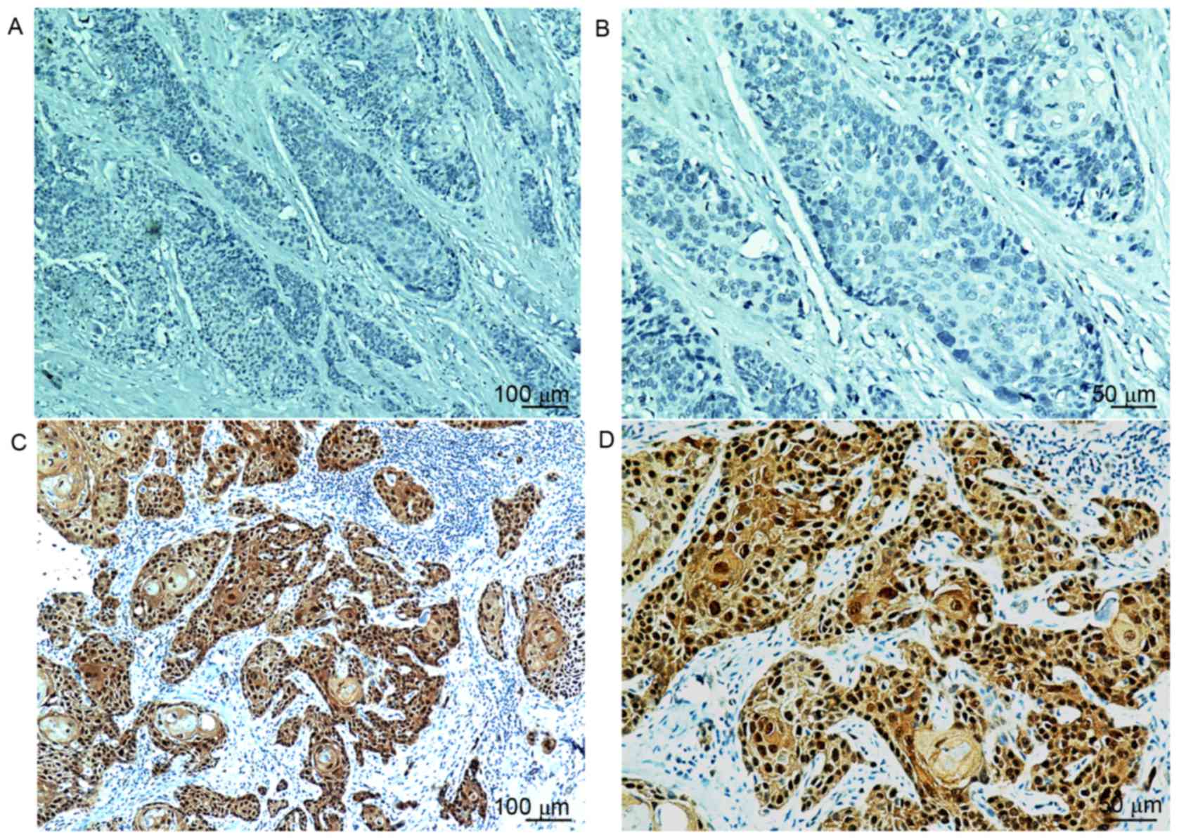
Staining and evaluation
The 4-mm thick paraffin-embedded slices were dewaxed
and rehydrated, and endogenous peroxidase activity was blocked with
0.3% H2O2. The slides were boiled in sodium
citrate for 5 min at 95°C and 20 min at 60°C in a microwave for
antigen retrieval. The primary mouse monoclonal antibody against
human p16INK4a (CINtec® p16INK4a
Histology kit; clone E6H4, prediluted, MTM-Laboratories; Roche
Diagnostics, Basel, Switzerland) was used for the detection of the
protein p16INK4a. Subsequently, the slides were
incubated with a horseradish peroxidase-conjugated goat anti-mouse
IgG secondary antibody (cat. no. 7076s; 1:2,000; Cell Signaling
Technology, Inc., Danvers, MA, USA) at 37°C for 30 min. Finally,
stained slides were counterstained with 5% Mayer's haematoxylin
solution at room temperature for 60 sec. All slides were
interpreted by two pathologists in a double-blinded manner.
Positive p16INK4a expression was defined as a strong and
diffuse nuclear and cytoplasmic staining in ≥70% of the tumor
cells, as described previously (6,21,27).
Statistical analysis
All data were analyzed with the SPSS 19.0
statistical software package (IBM Corp., Armonk, NY, USA). The
results of analysis were presented as the mean ± standard error of
the mean. The χ2 test was used to assess the
associations between p16INK4a expression and other
characteristics of patients. The disease-free survival (DFS) time
was defined as the time between diagnosis and loco-regional
recurrence or progression, distant metastasis. The overall survival
(OS) time was calculated from the time of diagnosis to the date of
mortality or the last follow-up visit. Survival was analyzed with
Kaplan-Meier survival curves and univariate analysis. Multivariable
hazard ratios were estimated using the Cox proportional hazards
model. A two-sided P<0.05 was considered to indicate a
statistically significant difference.
Results
Patient characteristics
The clinicopathological characteristics of the
patients are summarized in Table I.
The 183 eligible patients with stages I–IVb non-oropharyngeal
cancer had a median age of 53 years (range, 26–86 years), and males
comprised 75.4% of the patients, with 58.5% of all patients being
current or former smokers. Of the 183 patients, 145 exhibited oral
cancer types, 28 exhibited glottis and 10 were supraglottic larynx
cancer types. Adjuvant treatment involving radiotherapy was
administered to 45 (24.6%) patients. The median follow-up time was
95 months (range, 3–184 months). As of the last follow-up visit,
113 (61.7%) patients exhibited local or distant relapse events.
Only 5 (2.7%) patients were lost to follow-up. The disease-specific
mortality was 44.8% (82/183).
 | Table I.Associations between
p16INK4a expression and clinicopathological
characteristics of patients with oral cavity and larynx squamous
cell carcinoma (n=183). |
Table I.
Associations between
p16INK4a expression and clinicopathological
characteristics of patients with oral cavity and larynx squamous
cell carcinoma (n=183).
|
| p16INK4a
expressiona |
|
|---|
|
|
|
|
|---|
| Characteristic | Negative, n
(%) | Positive, n
(%) | P-value |
|---|
| Total | 137 (74.9) | 46 (25.1) |
|
| Age at diagnosis,
years |
|
| 0.560 |
|
<50 | 50 (36.5) | 19 (41.3) |
|
|
≥50 | 87 (63.5) | 27 (58.7) |
|
| Sex |
|
| 0.360 |
|
Male | 101 (73.7) | 37 (80.4) |
|
|
Female | 36 (26.3) | 9 (19.6) |
|
| Smoking
history |
|
| 0.156 |
| No | 61 (44.5) | 15 (32.6) |
|
|
Yes | 76 (55.5) | 31 (67.4) |
|
| Alcohol
consumption |
|
| 0.467 |
| No | 103 (75.2) | 37 (80.4) |
|
|
Yes | 34 (24.8) | 9 (19.6 |
|
| Tumor position |
|
| 0.633 |
| Oral
cavity | 109 (79.6) | 36 (78.3) |
|
| Buccal
mucosa | 8 | 2 |
|
| Floor
of mouth | 12 | 1 |
|
|
Anterior tongue | 71 | 28 |
|
|
Alveolar ridge | 18 | 5 |
|
|
Larynx | 28 (20.4) | 10 (21.7) |
|
|
Supraglottic | 8 | 2 |
|
|
Glottic | 20 | 8 |
|
| Histological
differentiation |
|
| 0.325 |
|
Well | 74 (54.0) | 30 (65.2) |
|
|
Moderate | 49 (35.8) | 11 (23.9) |
|
|
Poor | 14 (10.2) | 5 (10.9) |
|
| Clinical stage |
|
| 0.405 |
|
I/II | 86 (62.8) | 32 (69.6) |
|
|
III/IV | 51 (37.2) | 14 (30.4) |
|
| Disease
recurrence |
|
| 0.025 |
| No | 46 (33.6) | 24 (52.5) |
|
|
Yes | 91 (66.4) | 22 (47.8) |
|
| Recurrent tumor
sites |
|
| 0.143 |
|
Local | 36 (39.6) | 7 (30.4) |
|
| Lymph
node metastasis | 49 (53.8) | 16 (69.6) |
|
| Distant
metastasis | 6 (6.6) | 0 (0) |
|
Overall, 46 patients (25.1%) were scored as
p16INK4a-positive, as defined by moderate or strong
intensity staining in ≥70% of tumor cells (Fig. 1). The group of patients with larynx
cancer exhibited the highest rate of p16INK4a-positive
staining (26.3%), followed by the oral cavity (24.8%). Negative
p16INK4a expression was significantly associated with
recurrence or metastasis (66.4% vs. 47.8%; P=0.025). No other
significant clinicopathological differences were observed between
the two groups (Table I).
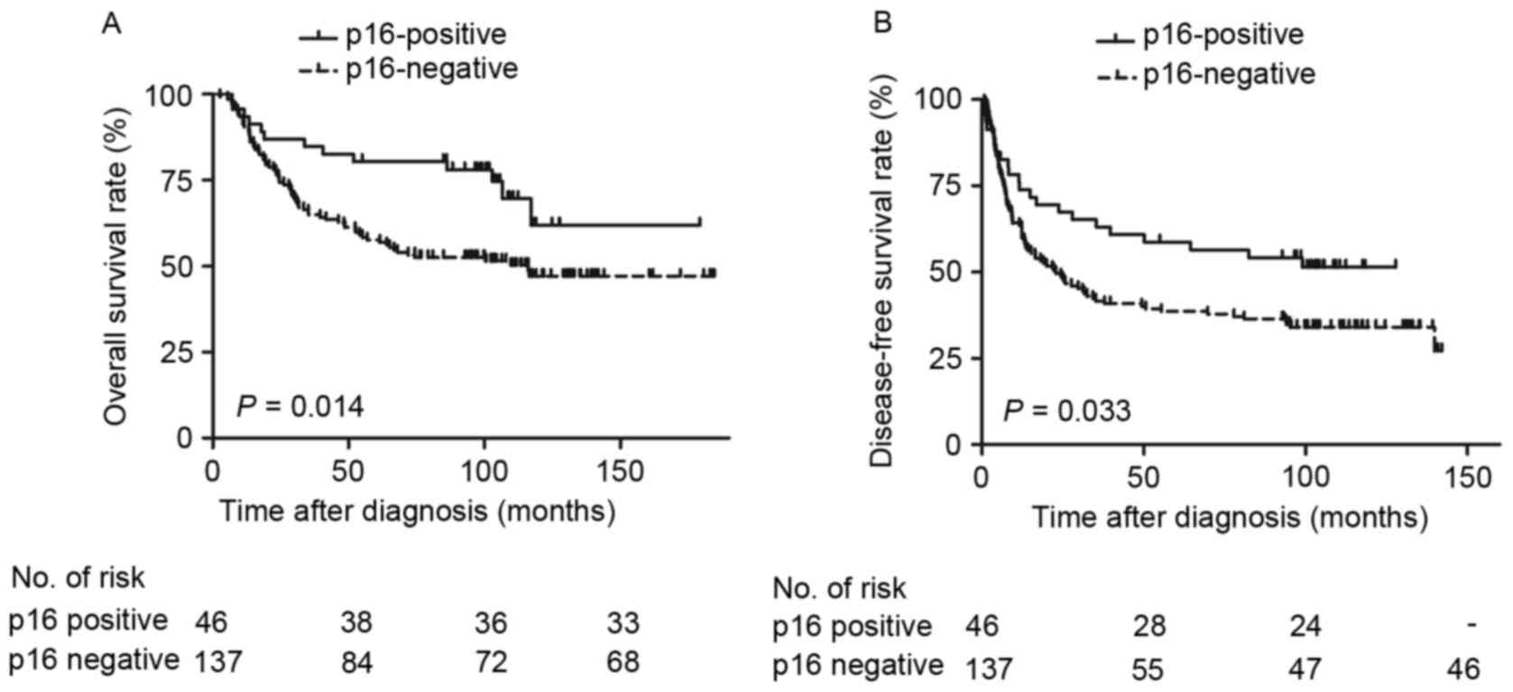
Survival outcomes based on
p16INK4a status
Survival outcomes based on p16INK4a
status in patients with non-OPSCC were examined. In the
p16INK4a-negative group, the 5-year OS and DFS rates
were 57.7% and 38.6%, respectively. The
p16INK4a-postitive expression group exhibited markedly
improved survival rates: The 5-year OS and DFS rates were 78.1% and
56.4%, respectively. Patients with p16INK4a-positive
expression exhibited improved DFS and OS rates compared with those
with negative expression (Fig. 2).
Furthermore, male patients (P=0.037), who smoked tobacco (P=0.016)
and alcohol (P=0.020), with poorly differentiated tumor types
(P=0.018) and stage III or IV disease (P<0.001) exhibited a
reduced OS rate, and patients with poorly differentiated tumor
types (P=0.004) and advanced disease (P=0.002) exhibited a reduced
DFS rate, compared with their counterparts (Table II). Cox proportional hazards analysis
revealed that p16INK4a-negative independently predicted
low OS rate (Table III; P=0.020)
but not DFS rate (P=0.096).
| Table II.Univariate survival analysis of
patients with oral cavity and larynx squamous cell carcinoma
(n=183). |
Table II.
Univariate survival analysis of
patients with oral cavity and larynx squamous cell carcinoma
(n=183).
|
| Overall survival
rate | Disease-free
survival rate |
|---|
|
|
|
|
|---|
| Variable | RR 95% CI | P-value | RR 95% CI | P-value |
|---|
| Male | 1.88
(1.04–3.41) | 0.037 | 1.53
(0.95–2.46) | 0.081 |
| Tobacco use | 1.78
(1.11–2.86) | 0.016 | 1.36
(0.93–2.00) | 0.118 |
| Alcohol use | 1.74
(1.09–2.77) | 0.020 | 1.25
(0.83–1.89) | 0.291 |
| Age <50
years | 1.13
(0.72–1.77) | 0.602 | 1.06
(0.72–1.55) | 0.779 |
| Poorer
differentiation | 1.69
(1.09–2.61) | 0.018 | 1.71
(1.18–2.48) | 0.004 |
| Cyclin-dependent
kinase inhibitor 2A -positive expression | 0.48
(0.27–0.87) | 0.014 | 0.61
(0.38–0.97) | 0.033 |
| Stage III/IV
disease | 2.26
(1.47–3.50) | <0.001 | 1.79
(1.23–2.60) | 0.002 |
| Table III.Multivariate survival analysis of
patients with oral cavity and larynx squamous cell carcinoma
(n=183). |
Table III.
Multivariate survival analysis of
patients with oral cavity and larynx squamous cell carcinoma
(n=183).
|
| Overall survival
rate | Disease-free
survival rate |
|---|
|
|
|
|
|---|
| Variable | RR | 95% CI | P-value | RR | 95% CI | P-value |
|---|
| Male | 0.95 | 0.42–2.15 | 0.909 | – | – | – |
| Tobacco use | 1.75 | 0.92–3.32 | 0.088 | – | – | – |
| Alcohol use | 1.53 | 0.92–2.52 | 0.099 | – | – | – |
| Poorer
differentiation | 1.28 | 0.81–2.04 | 0.294 | 1.47 | 1.00–2.17 | 0.051 |
| Cyclin-dependent
kinase inhibitor 2A -positive expression | 0.49 | 0.27–0.89 | 0.020 | 0.67 | 0.42–1.07 | 0.096 |
| Stage III/IV
disease | 2.01 | 1.25–3.23 | 0.004 | 1.53 | 1.03–2.27 | 0.034 |
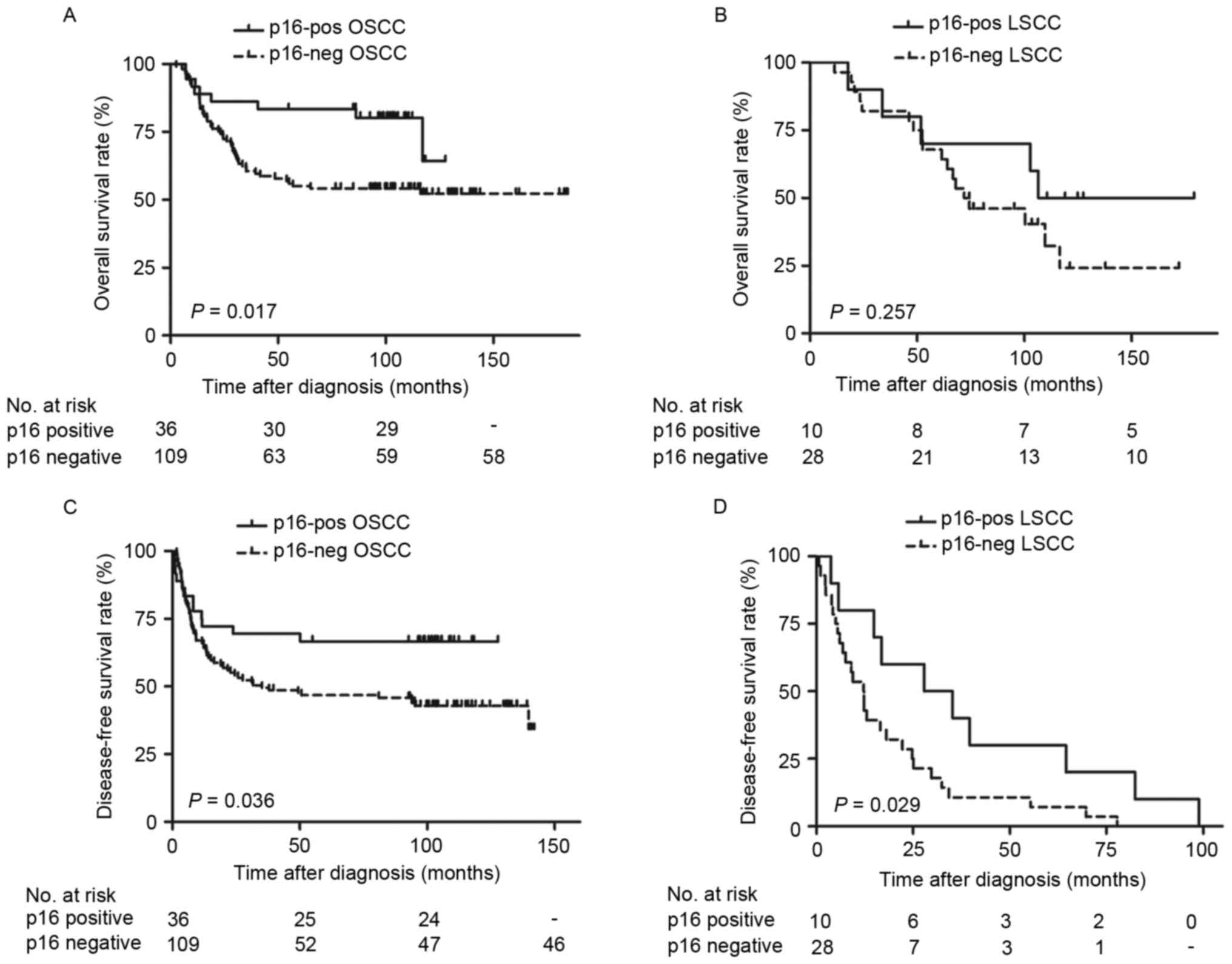
The survival outcomes of p16INK4a based
on reclassifying by primary site (oral cavity or larynx) and stage
(stage I/II or III/IV) were also examined. The patients with OSCC
with p16INK4a-positive expression exhibited
significantly improved OS (Fig. 3A;
P=0.017) and DFS (Fig. 3B; P=0.036)
rates, as determined by Kaplan-Meier survival curves. Additionally,
patients with LSCC with positive p16INK4a expression
exhibited no significant difference in OS rate (Fig. 3C; P=0.257), but significantly
increased DFS rate (Fig. 3D;
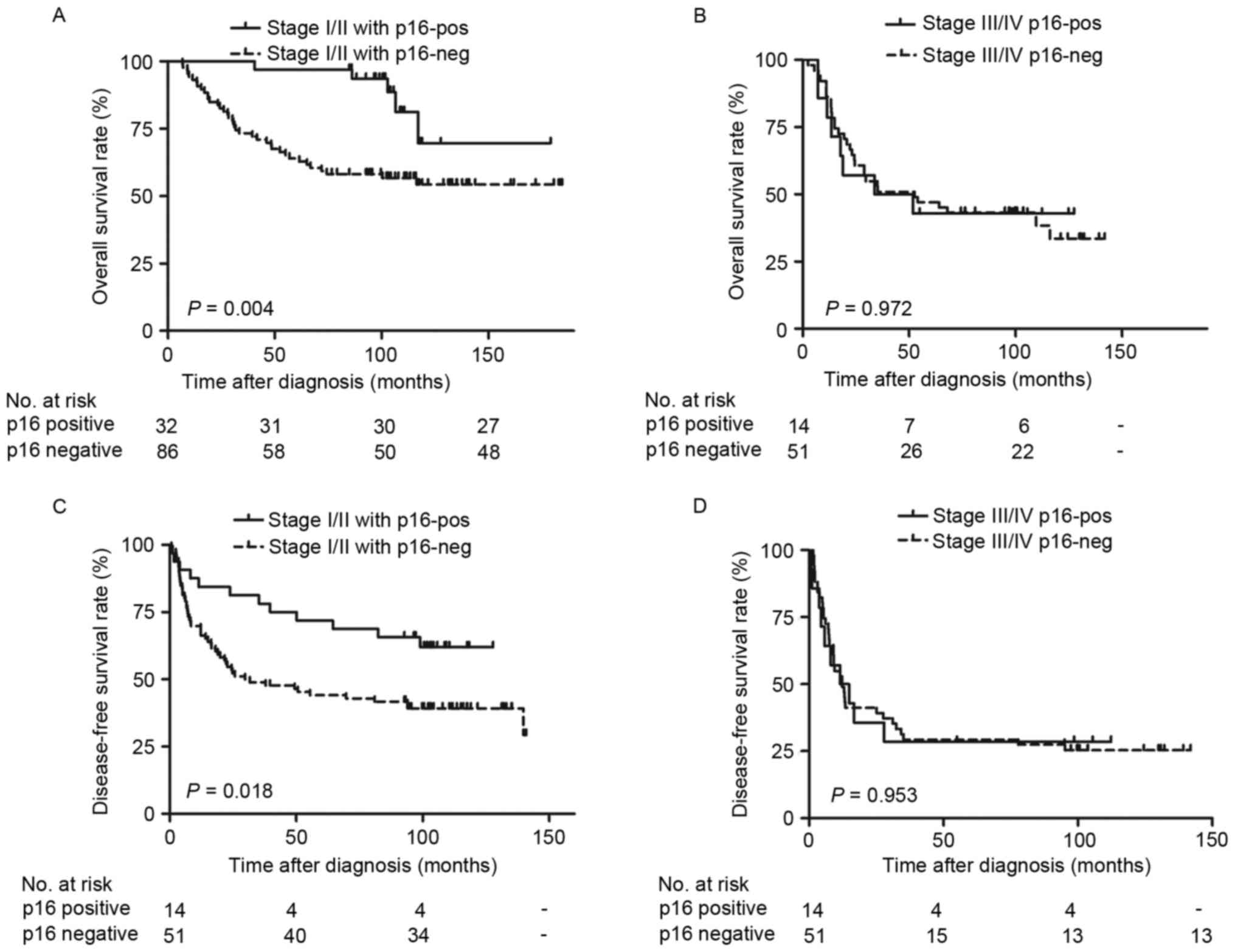
P=0.029). Furthermore, significantly improved OS (Fig. 4A; P=0.004) and DFS rates (Fig. 4B; P=0.018) were observed in patients
with stage I/II tumor types with positive p16INK4a
expression. However, there were no significant differences in stage
III/IV tumor types (Fig. 4C and D;
P>0.05). Additional univariate analysis confirmed that positive
p16INK4a status predicted favorable OS rate in oral
cancer [Table IV; relative risk
(RR)=0.42; P=0.017] and in the stage I/II disease (RR=0.25;
P=0.004) subgroups. Although patients who were
p16INK4a-positive exhibited increased DFS rates,
compared with those who were p16INK4a-negative (Fig. 2B; P=0.033), the statistical difference
calculated by multivariate analysis between groups was not
significant (Table III; RR=0.67;
P=0.096).
| Table IV.Univariate survival analysis of
cyclin-dependent kinase inhibitor 2A in patients stratified by
primary site and stage. |
Table IV.
Univariate survival analysis of
cyclin-dependent kinase inhibitor 2A in patients stratified by
primary site and stage.
|
| Overall survival
rate |
| Disease-free
survival rate |
|---|
|
|
|
|
|
|---|
| Stratification
factors | RR | 95% CI | P-value | RR | 95% CI | P-value |
|---|
| Primary site | – | – | – | – | – | – |
| Oral
cancer | 0.42 | 0.20–0.88 | 0.017 | 0.52 | 0.28–0.97 | 0.036 |
| Larynx
cancer | 0.56 | 0.20–1.54 | 0.257 | 0.42 | 0.19–0.93 | 0.029 |
| Primary stage | – | – | – | – | – | – |
|
I/II | 0.25 | 0.10–0.65 | 0.004 | 0.48 | 0.25–0.90 | 0.018 |
|
III/IV | 1.01 | 0.46–2.21 | 0.972 | 1.02 | 0.51–2.05 | 0.953 |
Discussion
HPV E6/E7 RNA expression detected using RT-qPCR, the
results of which indicate active viral gene transcription in a
tumor, is considered to be the gold standard (16). Although p16INK4a protein is
a reliable indicator of HPV infection in the oropharynx, the
sensitivity and specificity of p16INK4a in OSCC and LSCC
has not been conclusively determined (14,20,22,23,28–30).
It was demonstrated that the incidence of p16INK4a
positivity in southern Chinese patients with LSCC is 26.3%, and in
patients with OSCC is 24.8% (14).
Johns Hopkins University published a large-scale screening study
that evaluated p16INK4a expression by
immunohistochemistry, in which the patients with non-OPSCC were
collected from Radiation Therapy Oncology Group (RTOG) 0129, 0234
and 0522 studies (21). A total of
19.3% (62/322) patients were p16INK4a positive, and the
patients with OSCC exhibited the highest rate of
p16INK4a positivity [21/80 (26.3%)], followed by the
larynx [31/181 (17.1%)], which was similar to the present study.
However, Xu et al (23)
detected the status in Chinese patients with laryngeal cancer, and
revealed a positive rate of 7.57%. In general, previously
identified rates of p16INK4a protein expression in
patients with non-OPSCC are not concordant (20,22,23,25,30),
and multiple factors contribute to this discrepancy, for example:
Heterogeneous patient selection (geographical differences, tobacco
and alcohol status of the patient or the number of cases); or the
scoring system used for defining p16INK4a as
positive.
A debate exists with respect to the prognostic role
of p16INK4a in non-OPSCC. In the present study, the OS
and DFS rates among all 183 patients with non-OPSCC were improved
in p16INK4a-positive patients compared with negative
expression patients, although the multivariate survival analysis
suggested that the difference between DFS rate was not
statistically significant. The data from patients with non-OPSCC
collected from RTOG studies concluded that, similar to the results
in patients with OPSCC (6,17,31,32),
patients with p16INK4a-positive exhibit improved
outcomes compared with those with p16INK4a-negative
non-OPSCC (21). Nevertheless, other
studies did not observe a significant correlation between
p16INK4a expression and improved outcomes (14,22,33). These
differences may be due to the small number of samples, different
anatomic sites enrolled or tumor heterogeneity in each
retrospective cohort.
Additional stratified analysis identified that
patients who were p16INK4a-positive with stage I/II
tumor types or tumor types in oral cavity sites demonstrated
significantly improved OS rate compared with patients who were
negative expression. Patients who were p16INK4a-positive
in stage I/II or at larynx sites demonstrated markedly improved DFS
rates compared with patients who were p16INK4a-negative;
and this observation may be of clinical importance. The treatment
of an early stage tumor consists of surgery combined with
radiotherapy (34,35). Nevertheless, the toxicities of these
treatments may result in organ dysfunction and potentially
treatment-associated mortalities (36,37). It is
necessary to decrease the intensity of treatment for patients with
favorable prognosis. However, no specific indicator for this
subgroup was identified. In the present study, negative
p16INK4a status was considered as an indicator for a
requirement for aggressive therapy in patients with stage I/II
tumor types, to achieve survival benefit.
However, due to the small sample sizes (n=183), the
prognostic role of p16INK4a in LSCC demonstrated in the
present study should be interpreted with caution. The present study
aimed to detect the level of HPV DNA in the same set of samples,
but the outcome was not conclusive as the DNA quality in the
paraffin block was not adequate. Concomitantly, as a retrospective
investigation, the present study is limited by the lack of
large-scale screening.
The present study demonstrated an analysis of
diagnostic tests, the diagnostic accuracy and prognostic relevance
of p16INK4a in patients from South China. It was
identified that the p16INK4a positive rate in patients
with non-OPSCC was 25.1% (26.3% in LSCC and 24.8% in OSCC). It was
suggested that p16INK4a expression in patients with
non-OPSCC predicted favorable clinical outcomes, particularly in
OSCC and early stage non-OPSCC. If so, these patients with
p16INK4a-negative should receive more aggressive therapy
and a closer follow-up. It is hoped that the design of forthcoming
clinical trials in China, aimed at therapy in patients who are
p16INK4a-positive with non-OPSCC and perhaps aggressive
therapy for patients who are p16INK4a-negative, will be
informed by these results.
Acknowledgements
Not applicable.
Funding
The present study was supported by grants from the
Natural Science Foundation of China (grant no. 81201716).
Availability of data and materials
The datasets used and analyzed during the present
study are available from the corresponding author on reasonable
request.
Authors' contributions
WQJ and YXS participated in the design of the
research. HY and YC conducted the IHC studies, participated in the
collection of cases and drafted the manuscript. ZML and YJL helped
the statistical analysis and participated in the IHC studies. WQJ
and YXS helped to revise the manuscript. All authors read and
approved the final manuscript.
Ethics approval and consent to
participate
The current study was approved by the Ethics Review
Board of Sun Yat-Sen University Cancer Center. All activities were
in accord with the 1964 Declaration of Helsinki. Each patient
signed informed consent for participate in the study and collect
specimens.
Patient consent for publication
The patients involved in the present study signed
informed consent for the publication of any associated data and
accompanying images.
Competing interests
The authors declared that they have no competing
interests.
References
|
1
|
Wong IC, Ng YK and Lui VW: Cancers of the
lung, head and neck on the rise: Perspectives on the genotoxicity
of air pollution. Chin J Cancer. 33:476–480. 2014.PubMed/NCBI
|
|
2
|
Siegel RL, Miller KD and Jemal A: Cancer
statistics, 2015. CA Cancer J Clin. 65:5–29. 2015. View Article : Google Scholar : PubMed/NCBI
|
|
3
|
Torre LA, Bray F, Siegel RL, Ferlay J,
Lortet-Tieulent J and Jemal A: Global cancer statistics, 2012. CA
Cancer J Clin. 65:87–108. 2015. View Article : Google Scholar : PubMed/NCBI
|
|
4
|
Atienza JA and Dasanu CA: Incidence of
second primary malignancies in patients with treated head and neck
cancer: A comprehensive review of literature. Curr Med Res Opin.
28:1899–1909. 2012. View Article : Google Scholar : PubMed/NCBI
|
|
5
|
Mork J, Lie AK, Glattre E, Hallmans G,
Jellum E, Koskela P, Møller B, Pukkala E, Schiller JT, Youngman L,
et al: Human papillomavirus infection as a risk factor for
squamous-cell carcinoma of the head and neck. N Engl J Med.
344:1125–1131. 2001. View Article : Google Scholar : PubMed/NCBI
|
|
6
|
Ang KK, Harris J, Wheeler R, Weber R,
Rosenthal DI, Nguyen-Tân PF, Westra WH, Chung CH, Jordan RC, Lu C,
et al: Human papillomavirus and survival of patients with
oropharyngeal cancer. N Engl J Med. 363:24–35. 2010. View Article : Google Scholar : PubMed/NCBI
|
|
7
|
Chaturvedi AK, Engels EA, Pfeiffer RM,
Hernandez BY, Xiao W, Kim E, Jiang B, Goodman MT, Sibug-Saber M,
Cozen W, et al: Human papillomavirus and rising oropharyngeal
cancer incidence in the united states. J Clin Oncol. 29:4294–4301.
2011. View Article : Google Scholar : PubMed/NCBI
|
|
8
|
Ndiaye C, Mena M, Alemany L, Arbyn M,
Castellsagué X, Laporte L, Bosch FX, de Sanjosé S and Trottier H:
HPV DNA, E6/E7 mRNA, and p16INK4a detection in head and neck
cancers: A systematic review and meta-analysis. Lancet Oncol.
15:1319–1331. 2014. View Article : Google Scholar : PubMed/NCBI
|
|
9
|
Bouda M, Gorgoulis VG, Kastrinakis NG,
Giannoudis A, Tsoli E, Danassi-Afentaki D, Foukas P, Kyroudi A,
Laskaris G, Herrington CS and Kittas C: ‘High risk’ HPV types are
frequently detected in potentially malignant and malignant oral
lesions, but not in normal oral mucosa. Mod Pathol. 13:644–653.
2000. View Article : Google Scholar : PubMed/NCBI
|
|
10
|
Paradiso A, Ranieri G, Stea B, Zito A,
Zehbe I, Tommasino M, Grammatica L and De Lena M: Altered p16INK4a
and Fhit expression in carcinogenesis and progression of human oral
cancer. Int J Oncol. 24:249–255. 2004.PubMed/NCBI
|
|
11
|
Bradley KT, Budnick SD and Logani S:
Immunohistochemical detection of p16INK4a in dysplastic lesions of
the oral cavity. Mod Pathol. 19:1310–1316. 2006. View Article : Google Scholar : PubMed/NCBI
|
|
12
|
Baumann JL, Cohen S, Evjen AN, Law JH,
Vadivelu S, Attia A, Schindler JS, Chung CH, Wirth PS, Meijer CJ,
et al: Human papillomavirus in early laryngeal carcinoma.
Laryngoscope. 119:1531–1537. 2009. View Article : Google Scholar : PubMed/NCBI
|
|
13
|
Halec G, Holzinger D, Schmitt M,
Flechtenmacher C, Dyckhoff G, Lloveras B, Höfler D, Bosch FX and
Pawlita M: Biological evidence for a causal role of HPV16 in a
small fraction of laryngeal squamous cell carcinoma. Br J Cancer.
109:172–183. 2013. View Article : Google Scholar : PubMed/NCBI
|
|
14
|
Xu Y, Liu S, Yi H, Wang J, Dong P, Li X
and Yin S: Human papillomavirus infection in 674 Chinese patients
with laryngeal squamous cell carcinoma. PLoS One. 9:e1159142014.
View Article : Google Scholar : PubMed/NCBI
|
|
15
|
Young RJ, Urban D, Angel C, Corry J, Lyons
B, Vallance N, Kleid S, Iseli TA, Solomon B and Rischin D:
Frequency and prognostic significance of p16(INK4A) protein
overexpression and transcriptionally active human papillomavirus
infection in laryngeal squamous cell carcinoma. Br J Cancer.
112:1098–1104. 2015. View Article : Google Scholar : PubMed/NCBI
|
|
16
|
Shi W, Kato H, Perez-Ordonez B, Pintilie
M, Huang S, Hui A, O'Sullivan B, Waldron J, Cummings B, Kim J, et
al: Comparative prognostic value of HPV16 E6 mRNA compared with in
situ hybridization for human oropharyngeal squamous carcinoma. J
Clin Oncol. 27:6213–6221. 2009. View Article : Google Scholar : PubMed/NCBI
|
|
17
|
Rischin D, Young RJ, Fisher R, Fox SB, Le
QT, Peters LJ, Solomon B, Choi J, O'Sullivan B, Kenny LM and
McArthur GA: Prognostic significance of p16INK4A and human
papillomavirus in patients with oropharyngeal cancer treated on
TROG 02.02 phase III trial. J Clin Oncol. 28:4142–4148. 2010.
View Article : Google Scholar : PubMed/NCBI
|
|
18
|
Jordan RC, Lingen MW, Perez-Ordonez B, He
X, Pickard R, Koluder M, Jiang B, Wakely P, Xiao W and Gillison ML:
Validation of methods for oropharyngeal cancer HPV status
determination in US cooperative group trials. Am J Surg Pathol.
36:945–954. 2012. View Article : Google Scholar : PubMed/NCBI
|
|
19
|
Buajeeb W, Poomsawat S, Punyasingh J and
Sanguansin S: Expression of p16 in oral cancer and premalignant
lesions. J Oral Pathol Med. 38:104–108. 2009. View Article : Google Scholar : PubMed/NCBI
|
|
20
|
Bussu F, Sali M, Gallus R, Vellone VG,
Zannoni GF, Autorino R, Dinapoli N, Santangelo R, Martucci R,
Graziani C, et al: HPV infection in squamous cell carcinomas
arising from different mucosal sites of the head and neck region.
Is p16 immunohistochemistry a reliable surrogate marker? Br J
Cancer. 108:1157–1162. 2013.PubMed/NCBI
|
|
21
|
Chung CH, Zhang Q, Kong CS, Harris J,
Fertig EJ, Harari PM, Wang D, Redmond KP, Shenouda G, Trotti A, et
al: p16 protein expression and human papillomavirus status as
prognostic biomarkers of nonoropharyngeal head and neck squamous
cell carcinoma. J Clin Oncol. 32:3930–3938. 2014. View Article : Google Scholar : PubMed/NCBI
|
|
22
|
Lim AM, Do H, Young RJ, Wong SQ, Angel C,
Collins M, Takano EA, Corry J, Wiesenfeld D, Kleid S, et al:
Differential mechanisms of CDKN2A (p16) alteration in oral tongue
squamous cell carcinomas and correlation with patient outcome. Int
J Cancer. 135:887–895. 2014. View Article : Google Scholar : PubMed/NCBI
|
|
23
|
Xu Y, Liu S, Yi H, Wang J, Luo Y and Yin
S: Low prevalence of human papillomavirus in head and neck squamous
cell carcinoma in chinese patients. J Med Virol. 87:281–286. 2015.
View Article : Google Scholar : PubMed/NCBI
|
|
24
|
Fu ZJ, Ma ZY, Wang QR, Lei DP, Wang R, Liu
CX and Pan XL: Overexpression of cyclind1 and underexpression of
p16 correlate with lymph node metastases in laryngeal squamous cell
carcinoma in chinese patients. Clin Exp Metastasis. 25:887–892.
2008. View Article : Google Scholar : PubMed/NCBI
|
|
25
|
Li D, Zhang Q and Zhang X: Expression and
clinical significance of MDM2 and P16 in laryngeal squamous cell
carcinoma. Lin Chung Er Bi Yan Hou Tou Jing Wai Ke Za Zhi.
26:802–805. 2012.(In Chinese). PubMed/NCBI
|
|
26
|
Edge SB and Compton CC: The american joint
committee on cancer: The 7th edition of the AJCC cancer staging
manual and the future of TNM. Ann Surg Oncol. 17:1471–1474. 2010.
View Article : Google Scholar : PubMed/NCBI
|
|
27
|
Schache AG, Liloglou T, Risk JM, Filia A,
Jones TM, Sheard J, Woolgar JA, Helliwell TR, Triantafyllou A,
Robinson M, et al: Evaluation of human papilloma virus diagnostic
testing in oropharyngeal squamous cell carcinoma: Sensitivity,
specificity, and prognostic discrimination. Clin Cancer Res.
17:6262–6271. 2011. View Article : Google Scholar : PubMed/NCBI
|
|
28
|
Schlecht NF, Brandwein-Gensler M, Nuovo
GJ, Li M, Dunne A, Kawachi N, Smith RV, Burk RD and Prystowsky MB:
A comparison of clinically utilized human papillomavirus detection
methods in head and neck cancer. Mod Pathol. 24:1295–1305. 2011.
View Article : Google Scholar : PubMed/NCBI
|
|
29
|
Doxtader EE and Katzenstein AL: The
relationship between p16 expression and high-risk human
papillomavirus infection in squamous cell carcinomas from sites
other than uterine cervix: A study of 137 cases. Hum Pathol.
43:327–332. 2012. View Article : Google Scholar : PubMed/NCBI
|
|
30
|
van Monsjou HS, van Velthuysen ML, van den
Brekel MW, Jordanova ES, Melief CJ and Balm AJ: Human
papillomavirus status in young patients with head and neck squamous
cell carcinoma. Int J Cancer. 130:1806–1812. 2012. View Article : Google Scholar : PubMed/NCBI
|
|
31
|
Posner MR, Lorch JH, Goloubeva O, Tan M,
Schumaker LM, Sarlis NJ, Haddad RI and Cullen KJ: Survival and
human papillomavirus in oropharynx cancer in TAX 324: A subset
analysis from an international phase III trial. Ann Oncol.
22:1071–1077. 2011. View Article : Google Scholar : PubMed/NCBI
|
|
32
|
Rietbergen MM, Brakenhoff RH, Bloemena E,
Witte BI, Snijders PJ, Heideman DA, Boon D, Koljenovic S,
Baatenburg-de Jong RJ and Leemans CR: Human papillomavirus
detection and comorbidity: Critical issues in selection of patients
with oropharyngeal cancer for treatment De-escalation trials. Ann
Oncol. 24:2740–2745. 2013. View Article : Google Scholar : PubMed/NCBI
|
|
33
|
Kalfert D, Celakovsky P, Laco J and
Ludvikova M: The role of protein p16(INK4a) in glottic laryngeal
squamous cell carcinoma. Pathol Oncol Res. 20:909–915. 2014.
View Article : Google Scholar : PubMed/NCBI
|
|
34
|
Forastiere AA, Goepfert H, Maor M, Pajak
TF, Weber R, Morrison W, Glisson B, Trotti A, Ridge JA, Chao C, et
al: Concurrent chemotherapy and radiotherapy for organ preservation
in advanced laryngeal cancer. N Engl J Med. 349:2091–2098. 2003.
View Article : Google Scholar : PubMed/NCBI
|
|
35
|
Adelstein DJ, Saxton JP, Rybicki LA,
Esclamado RM, Wood BG, Strome M, Lavertu P, Lorenz RR and Carroll
MA: Multiagent concurrent chemoradiotherapy for locoregionally
advanced squamous cell head and neck cancer: Mature results from a
single institution. J Clin Oncol. 24:1064–1071. 2006. View Article : Google Scholar : PubMed/NCBI
|
|
36
|
Machtay M, Moughan J, Trotti A, Garden AS,
Weber RS, Cooper JS, Forastiere A and Ang KK: Factors associated
with severe late toxicity after concurrent chemoradiation for
locally advanced head and neck cancer: An RTOG analysis. J Clin
Oncol. 26:3582–3589. 2008. View Article : Google Scholar : PubMed/NCBI
|
|
37
|
Corry J, Peters LJ and Rischin D:
Optimising the therapeutic ratio in head and neck cancer. Lancet
Oncol. 11:287–291. 2010. View Article : Google Scholar : PubMed/NCBI
|