Introduction
In 2012, liver cancer was reported as a significant
cause of tumor-associated mortality worldwide with 746,000 cases
and a mortality rate of 9.1% (1).
Various treatments for liver cancer are available, including
surgery, radiotherapy, systemic chemotherapy and transcatheter
arterial chemoembolization (TACE). However, liver cancer is
resistant to a number of chemotherapy and radiotherapy regimes
(2). TACE, combined arterial
embolization with chemotherapy for liver cancer treatment, is
typically used as a treatment for advanced liver tumor (3). However, patient responses to TACE
therapy are varied. It is known that hepatitis B virus (HBV) is a
risk factor for hepatic carcinoma and that it is associated with
liver cirrhosis and liver cancer development (4). It has been reported that HBV may affect
the efficacy of chemotherapy by mediating gene mutation and
intratumor heterogeneity in tumor livers (5). Furthermore, it has been reported that
the apoptosis-inducing effects of chemotherapy are different in
HBV-associated liver cancer cells compared with that in
HBV-negative hepatic tumor cells (6).
The aim of the present study was to use clinical data to assess
differences in treatment response to TACE in patients with
HBV-associated liver cancer and those with non-HBV liver cancer,
and to elucidate the possible mechanisms.
Exosomes are membrane-bound microvesicles
(50–150-nm) that are released by various cells under normal and
pathological conditions (7,8). Exosomes are established as participating
in a number of biological functions and contain various cargos,
including proteins and RNAs (9). It
has been demonstrated that exosomes serve an important role in the
formation and progression of tumors, with anti-apoptotic effects in
malignant cancer (10–12). For instance, extracellular vesicles
released from ovarian cancer cells stimulated by cisplatin
treatment may induce invasiveness and bystander cell drug
resistance via p38 and c-Jun N-terminal kinase signaling (13). In addition, it has been reported that
exosomes are able to induce chemoresistance in liver cancer treated
with sorafenib (14).
Chaperone-mediated autophagy (CMA) is a selective
form of autophagy in which cytosolic proteins bearing a
pentapeptide motif biochemically associated with the KFERQ sequence
are recognized by a cytosolic heat shock cognate protein, delivered
to the lysosomal membrane and directly translocated across it by a
protein complex containing lysosome-associated membrane protein 2a
(Lamp2a) (15). CMA is associated
with tumor growth, metastasis (16)
and resistance to anticancer therapy (17). Lamp2a overexpression in breast tumors
increases overall cell survival via the CMA pathway, while Lamp2a
inhibition causes glyceraldehyde 3-phosphate dehydrogenase (GAPDH)
accumulation, Protein kinase B (AKT1) phosphorylation, reactive
oxygen species generation and increased cellular apoptosis in
breast cancer cells (18). A previous
study revealed that autophagy also serves an essential role in
neuroblastoma cells drug resistance and cell survival (19). We therefore speculated that CMA may be
able to induce chemoresistance in HBV-associated liver cancer.
In the present study, in order to investigate the
underlying mechanisms of different responses to TACE in HBV and
non-HBV patients with liver cancer, exosomes derived from
HBV-associated liver cancer cells were isolated and the crucial
roles of exosomes in chemoresistance were further investigated. The
research of the present study would provide novel insight in the
underlying mechanisms in liver tumors diagnostics and
therapeutics.
Materials and methods
Patient data and samples
Data was collected from patients with hepatic
carcinoma who were treated with TACE therapy (oxaliplatin 130
mg/m2) at the Department of Hepatobiliary Surgery
between January 2014 and June 2017 in North China University of
Science and Technology Affiliated Hospital (Tangshan, China).
Inclusion criteria were as follows: Based Specification for
Diagnosis and Treatment of Primary Liver Cancer (20); liver function A-B level (Child-Pugh
Classification) (21,22); Barcelona Clinical Liver Cancer Staging
System (BCLC) B stage (23); patients
with no surgical history and other therapy treatments, and patients
with no other primary tumor. The exclusion criteria were as
follows: Patients suffering from other severe diseases, including
heart and renal dysfunction, diffuse liver cancer, coagulation
disorders, and patients infected with hepatitis C. Patients were
sorted into HBV-associated liver cancer and non-HBV-associated
liver cancer groups depending on whether they were positive for
hepatitis B surface antigen, hepatitis B e-antigen (HBeAg) and
hepatitis B core antibody. In the HBV-associated liver cancer
group, there were 3 female and 15 male patients, with a sex ratio
(male:female) of 15:3 and a mean age of 54.63±6.21 years (range,
39–68 years). In the non HBV-associated liver cancer group, there
were 4 female and 11 male patients, with a sex ratio (male:female)
of 11:4 and a mean age of 56.25±7.57 years (range, 38–74 years).
Data collection from the patients was approved by the Institutional
Review Board of North China University of Science and Technology
Affiliated Hospital (Tangshan, Hebei, China). Serum samples were
collected from HBV-DNA-positive and healthy individuals at the
Clinical Laboratory of North China University of Science and
Technology Affiliated Hospital. All specimens were flash-frozen
upon collection and stored at −80°C until further use. The present
study was approved by the Ethics Review Committee of North China
University of Science and Technology Affiliated Hospital (approval
no. 17014).
Cell lines, reagents and
antibodies
The hepatoblastoma HepG2 cell line (24) (supplied by Cell Resource Center,
Shanghai Institute of Life Sciences, Chinese Academy of Sciences,
Shanghai, China) was cultured at 37°C in a humidified atmosphere
containing 5% CO2 in high-glucose Dulbecco's modified
Eagle's medium (DMEM) supplemented with 10% heat-inactivated FBS
(both Gibco; Thermo Fisher Scientific, Inc., Waltham, MA, USA), 100
U/ml penicillin and 100 mg/ml streptomycin (Beijing Solarbio
Technology Co., Ltd., Beijing, China). The primary antibody against
cleaved caspase-3 (dilution, 1:500; catalog no. PB0183) was
obtained from Wuhan Boster Biological Technology, Ltd. (Wuhan,
China), the anti-B-cell lymphoma-2 (Bcl-2) antibody (dilution,
1:500; catalog no. WL01556) was obtained from Wanleibio Co., Ltd.
(Shanghai, China) and the antibody against GAPDH (dilution,
1:5,000; catalog no. 5174S) was obtained from Cell Signaling
Technology, Inc. (Danvers, MA, USA). The antibody against Lamp2a
(dilution, 1:1,000; catalog no. ab125068) was obtained from Abcam
(Cambridge, MA, USA). The Goat anti-rabbit IgG secondary antibody
with the (dilution, 1:4,000; catalog no. BA1039) was obtained from
Wuhan Boster Biological Technology, Ltd. (Wuhan, China).
HBV virus infection
Cells were seeded at a concentration of
1×10⁵ cells/dish. At 2 days post-plating, the cells were
incubated at 4°C for 2 h, then at 37°C for 6 h in an atmosphere
containing 5% CO2. The HepG2 cells were infected with
HBV-positive serum (HBV-particles 1×10¹⁰ copies/ml) and
cocultured with DMEM for 48 h. The HBV-positive serum was then
removed, the cells were washed with phosphate-buffered saline (PBS)
8 times and, in order to ensure no residual HBV virus was present
in the supernatants, the last washing PBS was restored for
polymerase chain reaction analysis to detect HBV virus DNA
replication. The cells were subsequently incubated with pure DMEM
for 24, 48, 72 and 96 h. Culture supernatants were collected after
these different incubation durations and stored at −80°C for later
use.
PCR analysis
HBV DNA copies of cell supernatant were quantified
by a SLAN-96P Real-time PCR system (Shanghai Hongshi Medical
Technology, Co., Ltd., Shanghai, China), according to the Hepatitis
B Viral DNA Quantitative Fluorescence Diagnostic Kit
(PCR-Fluorescence Probing kit; cat. no., 20153400083; Sansure
Biotech Inc., Hunan, China.) instructions. The thermocycling
conditions were as follows: 50°C for 2 min, 94°C for 5 min, 45
cycles of 94°C for 15 sec and 57°C for 30 sec, and a final cooling
at 25°C for 10 sec.
Isolation and identification of
exosomes
HBV-associated exosomes derived from HepG2 cells
infected with HBV serum and non-HBV exosomes derived from HepG2
cells incubated with HBV-negative serum were collected from the
supernatant. Exosomes were isolated according to Exosome Isolation
Reagent protocols (GS™ Exosome Isolation Reagent, cat. no., E1002;
Geneseed Biotech, Co., Ltd, Guangzhou, China). Exosomes Vesicles
were resuspended in 100–200 µl PBS and stored at −80°C for further
use. The biomarkers of exosomes, including cluster of
differentiation (CD)9 and CD63, were identified using western
blotting and the protein in HepG2 cells was used as a positive
control. Exosome pellets were resuspended in PBS and placed onto
Formvor carbon-coated electron microscope grids (Electron
Microscope Sciences, Hatfield, PA, USA). Following incubation for 5
min at room temperature, exosomes were fixed in 2% paraformaldehyde
at room temperature for 4 h, and washed twice with water. The grids
were then negatively stained with 10% uranyl acetate for 10 min.
The preparations were examined and images were captured using
transmission electron microscopy (TEM; JEM-2100; JEOL, Ltd., Tokyo,
Japan). Each isolation was verified by nanoparticle tracking
analysis using a Nanosight N-300 (Nanosight Ltd., Amesbury, UK) to
determine the size and quantity of EVs extracted.
Silencing
HepG2 cells were seeded in 6-well plates with
complete medium for 24 h, following which they were transfected
with short hairpin RNA (shRNA) targeting Lamp2a (targeted sequence
5′-TCTTATGCATTGGAACTTAATTTGACATCT0-3′; LAMP2 siRNA/shRNA/RNAi
lentivirus Human Target A; cat. no., iV012052a; ABM lnc, China) and
negative control (NC) shRNA (targeted sequence
5′-GGGTGAACTCACGTCAGAA-3′; Scrambled shRNA GFP lentivirus; cat.
no., LVP015-G; ABM lnc.). The concentration of lentivirus was
>107 IU/ml. The transfection reagent EndoFectin™-Max
(iGeneBio, Guangzhou, China) was used to transfect HepG2 cells with
Lamp2a shRNA lentivirus. At 96 h post-transfection, the cells were
washed twice and treated with 0.1 µg/µl puromycin for screening.
Transfection efficiency was determined immediately using
florescence microscopy and the effects of transfection were
assessed using western blotting.
Western blotting
HepG2 cells and cells treated with exosomes or PBS
were harvested and were lysed for 50 min on ice in 100 ml of lysis
buffer (containing phenylmethanesulfonyl fluoride and a phosphatase
inhibitor). Lysates were centrifuged at 12,000 × g for 15 min at
4°C. Proteins were quantified by using the bincinchoninic acid
protein assay kit (Thermo Fisher Scientific, Inc.), according to
the manufacturer's protocols. Equal amounts (60 µg) of cell lysates
were loaded and separated by 10% SDS-PAGE, following which proteins
were electrotransferred to polyvinylidene difluoride membranes
(Bio-Rad Laboratories, Inc., Hercules, CA, USA). The membranes were
blocked for 60 min at 37°C with 5% skimmed milk in the 1×TBST
(Beijing Solarbio Technology Co., Ltd., Beijing, China) suspension
medium and incubated with the aforementioned primary antibodies at
4°C overnight. After three washes with TBST, the membranes were
incubated with secondary antibodies for 60 min at room temperature
and washed again. Proteins were detected using an enhanced
chemiluminescence system (Pierce; Thermo Fisher Scientific, Inc.).
Each experiment was repeated three times and similar results were
obtained.
Flow cytometry
Cell samples were analyzed using flow cytometry
analysis. Cells were stained with Annexin-V and propidium iodide
reagents (Annexin V-FITC/PI Apoptosis Detection kit; catalog no.
BB-4101-1; Bestbio, Co., Shanghai, China) to assess apoptosis. Data
were analyzed using a FACSCalibur flow cytometer and BD CellQuest
Pro software 5.1 (BD Biosciences, Franklin Lakes, NJ, USA). The
analysis was performed three times.
Statistical analysis
Graphs were created using Image Lab system (v4.1;
Bio-Rad Laboratories, Inc.). Student's t-test or one-way analysis
of variance with Scheffe's F post hoc test were performed using
SPSS 17.0 software (SPSS, Inc., Chicago, IL, USA). Data were
presented as mean ± standard deviation. P<0.05 was considered to
indicate a statistically significant difference.
Results
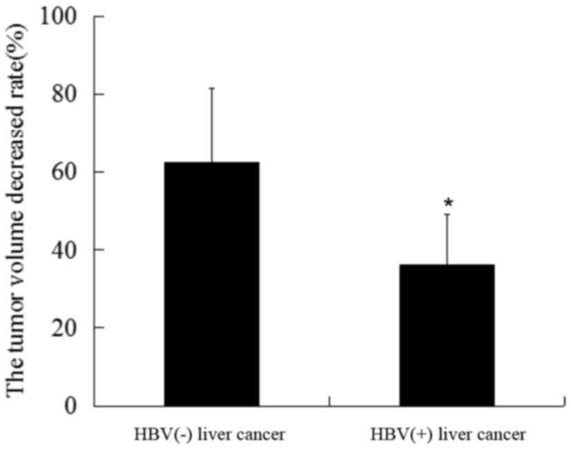
Tumor volume reduction is lower in
HBV-associated hepatic carcinoma patients compared with that in
non-HBV-associated liver cancer patients following TACE
treatment
To determine the effects of HBV on the therapeutic
efficacy of TACE, pre- and post-treatment tumor volume data were
collected for HBV-positive and -negative liver cancer patients
treated with TACE. In addition, a comparison of general patient
characteristics was performed, as shown in Table I. The data results revealed that the
reduction in tumor volume following TACE was significantly smaller
in the HBV-associated liver cancer group compared with the
HBV-negative liver cancer group (Fig.
1). These results indicated that HBV infection was associated
with a reduced response to TACE therapy.
 | Table I.Comparison of patient characteristics
between the experimental and control groups. |
Table I.
Comparison of patient characteristics
between the experimental and control groups.
| Clinical
features | HBV-associated
liver cancer | Non-HBV-associated
liver cancer |
|---|
| Sex
(male/female) | 15/3 | 11/4 |
| Mean age, years
(mean ± standard deviation) | 54.63±6.21 | 56.25±7.57 |
| Liver function
(Child-Pugh classification) | A-B | A-B |
| BCLC staging system
(23) | B | B |
Exosomes derived from HBV-associated
HepG2 cells modulate chemoresistance in liver cancer cells
A cell model infected with HBV serum was established
as previously described (25). PCR
analysis was used to confirm that the last PBS wash was negative
for HBV-DNA. Therefore, no residual HBV virus existed in the
washing supernatants. As indicated in Fig. 2A, the copies of HBV DNA in the 96 h
duration was the highest compared with 12, 24 and 48 h durations.
Cell supernatant containing pure DMEM for 96 h were selected for
use in further experiments.
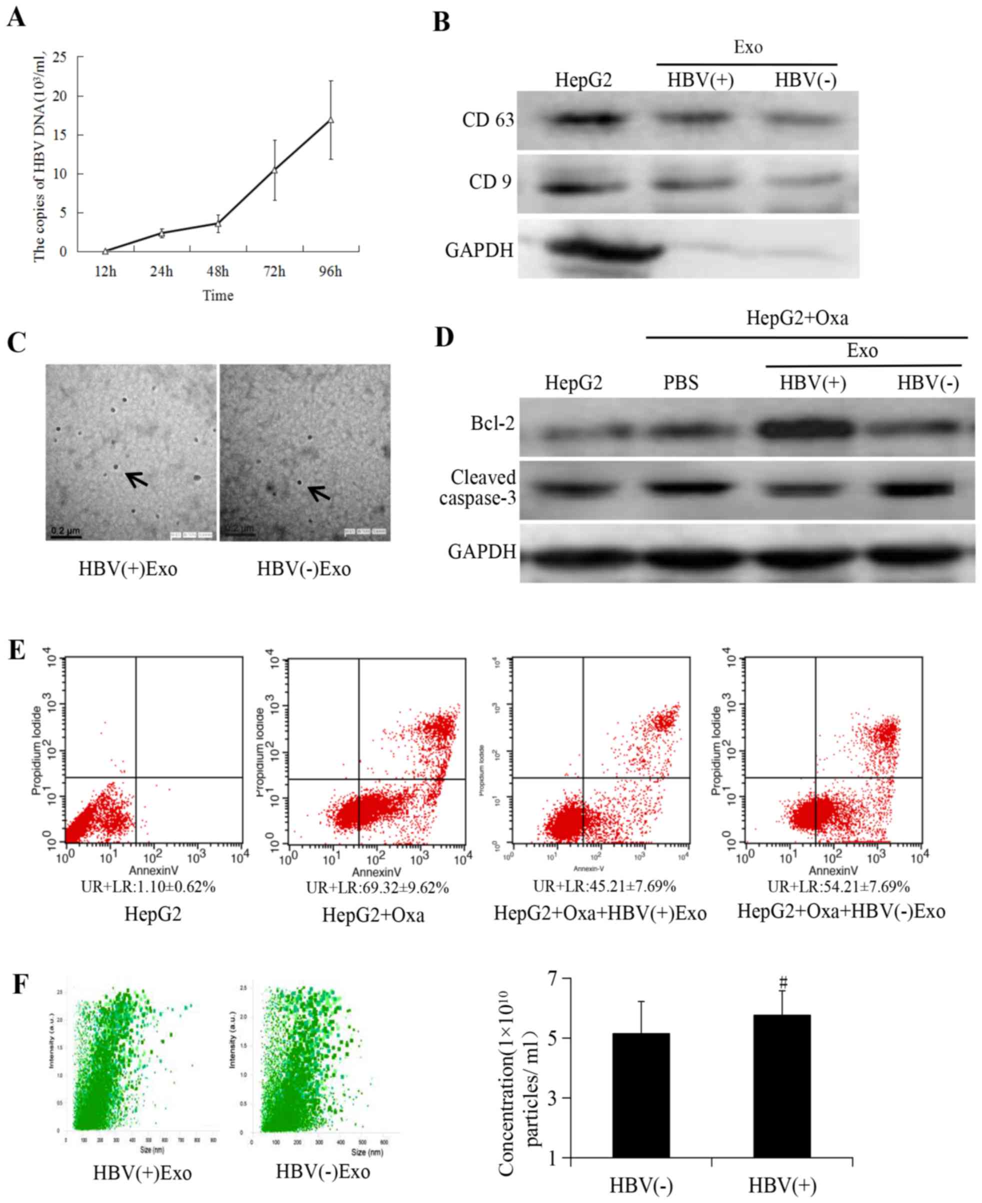 | Figure 2.Exosomes derived from HBV-positive
liver cancer cells promote cell chemoresistance. (A) HBV-DNA
contents in culture supernatant reached a peaked at 96 h. Exosomes
were isolated according to manufacturer's instructions (47). HepG2 cells were treated with
HBV-positive serum (HBV-DNA 1×10¹⁰ copies/ml) and
HBV-DNA copy of the cell supernatant containing pure DMEM was
detected at different times following removal of serum; experiments
demonstrated that HBV-DNA copies reached a peaked
(16.89±5.02×10³ copies/ml) at 96 h comparing with
HBV-DNA copies in 12, 24, 48 h, which provided the basis for
subsequent experiments. (B) Exosome markers CD63 and CD9 were
assessed by western blotting. GAPDH was used as an internal
reference. (C) Exosomes were characterized as round vesicles when
examined using transmission electron microscopy (magnification,
×40,000). (D) Detection of cell apoptosis on treatment with
1×10¹⁰ particles of HBV-associated exosomes in the
experimental group and an equal amount of particles of
non-HBV-associated exosomes in the negative group. (E) Flow
cytometry analysis of cell apoptosis. (F) Analysis of size
distribution and exosome concentration in purified exosomes using
Nanosight technology. No statistically significant differences were
indicated between HBV-associated and non-HBV-associated liver
cancer cells concerning exosomes concentration.
#P>0.05 vs. exosomes from non-HBV-associated liver
cancer cells. HBV, hepatitis B virus; Bcl-2, B-cell lymphoma; CD,
cluster of differentiation; GAPDH, glyceraldehyde 3-phosphate
dehydrogenase; Exo, exosome; Oxa, oxaliplatin; UR, upper right; LR,
lower right. |
To confirm that the pellets were exosomes, their
characteristics were determined via various methods, including
western blotting, TEM and Nanosight tracking analysis. Exosome
markers CD63 and CD9 were identified using western blotting
(Fig. 2B). Exosomes were also
confirmed to have round vesicular morphology, as observed in the
pellets (Fig. 2C). It has been
reported that exosomes released from liver cancer cells induce cell
resistance to sorafenib in vivo and in vitro
(14). However, the effects of
exosomes derived from HBV-associated hepatic tumors on liver cell
chemoresistance remain unknown. In the present study, it was
demonstrated that different apoptotic effects were achieved when
cells were treated with equivalent HBV-positive or -negative
exosomes (1×10¹⁰ particles). Cleaved caspase-3
expression was decreased and Bcl-2 expression was markedly
increased in HepG2 cells treated with HBV-associated exosomes
compared with that in the negative and blank control groups
(Fig. 2D and E). Exosome
concentration and distribution were analyzed using Nanosight
tracking analysis following their isolation from the supernatant.
The results revealed that there was no significant difference in
exosome concentration between cells treated with HBV-positive and
HBV-negative serum (Fig. 2F). As
such, the possibility that exosomes released from liver cancer
cells infected with HBV induce chemoresistance due to an increase
in exosome secretion was eliminated. It has previously been
demonstrated in certain tumors that specific exosomes containing
RNAs can be transferred to specific target cells, in which shuttled
RNA induces functional chemoresistance (12,13). We
therefore concluded that HBV-associated exosomes significantly
downregulate apoptosis in liver cancer cells by affecting the CMA
pathway.
HBV-associated exosomes downregulate
chemosensitivity in a concentration-dependent manner
To further identify the role of HBV-associated
exosomes in liver tumor chemoresistance, apoptosis was assessed in
cells transfected with HBV-associated exosomes at different
concentrations (4×10⁹, 8×10⁹ and
1.6×10¹⁰ particles/ml). The results revealed that
apoptosis was negatively associated with the concentration of
HBV-associated exosomes (Fig. 3A and
B). The data therefore confirmed that exosomes released from
HBV-associated liver cancer cells could downregulate cell
sensitivity to oxaliplatin therapy in a concentration-dependent
manner. Previous studies have reported that CMA serves a role in
tumor anti-apoptosis (17). Based on
this, the following experiments were performed.
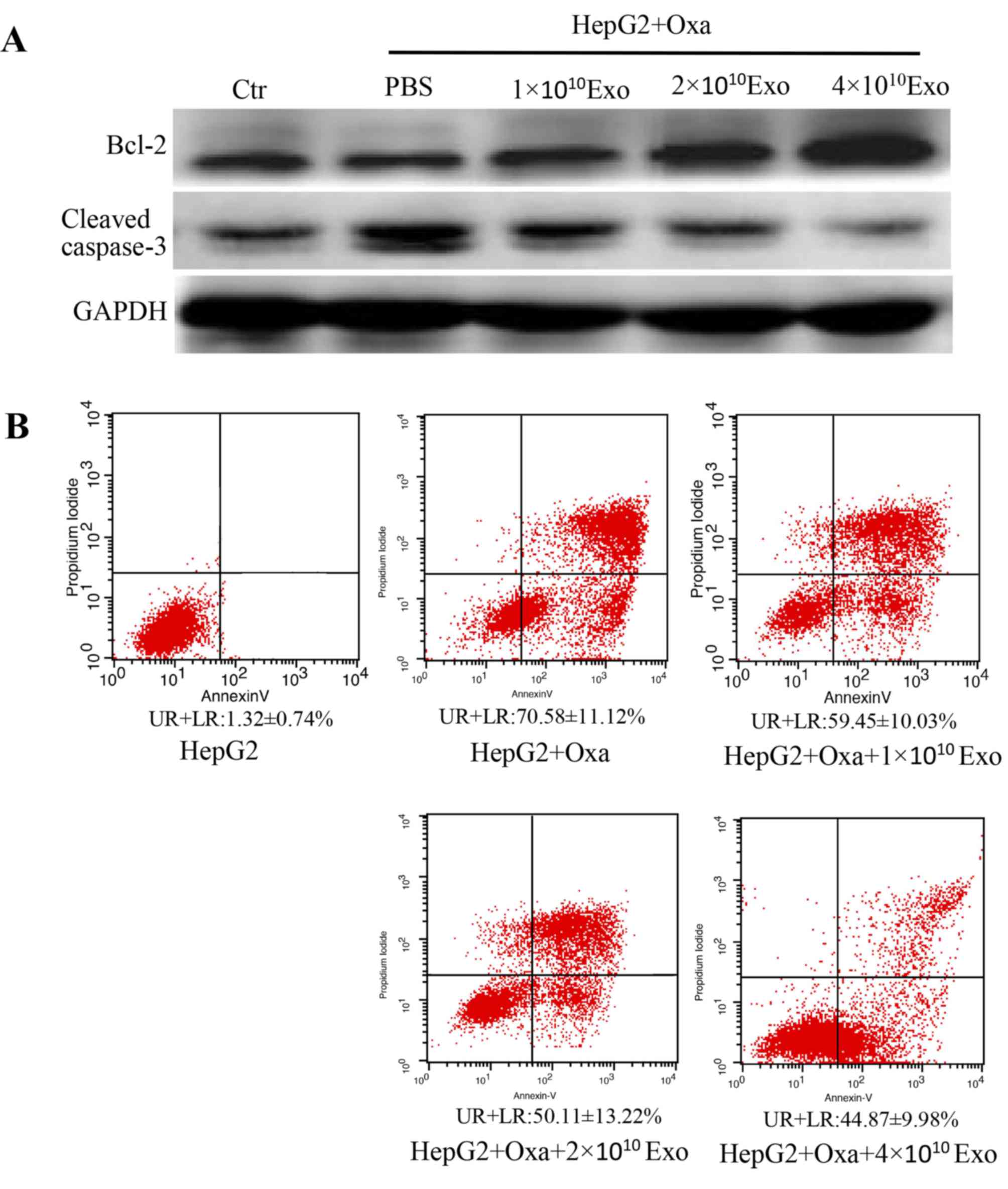 | Figure 3.Cell apoptosis was decreased by
HBV-associated exosomes in a concentration-dependent manner. (A)
Cells were treated with 1×10¹⁰, 2×10¹⁰ or
4×10¹⁰ HBV-associated exosomes, and the expression of
cleaved caspase-3 and Bcl-2 was assessed by western blotting. (B)
Flow cytometry results indicated that cell apoptosis was negatively
associated with the concentration of HBV-associated exosomes. HBV,
hepatitis B virus; Bcl-2, B-cell lymphoma; GAPDH, glyceraldehyde
3-phosphate dehydrogenase; Exo, exosome; Oxa, oxaliplatin; UR,
upper right; LR, lower right; Ctr, control; PBS, phosphate-buffered
saline. |
CMA activation decreases
chemosensitivity in cells treated with HBV-associated exosomes
CMA serves important roles in tumor progression and
inhibiting apoptosis (16,17). Autophagy has previously been reported
to be involved in cancer drug resistance (19). The aim of the present study was to
investigate whether the interaction between exosomes and CMA
influenced HBV-associated liver cancer chemoresistance. The
expression of Lamp2a, a key molecule in the CMA pathway, was
therefore investigated. Lamp2a expression was negatively associated
with cell apoptosis. In HepG2 cells treated with HBV-associated
exosomes and oxaliplatin, Lamp2a expression was upregulated in a
concentration-dependent manner (Fig. 4A
and B). These results revealed that HBV-associated exosomes
decreased cell chemosensitivity by activating the CMA pathway.
Furthermore, in order to investigate whether the apoptotic rate of
cells treated with HBV-associated exosomes was associated with CMA
activation, Lamp2a was knocked down in the present study. There was
no indication of Lamp2a protein expression on sh-Lamp2a HepG2
cells. (Fig. 4C and D). When cells
were treated with HBV-associated exosomes, apoptosis was increased
in HepG2-Lamp2a shRNA cells compared with in HepG2-control shRNA
cells (Fig. 4E). Therefore,
HBV-associated exosomes could influence cell viability via
regulating the CMA pathway in liver cancer chemotherapy.
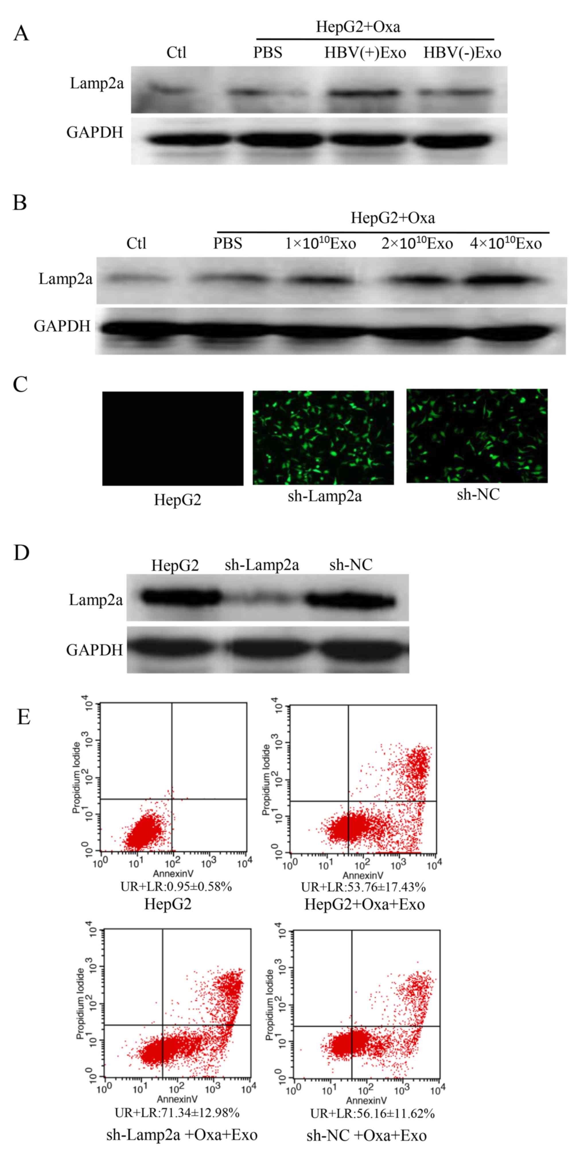 | Figure 4.HBV-associated exosomes modulate drug
resistance via CMA pathway activation. (A) Western blotting
revealed that Lamp2a was highly expressed following treatment with
oxaliplatin in the HBV-associated exosome group compared with in
the non-HBV exosome and blank control groups, and (B) that Lamp2a
was upregulated by HBV-associated exosomes in a
concentration-dependent manner. (C) Transfection efficiency of
shRNA as assessed using fluorescence microscopy. (D) Interference
efficiency of shRNA in HepG2 cells as assessed by western blotting.
(E) The anti-apoptotic effects of HBV-associated exosomes in HepG2
cells were reversed by Lamp2a silencing. Lamp2a,
lysosome-associated membrane protein; HBV, hepatitis B virus; oxa,
oxaliplatin; CMA, chaperone-mediated autophagy; shRNA, short
hairpin RNA; UR, upper right; LR, lower right; Ctr, control; PBS,
phosphate-buffered saline; NC, negative control; GAPDH,
glyceraldehyde 3-phosphate dehydrogenase. |
Discussion
The present study demonstrated the effect of
exosomes derived from HBV-associated HepG2 cells on liver cancer
chemoresistance. Hepatoblastoma cell HepG2 cells has been reported
could generate resistance to chemotherapeutic drugs in vivo
and in vitro (26–28). Previous studies reported that exosomes
derived from hepatic carcinoma cells may generate resistance to
sorafenib in mice and induce sorafenib resistance via the
HGF/c-Met/Akt pathway (14). It has
been reported that HBV infection increases the risk of hepatic
carcinoma development and progression (29,30).
Furthermore, HBV-associated liver cancer cells and
non-HBV-associated liver cancer cells respond differently to
chemotherapy (6). The focus of the
current study was the effects of exosomes derived from HepG2 cells
infected with HBV-positive serum on liver cancer chemoresistance.
It has been reported that the HBV virus affects tumor cell growth
and associated microRNA (miR) production (31), and it has been revealed that
HBeAg-induced miR-106b expression contributes to the pathogenesis
of HBV-associated liver cancer by downregulating the retinoblastoma
gene (32). Furthermore, various long
non-coding RNAs serve functional roles in HBV-associated hepatic
carcinoma by regulating biological processes (33,34).
Exosomes are 50- to 150-nm extracellular vesicles
released from cells that deliver cell-to-cell communications in
diverse conditions (7,35). It has been indicated that miR-21 in
exosomes derived from neuroblastoma (NBL) is transferred to human
monocytes, while miR-155 in exosomes released from human monocytes
is transferred to NBL cells, and that their interaction may mediate
cisplatin resistance (36). miR-155
released by exosomes derived from cancer stem cells mediates
chemoresistance and migration in breast cancer cells (37). Indeed, in various tumors, the cargo
transferred by exosomes clearly serves a role in chemoresistance
(38). CMA has been reported to serve
a role in the development of various cancer types, including breast
cancer (16,39) and gastric cancer (40). CMA activation has been assessed by
measuring the expression of Lamp2a, which is required for the
growth of breast tumors (18). We
previously demonstrated that CMA pathway activation serves an
important role in liver cancer chemoresistance and radioresistance
(41). However, to the best of our
knowledge, no evidence exists regarding the combined effect of
exosomes and the CMA pathway on liver cancer chemoresistance. In
the present study, in order to specify the effects of CMA
activation on liver cancer chemoresistance, the expression of
Lamp2a was investigated using western blotting following incubation
with HBV-associated exosomes and oxaliplatin. The results revealed
that Lamp2a was highly expressed in cells treated with
HBV-associated exosomes, and that it was negatively associated with
apoptosis. These results indicate that exosomes derived from
HBV-associated liver cancer cells promote chemoresistance by
modulating the CMA pathway. These findings may have clinical
relevance for HBV-associated liver cancer resistance to TACE
treatment.
However, the mechanisms by which HBV-associated
exosomes regulate the CMA pathway to induce chemoresistance in
liver cancer cells were not directly addressed in the current
study. Exosomes serve a role in a range of biological processes and
it has been revealed that miR-1246 carried by exosomes may induce
breast cancer progression and chemoresistance via targeting of
cyclin-G2 (42). More notably, it has
been reported that HBV-encoded X protein induces miR-21 expression
in hepatic tumor cells, and upregulates levels of exosomal miR-21
in hepatic carcinoma cells (43,44).
miR-21 upregulation may repress the tumor-suppressor function of
programmed cell death protein 4, leading to the proliferation of
hepatic carcinoma cells (45). In
addition, miR-21 has been indicated to modulate systemic therapy
resistance via autophagy in breast cancer cells (46). It was therefore assumed that exosomal
miR-21 may modulate HBV-associated liver cancer chemoresistance via
CMA activation. Further studies are required to elucidate the exact
role of exosomes released from HBV-associated HepG2 cells on the
efficacy of chemotherapy in liver cancer, and to identify novel
treatment targets for liver cancer patients with HBV-infection.
In summary, the results of the present study
revealed that tumor sensitivity to TACE differed greatly between
HBV and non-HBV liver cancer patients. To the best of our
knowledge, the present study is the first to investigate the
interaction between exosomes and CMA pathway activation in liver
cancer chemoresistance, and may provide a basis for targeting
exosomes to increase chemosensitivity in patients with liver cancer
and HBV infection.
Acknowledgements
Not applicable.
Funding
The present study was supported by the Natural
Science Foundation of Hebei Province (grant no. H2016209007), the
Project for Scientific Research Program of Health and Family
Planning Commission of Hebei (grant no. 20180754), the State
Administration of Traditional Chinese Medicine of Hebei (grant no.
2018182) and the Innovation Project for Postgraduate of North China
University of Science and Technology, Hebei Province, China (grant
no. 2018S39).
Availability of data and material
The datasets used and/or analyzed during the present
study are available from the corresponding author on reasonable
request.
Authors' contributions
DXL and PPL participated in the literature search,
study design, data collection, data analysis and data
interpretation, and wrote the manuscript. JPG, LLL and BG performed
data collection and analysis, and provided critical revision. HBJ
conceived the study and participated in its design and
coordination. JHW and JMC participated in study design and provided
the critical revision.
Ethics approval and consent to
participate
The present study was approved by the Institutional
Review Board of North China University of Science and Technology
(approval no. 17014). Written consent for enrollment was provided
by all patients.
Patient consent for publication
Not applicable.
Competing interests
All authors declare that there they have no
competing interests.
References
|
1
|
Wallace MC, Preen D, Jeffrey GP and Adams
LA: The evolving epidemiology of hepatocellular carcinoma: A global
perspective. Expert Rev Gastroenterol Hepatol. 9:765–779. 2015.
View Article : Google Scholar : PubMed/NCBI
|
|
2
|
Wahid B, Ali A, Rafique S and Idrees M:
New insights into the epigenetics of hepatocellular carcinoma.
Biomed Res Int. 2017:16095752017. View Article : Google Scholar : PubMed/NCBI
|
|
3
|
Wang Y, Ma L, Sheng S, Yuan Z, Zheng J and
Li W: Combination therapy of TACE and CT-guided partial hepatic
segment ablation for liver cancer. Minim Invasive Ther Allied
Technol. 1–10. 2018.(Epub ahead of print). View Article : Google Scholar
|
|
4
|
Feng GX, Li J, Yang Z, Zhang SQ, Liu YX,
Zhang WY, Ye LH and Zhang XD: Hepatitis B virus X protein promotes
the development of liver fibrosis and hepatoma through
downregulation miR-30e targeting P4HA2 mRNA. Oncogene.
36:6895–6905. 2017. View Article : Google Scholar : PubMed/NCBI
|
|
5
|
Castelli G, Pelosi E and Testa U: Liver
cancer: Molecular characterization, clonal evolution and cancer
stem cells. Cancers (Basel). 9(pii): E1272017. View Article : Google Scholar : PubMed/NCBI
|
|
6
|
Li XY, Wen JY, Jia CC, Wang TT, Li X, Dong
M, Lin QU, Chen ZH, Ma XK, Wei LI, et al: MicroRNA-34a-5p enhances
sensitivity to chemotherapy by targeting AXL in hepatocellular
carcinoma MHCC-97L cells. Oncol Lett. 10:2691–2698. 2015.
View Article : Google Scholar : PubMed/NCBI
|
|
7
|
Raposo G and Stoorvogel W: Extracellular
vesicles: Exosomes, microvesicles, and friends. J Cell Biol.
200:373–383. 2013. View Article : Google Scholar : PubMed/NCBI
|
|
8
|
Pugholm LH, Revenfeld AL, Søndergaard EK
and Jørgensen MM: Antibody-based assays for phenotyping of
extracellular vesicles. Biomed Res Int. 2015:5248172015. View Article : Google Scholar : PubMed/NCBI
|
|
9
|
Geis-Asteggiante L, Belew AT, Clements VK,
Edwards NJ, Ostrand-Rosenberg S, El-Sayed NM and Fenselau C:
Differential content of proteins, mRNAs, and miRNAs suggests that
MDSC and their exosomes may mediate distinct immune suppressive
functions. J Proteome Res. 17:486–498. 2018. View Article : Google Scholar : PubMed/NCBI
|
|
10
|
Oushy S, Hellwinkel JE, Wang M, Nguyen GJ,
Gunaydin D, Harland TA, Anchordoquy TJ and Graner MW: Glioblastoma
multiforme-derived extracellular vesicles drive normal astrocytes
towards a tumour-enhancing phenotype. Philos Trans R Soc Lond B
Biol Sci. 373(pii): 201604772018. View Article : Google Scholar : PubMed/NCBI
|
|
11
|
Kawakubo-Yasukochi T, Morioka M, Hazekawa
M, Yasukochi A, Nishinakagawa T, Ono K, Kawano S, Nakamura S and
Nakashima M: miR-200c-3p spreads invasive capacity in human oral
squamous cell carcinoma microenvironment. Mol Carcinog. 57:295–302.
2018. View
Article : Google Scholar : PubMed/NCBI
|
|
12
|
Min QH, Wang XZ, Zhang J, Chen QG, Li SQ,
Liu XQ, Li J, Liu J, Yang WM, Jiang YH, et al: Exosomes derived
from imatinib-resistant chronic myeloid leukemia cells mediate a
horizontal transfer of drug-resistant trait by delivering miR-365.
Exp Cell Res. 362:386–393. 2018. View Article : Google Scholar : PubMed/NCBI
|
|
13
|
Samuel P, Mulcahy LA, Furlong F, McCarthy
HO, Brooks SA, Fabbri M, Pink RC and Carter DRF: Cisplatin induces
the release of extracellular vesicles from ovarian cancer cells
that can induce invasiveness and drug resistance in bystander
cells. Philos Trans R Soc Lond B Biol Sci. 373(pii): 201700652018.
View Article : Google Scholar : PubMed/NCBI
|
|
14
|
Qu Z, Wu J, Wu J, Luo D, Jiang C and Ding
Y: Exosomes derived from HCC cells induce sorafenib resistance in
hepatocellular carcinoma both in vivo and in vitro. J Exp Clin
Cancer Res. 35:1592016. View Article : Google Scholar : PubMed/NCBI
|
|
15
|
Catarino S, Pereira P and Girão H:
Molecular control of chaperone-mediated autophagy. Essays Biochem.
61:663–674. 2017. View Article : Google Scholar : PubMed/NCBI
|
|
16
|
Han Q, Deng Y, Chen S, Chen R, Yng M,
Zhang Z, Sun X, Wang W, He Y, Wang F, et al: Downregulation of
ATG5-dependent macroautophagy by chaperone-mediated autophagy
promotes breast cancer cell metastasis. Sci Rep. 7:47592017.
View Article : Google Scholar : PubMed/NCBI
|
|
17
|
Dewaele M, Martinet W, Rubio N, Verfaillie
T, de Witte PA, Piette J and Agostinis P: Autophagy pathways
activated in response to PDT contribute to cell resistance against
ROS damage. J Cell Mol Med. 15:1402–1414. 2011. View Article : Google Scholar : PubMed/NCBI
|
|
18
|
Saha T: LAMP2A overexpression in breast
tumors promotes cancer cell survival via chaperone-mediated
autophagy. Autophagy. 8:1643–1656. 2012. View Article : Google Scholar : PubMed/NCBI
|
|
19
|
Oehme I, Linke JP, Böck BC, Milde T,
Lodrini M, Hartenstein B, Wiegand I, Eckert C, Roth W, Kool M, et
al: Histone deacetylase 10 promotes autophagy-mediated cell
survival. Proc Natl Acad Sci USA. 110:E2592–E2601. 2013. View Article : Google Scholar : PubMed/NCBI
|
|
20
|
Qin S; Primary Liver Cancer Diagnosis and
Treatment Expert Panel of the Chinese Ministry of Health, .
Guidelines on the diagnosis and treatment of primary liver cancer
(2011 edition). Chin Clin Oncol. 1:102012.PubMed/NCBI
|
|
21
|
Lee TY, Lin CC, Chen CY, Wang TE, Lo GH,
Chang CS and Chao Y; Combination of transcatheter arterial
chemoembolization and interrupted dosing sorafenib improves patient
survival in early-intermediate stage hepatocellular carcinoma, . A
post hoc analysis of the START trial. Medicine (Baltimore).
96:e76552017. View Article : Google Scholar : PubMed/NCBI
|
|
22
|
Yamakado K, Miyayama S, Hirota S, Mizunuma
K, Nakamura K, Inaba Y, Maeda H, Matsuo K, Nishida N, Aramaki T, et
al: Subgrouping of intermediate-stage (BCLC stage B) hepatocellular
carcinoma based on tumor number and size and Child-Pugh grade
correlated with prognosis after transarterial chemoembolization.
Jpn J Radiol. 32:260–265. 2014. View Article : Google Scholar : PubMed/NCBI
|
|
23
|
European Association For The Study Of The
Liver1; European Organisation For Research And Treatment Of Cancer.
EASL-EORTC clinical practice guidelines: Management of
hepatocellular carcinoma. J Hepatol. 56:908–943. 2012. View Article : Google Scholar : PubMed/NCBI
|
|
24
|
López-Terrada D, Cheung SW, Finegold MJ
and Knowles BB: Hep G2 is a hepatoblastoma-derived cell line. Hum
Pathol. 40:1512–1515. 2009. View Article : Google Scholar
|
|
25
|
Paran N, Geiger B and Shaul Y: HBV
infection of cell culture: Evidence for multivalent and cooperative
attachment. EMBO J. 20:4443–4453. 2001. View Article : Google Scholar : PubMed/NCBI
|
|
26
|
Luo D, Cheng SC, Xie H and Xie Y: Effects
of Bcl-2 and Bcl-XL protein levels on chemoresistance of
hepatoblastoma HepG2 cell line. Biochem Cell Biol. 78:119–126.
2000. View Article : Google Scholar : PubMed/NCBI
|
|
27
|
Hsiao CC, Chen PH, Cheng CI, Tsai MS,
Chang CY, Lu SC, Hsieh MC, Lin YC, Lee PH and Kao YH: Toll-like
receptor-4 is a target for suppression of proliferation and
chemoresistance in HepG2 hepatoblastoma cells. Cancer Lett.
368:144–152. 2015. View Article : Google Scholar : PubMed/NCBI
|
|
28
|
Vander Borght S, van Pelt J, van
Malenstein H, Cassiman D, Renard M, Verslype C, Libbrecht L and
Roskams TA: Up-regulation of breast cancer resistance protein
expression in hepatoblastoma following chemotherapy: A study in
patients and in vitro. Hepatol Res. 38:1112–1121. 2008. View Article : Google Scholar : PubMed/NCBI
|
|
29
|
Rapti I and Hadziyannis S: Risk for
hepatocellular carcinoma in the course of chronic hepatitis B virus
infection and the protective effect of therapy with nucleos(t)ide
analogues. World J Hepatol. 7:1064–1073. 2015. View Article : Google Scholar : PubMed/NCBI
|
|
30
|
Shirvani-Dastgerdi E, Winer BY,
Celià-Terrassa T, Kang Y, Tabernero D, Yagmur E, Rodríguez-Frías F,
Gregori J, Luedde T, Trautwein C, et al: Selection of the highly
replicative and partially multidrug resistant rtS78T HBV polymerase
mutation during TDF-ETV combination therapy. J Hepatol. 67:246–254.
2017. View Article : Google Scholar : PubMed/NCBI
|
|
31
|
Lamontagne J, Steel LF and Bouchard MJ:
Hepatitis B virus and microRNAs: Complex interactions affecting
hepatitis B virus replication and hepatitis B virus-associated
diseasses. World J Gastroenterol. 21:7375–7399. 2015. View Article : Google Scholar : PubMed/NCBI
|
|
32
|
Samal J, Kandpal M and Vivekanandan P:
HBeAg-induced miR-106b promotes cell growth by targeting the
retinoblastoma gene. Sci Rep. 7:143712017. View Article : Google Scholar : PubMed/NCBI
|
|
33
|
Fan H, Zhang Q, Zhao X, Lv P, Liu M and
Tang H: Transcriptomic profiling of long non-coding RNAs in
hepatitis B virus-related hepatocellular carcinoma. Oncotarget.
8:65421–65434. 2017. View Article : Google Scholar : PubMed/NCBI
|
|
34
|
Moyo B, Nicholson SA and Arbuthnot PB: The
role of long non-coding RNAs in hepatitis B virus-related
hepatocellular carcinoma. Virus Res. 212:103–113. 2016. View Article : Google Scholar : PubMed/NCBI
|
|
35
|
Bayraktar R, Van Roosbroeck K and Calin
GA: Cell to cell communication: microRNAs as hormones. Mol Oncol.
11:1673–1686. 2017. View Article : Google Scholar : PubMed/NCBI
|
|
36
|
Challagundla KB, Wise PM, Neviani P, Chava
H, Murtadha M, Xu T, Kennedy R, Ivan C, Zhang X, Vannini I, et al:
Exosome-mediated transfer of microRNAs within the tumor
microenvironment and neuroblastoma resistance to chemotherapy. J
Natl Cancer Inst. 107(pii): djv1352015.PubMed/NCBI
|
|
37
|
Santos JC, Lima NDS, Sarian LO, Matheu A,
Ribeiro ML and Derchain SFM: Exosome-mediated breast cancer
chemoresistance via miR-155 transfer. Sci Rep. 8:8292018.
View Article : Google Scholar : PubMed/NCBI
|
|
38
|
Boelens MC, Wu TJ, Nabet BY, Xu B, Qiu Y,
Yoon T, Azzam DJ, Twyman-Saint Victor C, Wiemann BZ, Ishwaran H, et
al: Exosome transfer from stromal to breast cancer cells regulates
therapy resistance pathways. Cell. 159:499–513. 2014. View Article : Google Scholar : PubMed/NCBI
|
|
39
|
Li L, Fang R, Liu B, Shi H, Wang Y, Zhang
W, Zhang X and Ye L: Deacetylation of tumor-suppressor MST1 in
Hippo pathway induces its degradation through HBXIP-elevated HDAC6
in promotion of breast cancer growth. Oncogene. 35:4048–4057. 2016.
View Article : Google Scholar : PubMed/NCBI
|
|
40
|
Melo SA, Luecke LB, Kahlert C, Fernandez
AF, Gammon ST, Kaye J, LeBleu VS, Mittendorf EA, Weitz J5, Rahbari
N, et al: Glypican-1 identifies cancer exosomes and detects early
pancreatic cancer. Nature. 523:177–182. 2015. View Article : Google Scholar : PubMed/NCBI
|
|
41
|
Wu JH, Guo JP, Shi J, Wang H, Li LL, Guo
B, Liu DX, Cao Q and Yuan ZY: CMA down-regulates p53 expression
through degradation of HMGB1 protein to inhibit
irradiation-triggered apoptosis in hepatocellular carcinoma. World
J Gastroenterol. 23:2308–2317. 2017. View Article : Google Scholar : PubMed/NCBI
|
|
42
|
Li XJ, Ren ZJ, Tang JH and Yu Q: Exosomal
microRNA MiR-1246 promotes cell proliferation, invasion and drug
resistance by targeting CCNG2 in breast cancer. Cell Physiol
Biochem. 44:1741–1748. 2017. View Article : Google Scholar : PubMed/NCBI
|
|
43
|
Qiu X, Dong S, Qiao F, Lu S, Song Y, Lao
Y, Li Y, Zeng T, Hu J, Zhang L, et al: HBx-mediated miR-21
upregulation represses tumor-suppressor function of PDCD4 in
hepatocellular carcinoma. Oncogene. 32:3296–3305. 2013. View Article : Google Scholar : PubMed/NCBI
|
|
44
|
Kouwaki M, Fukushima Y, Daito T, Sanada T,
Yamamoto N, Mifsud EJ, Leong CR, Tsukiyama-Kohara K, Kohara M,
Matsumoto M, et al: Extracellular vesicles including exosomes
regulate innate immune responses to hepatitis B virus infection.
Front Immunol. 7:3352016. View Article : Google Scholar : PubMed/NCBI
|
|
45
|
Damania P, Sen B, Dar SB, Kumar S, Kumari
A, Gupta E, Sarin SK and Venugopal SK: Hepatitis B virus induces
cell proliferation via HBx-induced microRNA-21 in hepatocellular
carcinoma by targeting programmed cell death protein4 (PDCD4) and
phosphatase and tensin homologue (PTEN). PLoS One. 9:e917452014.
View Article : Google Scholar : PubMed/NCBI
|
|
46
|
Campos-Parra AD, Mitznahuat1 GC,
Pedroza-Torres A, Romo RV, Reyes FIP, López-Urrutia E and
Pérez-Plasencia C: Micro-RNAs as potential predictors of response
to breast cancer systemic therapy: Future clinical implications.
Int J Mol Sci. 18(pii): E11822017. View Article : Google Scholar : PubMed/NCBI
|
|
47
|
Malla B, Aebersold DM and Dal Pra A:
Protocol for serum exosomal miRNAs analysis in prostate cancer
patients treated with radiotherapy. J Transl Med. 16:2232018.
View Article : Google Scholar : PubMed/NCBI
|