Introduction
Gastric cancer (GC) is the third leading cause of
cancer-associated mortality, particularly in East Asia, Eastern
Europe and South America (1–3) and the 5-year overall survival rate
remains <25% (4). Therefore,
identification of molecular biomarkers, which contribute to the
early stratification of patients with poor prognosis, would aid in
selecting the most effective and appropriate therapeutic strategy
(3–5).
However, numerous GC-associated biomarkers, involving
carcinoembryonic antigen (CEA), carbohydrate antigen 19-9,
carbohydrate antigen 72-4 (CA72-4) and carbohydrate antigens 125,
frequently used for diagnosis, prognosis prediction and monitoring
of postoperative recurrence (6,7) are not
sensitive or specific for the detection of GC, particularly in
early diagnosis. Therefore, identification of novel biomarkers for
GC diagnosis have been a major focus of recent investigations
(8–10).
A previous study indicated that the release of
nucleic acids into the blood was associated with apoptosis and
necrosis of cancer cells, including thyroid cancer, nasopharyngeal
carcinoma and lung cancer, in the tumor microenvironment. In
addition, the study reported that secretion has also been indicated
to be a potential source of cell-free nucleic acids (11). Therefore, the detection of cell-free
(cf) RNAs in plasma or serum may serve as a ‘liquid biopsy’, which
would be useful for numerous diagnostic applications and would
reduce the requirement for tumor tissue biopsies (11,12). A
variety of extracellular RNAs have been detected and used as
diagnostic and prognostic indicators for early diagnosis and
disease outcomes (11–13). In contrast to miRNA, complete mRNA and
long non-coding RNA (lncRNA) molecules have been reported to be
limited in the plasma (14). Plasma
mRNAs and lncRNAs exist mainly in the form of RNA fragments.
Therefore, it is difficult to screen potential plasma RNA markers
using traditional lncRNA or mRNA microarray with a single probe
mapped to one gene. GeneChip® Human Transcriptome Array
2.0 (HTA2.0) contains >6.0 million distinct probes, covering
coding and non-coding transcripts, and was designed with ~10 probes
per exon, therefore ensuring that complete and accurate expression
data was obtained in the present study, even for fragmented RNA
molecules.
Therefore, in the present study, the Human
Transcriptome Array 2.0 was used to screen candidate RNAs that were
differentially expressed between plasma samples from healthy
participants and patients with GC. The differentially expressed RNA
molecules were validated in a training set and assessed in the
plasma samples of 81 patients with GC and 77 healthy participants
using reverse transcription-quantitative polymerase chain reaction
(RT-qPCR). The optimal candidate RNA biomarkers were isolated in a
test set of 36 patients with GC and 34 healthy participants and the
association between expression levels and patient
clinicopathological data was determined to assess the diagnostic
value of these biomarkers in patients with GC. The results of the
present study demonstrated that certain RNAs, including G-protein
signaling 18 (RGS18) and pro-platelet basic protein (PPBP), may be
significantly downregulated in the plasmas of patients with GC.
RGS18 has been reported to have a combined and cell-specific role
in regulating the function of cancer cells, along with the
progression of various types of cancer (15). PPBP has been reported to stimulate
various cellular processes, including regulating the growth and
metastasis of cholangiocarcinoma (16). The findings of the present study
indicate that a combination of RGS18 and PPBP mRNAs may be used as
a diagnostic or prognostic marker of GC.
Materials and methods
Tissue and blood samples
Peripheral blood samples were collected from 81
patients with GC and 77 healthy participants (Table I) at the Chinese People's Liberation
Army General Hospital (Beijing, China) and 36 patients with GC and
34 healthy participants (Table II)
at the China-Japan Union Hospital of Jilin University (Changchun,
China). GC tissues and paired para-tumorous tissues were collected
from 26 patients (Table III), who
underwent surgery for GC at the Chinese PLA General Hospital
(Beijing, China). None of the patients were administered
radiotherapy or chemotherapy treatment prior to the operation. All
tissue specimens and blood samples were immediately frozen in
liquid nitrogen and stored at −80°C. All samples were staged in
accordance with the Tumor-Node-Metastasis (TNM) classification and
the criteria of the Union for International Cancer Control (UICC)
and the tumor grade was assessed in accordance to the UICC criteria
(17,18). Written informed consent was obtained
from all patients. The Ethics Committee of the Chinese PLA General
Hospital (Beijing, China) and the China-Japan Union Hospital of
Jilin University (Changchun, China) approved the use of samples for
the present study.
 | Table I.Basic characteristics of enrolled
individuals in the training set. |
Table I.
Basic characteristics of enrolled
individuals in the training set.
|
| Control group
(n=77) | GC group
(n=81) |
|---|
|
|
|
|
|---|
| Clinical
parameter | Number of
cases | Number of
cases |
|---|
| Age (years) |
|
|
|
≤60 | 37 (48.1%) | 42 (51.9%) |
|
>60 | 40 (51.9%) | 39 (48.1%) |
| Mean age | 61 | 60 |
| Age range | 36–84 | 36–84 |
| Sex |
|
|
|
Male | 49 (63.6%) | 64 (79.0%) |
|
Female | 28 (36.4%) | 17 (21.0%) |
|
Differentiateda |
|
|
|
PDACb |
| 56 |
|
MDAC |
| 20 |
|
WDAC |
| 5 |
| Invasion depth |
|
|
| T1 |
| 10 |
| T2 |
| 19 |
| T3 |
| 7 |
| T4 |
| 37 |
|
Uncategorized |
| 8 |
| Regional lymph
nodes |
|
|
| N0 |
| 25 |
| N1 |
| 17 |
| N2 |
| 11 |
| N3 |
| 11 |
|
Uncategorized |
| 17 |
| Distant
metastasis |
|
|
|
Yes |
| 10 |
| No |
| 63 |
|
Uncategorized |
| 8 |
| TNM
stagingc |
|
|
| I |
| 19 |
|
II–IV |
| 52 |
| Size
(cm3) |
|
|
| ≥4 |
| 42 |
|
<4 |
| 24 |
|
Uncategorized |
| 15 |
| Table II.Basic characteristics of enrolled
individuals in testing set. |
Table II.
Basic characteristics of enrolled
individuals in testing set.
|
| Control group
(n=34) | GC group
(n=36) |
|---|
|
|
|
|
|---|
| Clinical
parameter | Number of
cases | Number of
cases |
|---|
| Age (years) |
|
|
|
≤60 | 19 (55.9%) | 11 (30.6%) |
|
>60 | 15 (44.1%) | 25 (69.4%) |
| Mean
age | 59 | 58 |
| Age
range | 36–78 | 36–83 |
| Sex |
|
|
|
Male | 16 (47.1%) | 20 (55.6%) |
|
Female | 18 (52.9) | 16 (44.4%) |
|
Differentiateda |
|
|
|
PDACb |
| 6 |
|
MDAC |
| 12 |
|
WDAC |
| 1 |
| Invasion depth |
|
|
|
T1-T2 |
| 2 |
|
T3-T4 |
| 7 |
| Regional lymph
nodes |
|
|
| N0 |
| 3 |
|
N1-N3 |
| 6 |
| Distant
metastasis |
|
|
|
Yes |
| 1 |
| No |
| 8 |
| TNM
stagingc |
|
|
| I |
| 2 |
|
II–IV |
| 7 |
| Size
(cm3) |
|
|
| ≥4 |
| 16 |
|
<4 |
| 4 |
| Table III.The clinical parameters of patients
with GC and GC tumors. |
Table III.
The clinical parameters of patients
with GC and GC tumors.
| Sample | Sex | Age (years) |
Differentiateda | TNM
stagingb | Depth of
invasion | LN metastasis |
|---|
| 1 | Male | 58 | MDACc | IIIB | T3 | N2 |
| 2 | Male | 74 | PDAC | II | T3 | N0 |
| 3 | Female | 79 | PDAC | III | T3 | N2 |
| 4 | Female | 76 | PDAC | IIIB | T3 | N2 |
| 5 | Male | 73 | MDAC | IV | T3 | N3 |
| 6 | Male | 87 | MDAC | IA | T1 | N0 |
| 7 | Female | 71 | MDAC | IV | T4 | N2 |
| 8 | Male | 73 | MDAC | II | T2 | N1 |
| 9 | Male | 57 | PDAC | IB | T2 | N0 |
| 10 | Female | 34 | PDAC | IA | T1 | N0 |
| 11 | Male | 75 | PDAC | IIIB | T3 | N2 |
| 12 | Male | 54 | MDAC | IB | T2 | N0 |
| 13 | Female | 70 | PDAC | IV | T3 | N3 |
| 14 | Male | 62 | MDAC | II | T2 | N1 |
| 15 | Female | 67 | MDAC | IV | T4 | N2 |
| 16 | Male | 26 | MDAC | IIIA | T3 | N1 |
| 17 | Female | 45 | MDAC | IIIB | T3 | N2 |
| 18 | Male | 59 | PDAC | IV | T3 | N3a |
| 19 | Male | 50 | MDAC | II | T2 | N1 |
| 20 | Male | 52 | MDAC | IV | T4a | N3b |
| 21 | Female | 42 | MDAC | II | T3 | N0 |
| 22 | Female | 67 | MDAC | IIIB | T3 | N2 |
| 23 | Female | 41 | PDAC | IV | T4a | N3b |
| 24 | Male | 53 | MDAC | IIIB | T3 | N2 |
| 25 | Male | 46 | PDAC | IV | T4a | N3b |
| 26 | Female | 34 | PDAC | IB | T2 | N0 |
Microarray analysis
Cell-free RNA was extracted from the mixed plasma
samples of patients with GC and healthy participants (n=20,
individual blood samples) using the miRNeasy Serum/Plasma kit
(Qiagen GmbH, Hilden, Germany), according to the manufacturer's
protocol, and treated with RNase-Free DNase set (Qiagen GmbH),
according to the manufacturer's protocol. The sample preparation
and microarray hybridization were performed by Shanghai Bohao
Biotechnology Co., Ltd. (Shanghai, China). Total RNA was amplified,
labelled and purified using an Affymetrix WT PLUS reagent kit
(Affymetrix; Thermo Fisher Scientific Inc., Waltham, MA, USA) to
obtain labelled cDNA. Samples were hybridized to a
GeneChip® Human Transcriptome Array 2.0 (Affymetrix;
Thermo Fisher Scientific Inc.), according to the manufacturer's
protocol. Arrays were scanned using the GeneChip®
Scanner 3000 (Affymetrix; Thermo Fisher Scientific, Inc.),
according to the manufacturer's protocol. Command Console software
4.10 (Affymetrix; Thermo Fisher Scientific Inc.) was used with
default settings to control the scanner and summarize probe cell
intensity data. Raw data were then normalized using Expression
Console software 4.10 (Affymetrix; Thermo Fisher Scientific,
Inc.).
Total RNA preparation and RT-qPCR
Total RNA was extracted from tissue samples using
TRIzol reagent (Sigma-Aldrich; Merck KGaA, Darmstadt, Germany),
according to the manufacturer's protocol. Plasma cfRNA was
extracted from 200 µl plasma using the RNeasy Serum/Plasma kit
(Qiagen GmbH), according to the manufacturer's protocols. cDNA was
synthesized and amplified using ImProm-II™ reverse
transcriptase (Promega Corporation, Madison, WI, USA), according to
the manufacturer's protocol. The primer sequences are indicated in
Table IV. qPCR was subsequently
performed using the SYBR qPCR mix (Takara Biotechnology Co., Ltd.,
Tokyo, Japan) with an Mx 3000p instrument (Agilent Technologies
Inc., Santa Clara, CA, USA), according to the manufacturer's
protocols. Amplification of the cDNA was confirmed using melting
curve analysis. cDNA was reverse transcribed from 20 µl plasma
cfRNA and the following thermocycling conditions were used for the
qPCR: Initial denaturation at 95°C for 5 min; 45 cycles of
denaturation at 95°C for 10 sec and annealing and extending at 60°C
for 20 sec. The relative expression level of each RNA was
quantified using the 2−∆∆Cq method (19) with the 18S rRNA gene as the endogenous
control for data normalization, since its expression level does not
significantly differ in the plasma samples of patients with GC and
healthy participants (20,21).
| Table IV.Sequences of primers used in the
present study. |
Table IV.
Sequences of primers used in the
present study.
| Gene | Forward
(5′-3′) | Reverse
(5′-3′) |
|---|
| RGS18 |
TGGACTAGAGGCTTTTAC |
ATTTGTTGAGGTCCCTTG |
| ITM2B |
TATTCAGAAACGTGAAGC |
CTTGACTGTTCAAGAAC |
| PPBP |
TGAGACAGAATGAAACAC |
AGGTGATGAATCTGCTG |
| NAP1L1 |
TGGCCAGCATCTGAGAAC |
CACGATGAACCTATTCTG |
| n324674 |
TGGATCACCTGAGGTCAG |
GGGTTCAAGCAATTCTCC |
|
ENST00000442382 |
TTCAGCATGGCTGGGAAG |
CTCTGTCCTGCTGCCTTG |
| 18S rRNA |
GTAACCCGTTGAACCCCATT |
CCATCCAATCGGTAGTAGCG |
Statistical analyses
Statistical analyses were performed using GraphPad
Prism 5.0 (GraphPad Software Inc., La Jolla, CA, USA) and SPSS 17.0
(SPSS, Inc., Chicago, IL, USA). Student's t-tests were used to
evaluate differences in the RNA expression levels of tissue and
plasma samples from patients with GC and healthy controls. The
specificity, sensitivity and area under the curve (AUC) of each RNA
sample was determined using receiver operating characteristic (ROC)
curve analysis. Using the binary outcome of patients with GC or
healthy control samples as dependent variables, a logistic
regression model was established using the stepwise model selection
method. All statistical tests were 2-tailed and P<0.05 was
considered to indicate a statistically significant difference.
Results
Plasma RNA screening and data
analysis
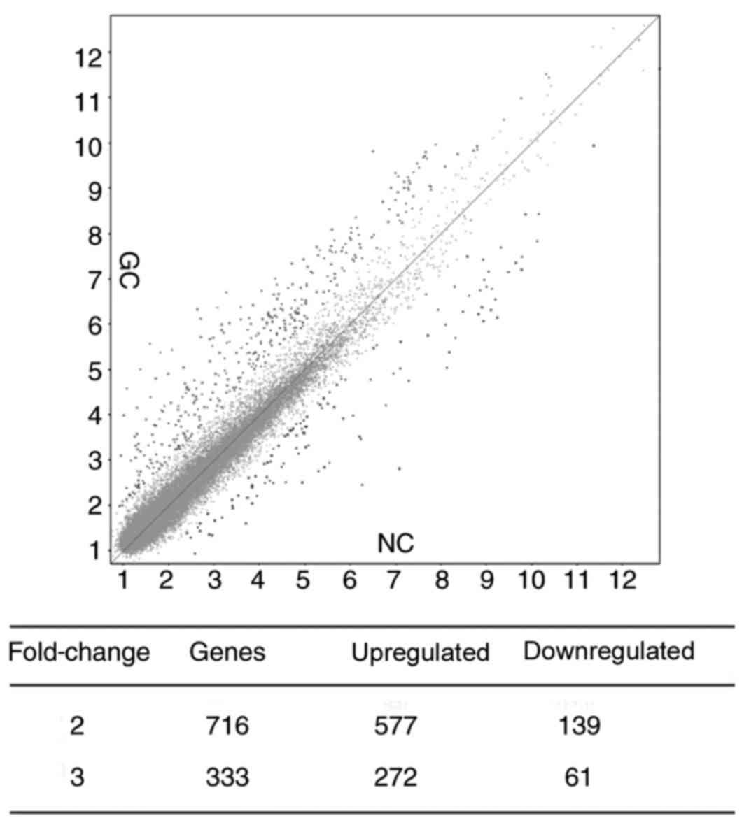
To investigate whether aberrantly expressed RNAs are
present in the plasma of patients with GC, 2 pools of plasma
specimens were generated from the GC group (n=16) and the control
group (n=16). Circulating RNAs in the 2 plasma pools were analyzed
using microarray [GeneChip® Human Transcriptome Array
2.0 (Affymetrix; Thermo Fisher Scientific Inc.)]. Numerous
significantly differentially expressed RNAs between the 2 pools
were detected, including 716 RNAs with a fold-change ≥2. Among
these, 577 RNAs were upregulated and 139 RNAs were downregulated in
patients with GC compared with the control group. There were 333
RNAs with a fold-change ≥3, including 272 RNAs that were
upregulated and 61 RNAs that were downregulated in patients with GC
compared with the control group (Fig.
1). Based on the expression levels of probe set regions and
integrated transcripts, 19 RNA molecules were selected for
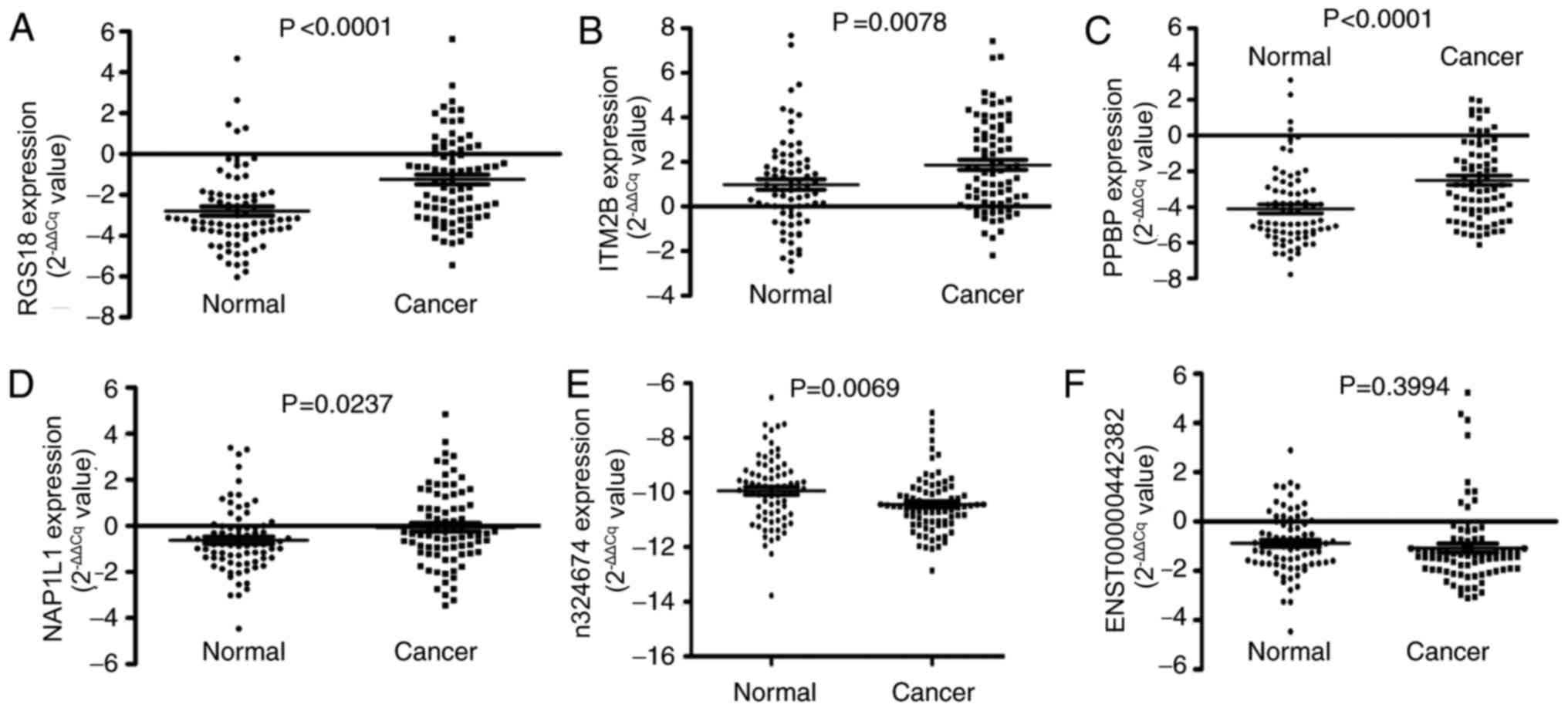
verification in the 2 plasma pools using RT-qPCR. It was indicated
that the expression levels of 2 non-coding RNAs (n324674 and
ENST00000442382) were significantly elevated, while a total of 4
mRNAs, regulator of G-protein signaling 18 (RGS18), integral
membrane protein 2B (ITM2B), pro-platelet basic protein (PPBP) and
nucleosome assembly protein1-like 1 (NAP1L1), were reduced in the
plasma of patients with GC (Figs. 2
and 4). Therefore, the expression
levels of these 6 RNAs were further verified in the large-scale
training sample of patients with GC.
Large-scale validation in plasma
samples
In the large-scale training sample of patients with
GC (n=81) and healthy participants (n=77), the expression level of
n324674 (P =0.0069) was indicated to be increased in the plasma of
patients with GC compared with that of healthy participants.
However, the expression level of RGS18 (P<0.0001), ITM2B
(P=0.0078), PPBP (P<0.0001) and NAP1L1 (P=0.0237) were reduced
in the plasma of patients with GC (Fig.
2). To evaluate whether plasma levels of the 5 candidate RNAs
could be used as diagnostic markers for GC, ROC curve analyses were
performed. It was indicated that the plasma levels of RGS18, ITM2B,
PPBP, NAP1L1 and n324674 discriminated patients with GC from
healthy participants, with AUCs of the ROC curves of 0.735 [95%
confidence interval (CI), 0.657–0.814], 0.635 (95% CI,
0.548–0.723), 0.721 (95% CI, 0.641–0.801), 0.629 (95% CI,
0.541–0.718) and 0.639 (95% CI, 0.550–0.727), respectively
(Fig. 3).
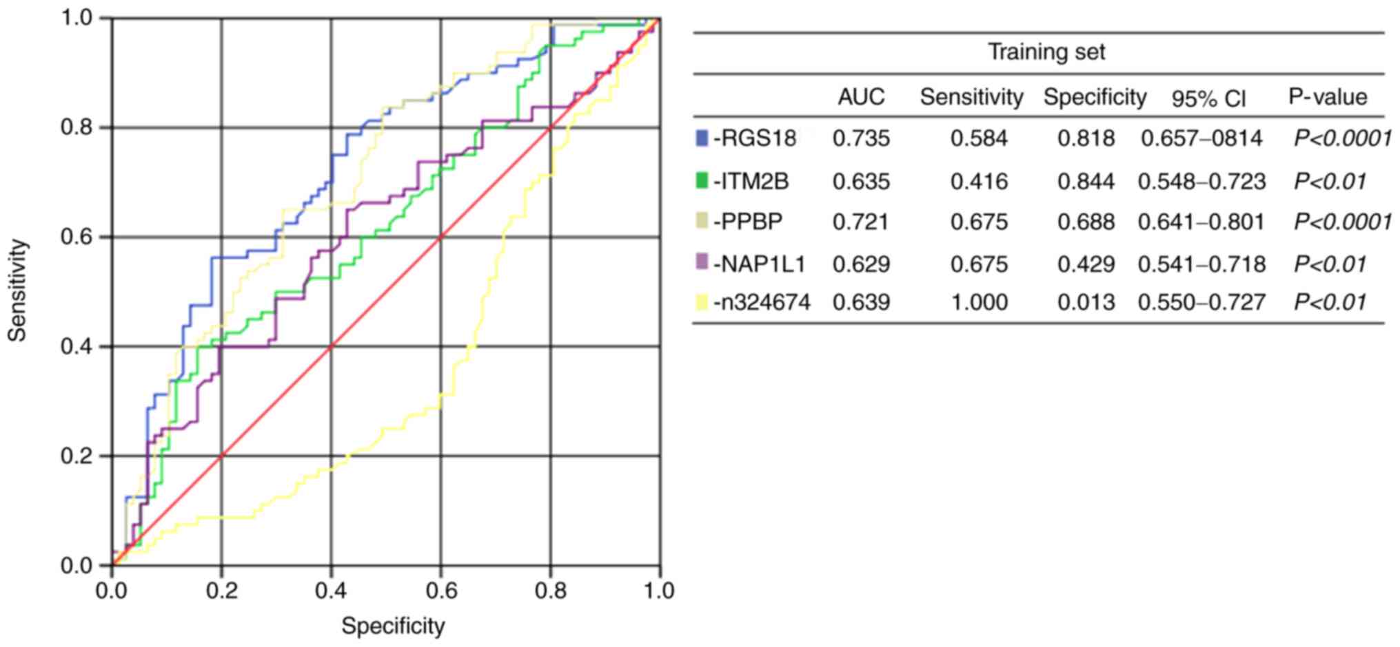 | Figure 3.Receiver operating characteristic
curve analysis of plasma RNAs: RGS18, ITM2B, PPBP, NAP1L1 and
n324674. RGS18, regulator of G-protein signaling 18; ITM2B,
integral membrane protein 2B; PPBP, pro-platelet basic protein;
NAP1L1, nucleosome assembly protein1-like 1; CI, confidence
interval, AUC, area under the curve. |
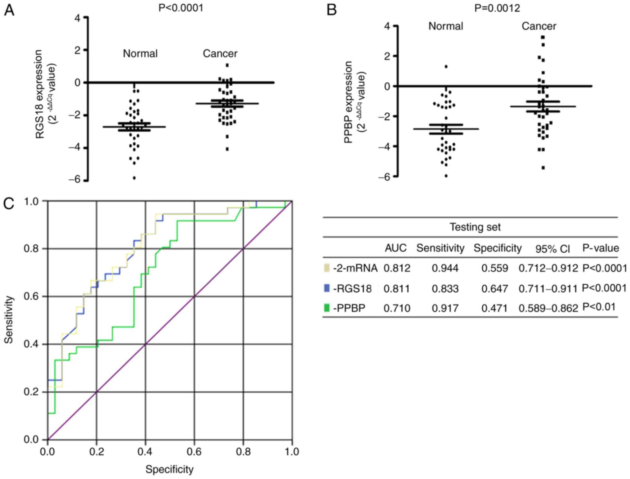
RGS18 and PPBP, the most significantly
differentially expressed RNAs, were subsequently selected for
further validation in an independent testing set of patients with
GC (n=36) and healthy participants (n=34) (Table II). The results confirmed that the
expression levels of RGS18 (P<0.0001) and PPBP (P=0.0012) were
reduced in the plasma samples from patients with GC compared with
those from healthy participants (Fig.
4). In addition, the AUC value of the 2-mRNA panel was 0.812,
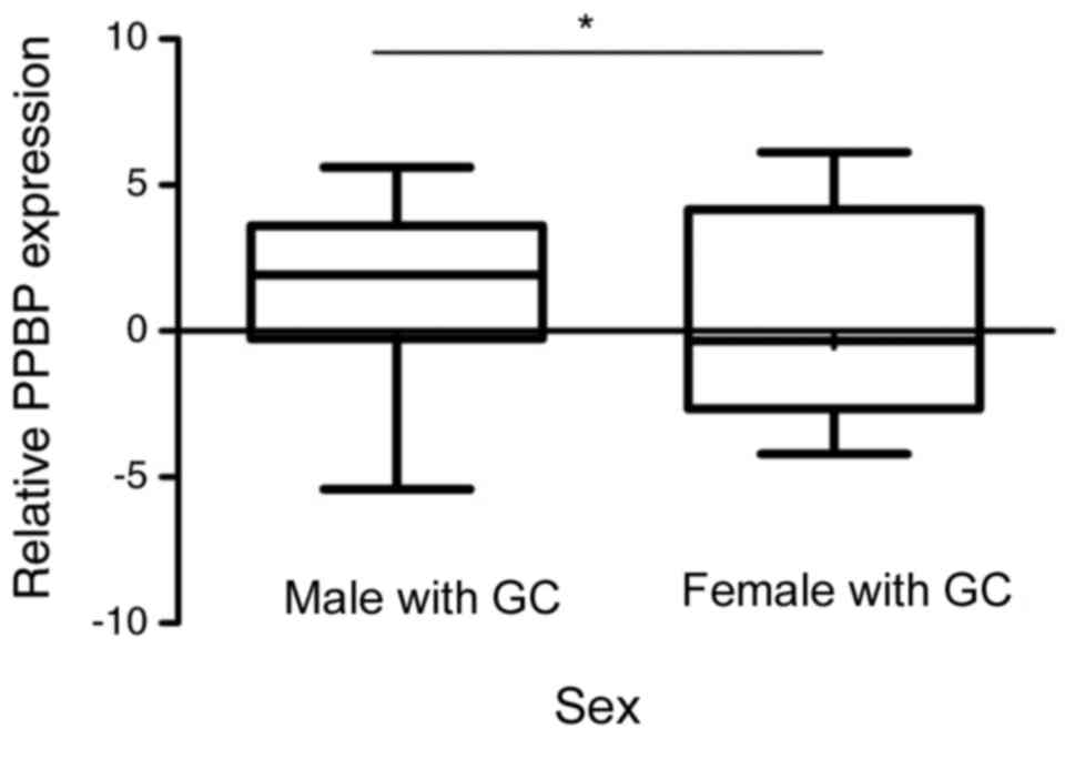
higher than that of the individual mRNAs. The association between
various clinicopathological features (age, sex, tumor size,
histological differentiation, invasion depth, regional lymph nodes,
distant metastasis and TNM stage) and expression levels of RGS18
and PPBP in GC plasma samples (n=117), including the training
(n=81) and testing sets (n=36), was subsequently investigated. It
was indicated that PPBP expression was reduced in female patients
with GC compared with male patients with GC (P=0.0328; Fig. 5).
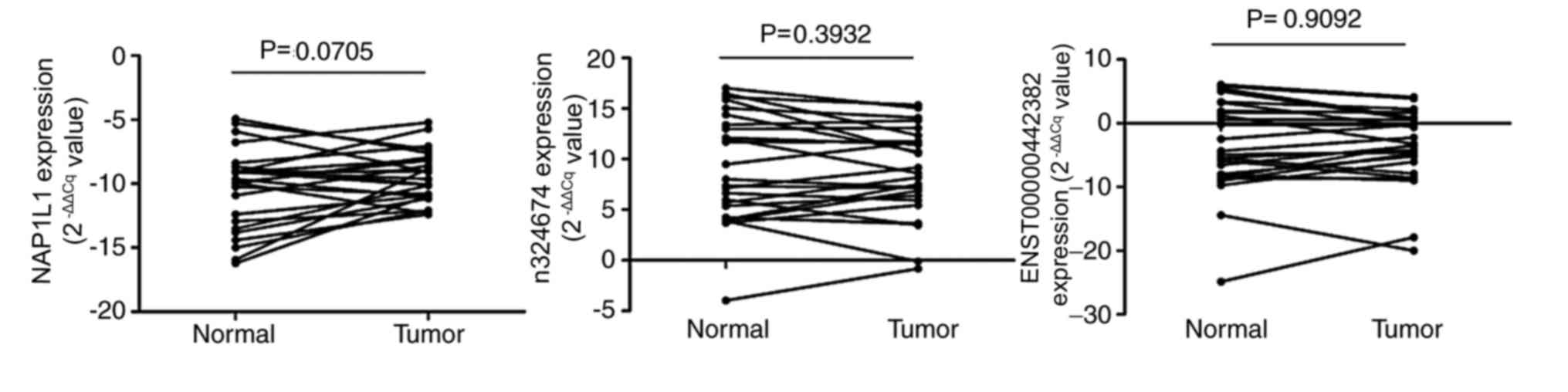
Detection of RNA expression in GC
tissues
The levels of the 6 candidate RNAs in 26 GC tissue
samples and paired adjacent histologically normal tissues were
assessed. RGS18, ITM2B and PPBP expression could not be detected
using RT-qPCR in GC tissues, whereas the expression of NAP1L1,
n324674 and ENST00000442382 was detected. However, their expression
patterns in the tissues were different from those in plasma
samples. NAP1L1 was downregulated in 9 tumor tissues compared with
levels in normal tissues, while n324674 was upregulated in 11
tissues and ENST00000442382 was upregulated in 12 tissues (Fig. 6). According to these results, it was
speculated that these plasma RNAs may not derive directly from
gastric carcinoma tissues.
Discussion
GC is the third leading cause of cancer-associated
mortality worldwide (1–3). GC tumor development and progression is a
multi-step processes, involving numerous genetic and epigenetic
alterations. However, only a few oncogenes and tumor suppressor
genes have been previously identified as being involved in gastric
carcinogenesis (22–25). Previous research has reported the use
of biomarkers for early detection and prediction of
chemotherapeutic sensitivity and prognosis in patients with various
types of cancer (11). Among the
various approaches reported in previous research, blood-based
testing has been indicated as the ideal method for identifying
biomarkers in cancer, due to its simplicity and minimal
invasiveness (26). However,
conventional plasma biomarkers, including CEA and CA72-4, lack
sufficient sensitivity and specificity (6,7). Similar
to other molecules, cfRNA has been stably detected in plasma or
serum samples (27–29). Previous studies have indicated that
plasma mRNAs and lncRNAs may be protected by exosome encapsulation
and complex formation with proteins, similar to plasma microRNAs
(27,30,31).
Genome-wide screening approaches have provided opportunities to
develop novel diagnostic or prognostic markers and to identify
novel therapeutic targets (32).
In the present study, the Human Transcriptome Array
was used to identify RNAs that are differentially expressed in
plasma samples from patients with GC and healthy participants. The
results indicated that the expression levels of RGS18, ITM2B, PPBP
and NAP1L1 were significantly decreased in GC plasma samples
compared with healthy participant plasma samples, whereas those of
n324674 were significantly increased, confirming the differential
expression of the aforementioned RNAs in 81 patients with GC and 77
healthy participants. The association between the plasma levels of
these 2 RNAs in 117 patients with GC (training and testing sets)
and various clinicopathological factors was analyzed. The results
indicated that the differential expression of PPBP between patients
with GC and healthy participants was more significant in females,
as females with GC exhibited decreased expression levels of PPBP
compared with male patients with GC. In terms of the utility of
these RNAs as biomarkers, it was indicated that plasma levels of
RGS18 and PPBP discriminated patients with GC from healthy
participants with a combined AUC of 0.812.
Among the 6 candidate RNAs, n324674 and
ENST00000442382 were lncRNAs. Numerous lncRNAs have been reported
to be associated with disease, particularly cancerous diseases
(33). lncRNAs, including H19,
imprinted maternally expressed transcript (non-protein coding)
(H19), hepatocellular carcinoma upregulated lncRNA, colon cancer
associated transcript 1 and run-related transcription factor 1,
have been reported to serve functional roles in GC (23,34,35). H19
has also been reported to circulate in the plasmas of patients with
GC (24). Therefore, n324674 may be a
novel lncRNA associated with GC.
The remaining RNAs are mRNAs. PPBP has been reported
to stimulate various cellular processes, including DNA synthesis,
mitosis, glycolysis and intracellular cyclic adenosine
monophosphate accumulation (16).
NAP1L1 participates in DNA replication and may serve a role in
modulating chromatin formation and regulating proliferation
(36). The underlying mechanisms of
PPBP and NAP1L1 in GC require further investigation. RGS proteins
have been reported to interact with and negatively regulate G
protein activation, and the RGS gene has been indicated to regulate
platelet aggregation, haemostasis and thrombosis (37). The ITM2B gene has been reported to be
a target of BCL6 repression in lymphomas and neurodegenerative
diseases and ITM2B protein generated by alternative splicing has
been reported to induce apoptosis in hematopoietic cell lines
(38).
Previous studies have demonstrated that the levels
of particular RNAs in body fluids were inconsistent with their
corresponding levels in tissues (39). It has been reported that one reason
for this is that cfRNAs may be transferred to body fluids by
exosomes (40,41). Previous studies have reported that
certain RNAs can be enriched in exosomes and selectively released
from healthy and malignant cells (42). In the present study, RGS18, ITM2B and
PPBP could not be detected in GC tissues by RT-qPCR and the
expression patterns of NAP1L1, n324674 and ENST00000442382 in GC
tissues were not consistent with those in plasma samples. This
unexpected result suggests that these RNAs in plasma may derive
from other tissues and not GC tissues.
In conclusion, the results of the present study
demonstrate that certain RNAs, including RGS18 and PPBP, were
significantly downregulated in the plasma of patients with GC
compared with healthy controls. These findings imply that a
combination of these 2 mRNAs could be used as a diagnostic or
prognostic marker for GC.
Acknowledgements
Not applicable.
Funding
The present study was supported partially by the
National Key Research and Development Program of China (grant no.
2016YFC1303600) and the Chinese National Natural Science Foundation
Projects (grant nos. 31370760, 91540202 and 31470782).
Availability of data and materials
The datasets used and/or analyzed during the present
study are available from the corresponding author on reasonable
request.
Authors' contributions
HF and XZ conceived the project and contributed to
the study design. CS, HL and ZP performed the data collection. CS,
HL and DK analyzed and interpreted the data. HL, CS and HF wrote
the manuscript. All authors read and approved the final version of
the manuscript.
Ethics approval and consent to
participate
Written informed consent was obtained from all
patients. The Ethics Committee of the Chinese PLA General Hospital
(Beijing, China) and the China-Japan Union Hospital of Jilin
University (Changchun, China) approved the use of samples for the
present study.
Patient consent for publication
Written informed consent for the publication of any
associated data was obtained from all the patient, or parent,
guardian or next of kin (in case of deceased patients).
Competing interests
The authors declare that they have no competing
interests.
References
|
1
|
Stewart BW and Wild C: International
Agency for Research on Cancer and World Health Organization. World
cancer report. WHO Press; 2014
|
|
2
|
Wu HH, Lin WC and Tsai KW: Advances in
molecular biomarkers for gastric cancer: miRNAs as emerging novel
cancer markers. Exp Rev Mol Med. 16:e12014. View Article : Google Scholar
|
|
3
|
Shao Y, Ye M, Jiang X, Sun W, Ding X, Liu
Z, Ye G, Zhang X, Xiao B and Guo J: Gastric juice long noncoding
RNA used as a tumor marker for screening gastric cancer. Cancer.
120:3320–3328. 2014. View Article : Google Scholar : PubMed/NCBI
|
|
4
|
Durães C, Almeida GM, Seruca R, Oliveira C
and Carneiro F: Biomarkers for gastric cancer: Prognostic,
predictive or targets of therapy? Virchows Arch. 464:367–378. 2014.
View Article : Google Scholar : PubMed/NCBI
|
|
5
|
Fassan M, Baffa R and Kiss A: Advanced
precancerous lesions within the GI tract: The molecular background.
Best Pract Res Clin Gastroenterol. 27:159–169. 2013. View Article : Google Scholar : PubMed/NCBI
|
|
6
|
Nakane Y, Okamura S, Akehira K, Boku T,
Okusa T, Tanaka K and Hioki K: Correlation of preoperative
carcinoembryonic antigen levels and prognosis of gastric cancer
patients. Cancer. 73:2703–2708. 1994. View Article : Google Scholar : PubMed/NCBI
|
|
7
|
Marrelli D, Pinto E, De Stefano A,
Farnetani M, Garosi L and Roviello F: Clinical utility of CEA CA
19-9 and CA 72-4 in the follow-up of patients with resectable
gastric cancer. Am J Surg. 181:16–19. 2001. View Article : Google Scholar : PubMed/NCBI
|
|
8
|
Feng F, Tian Y, Xu G, Liu Z, Liu S, Zheng
G, Guo M, Lian X, Fan D and Zhang H: Diagnostic and prognostic
value of CEA CA19-9, AFP and CA125 for early gastric cancer. BMC
Cancer. 17:7372017. View Article : Google Scholar : PubMed/NCBI
|
|
9
|
Matsuoka T and Yashiro M: Biomarkers of
gastric cancer: Current topics and future perspective. World J
Gastroenterol. 24:2818–2832. 2018. View Article : Google Scholar : PubMed/NCBI
|
|
10
|
Li Q, Shao Y, Zhang X, Zheng T, Miao M,
Qin L, Wang B, Ye G, Xiao B and Guo J: Plasma long noncoding RNA
protected by exosomes as a potential stable biomarker for gastric
cancer. Tumour Biol. 36:2007–2012. 2015. View Article : Google Scholar : PubMed/NCBI
|
|
11
|
Schwarzenbach H, Hoon DS and Pantel K:
Cell-free nucleic acids as biomarkers in cancer patients. Nat Rev
Cancer. 11:426–437. 2011. View
Article : Google Scholar : PubMed/NCBI
|
|
12
|
Viereck J and Thum T: Circulating
noncoding RNAs as biomarkers of cardiovascular disease and injury.
Circ Res. 120:381–399. 2017. View Article : Google Scholar : PubMed/NCBI
|
|
13
|
Zhou X, Yin C, Dang Y, Ye F and Zhang G:
Identification of the long non-coding RNA H19 in plasma as a novel
biomarker for diagnosis of gastric cancer. Sci Rep. 5:115162015.
View Article : Google Scholar : PubMed/NCBI
|
|
14
|
Tsui NB, Ng EK and Lo YM: Stability of
endogenous and added RNA in blood specimens, serum, and plasma.
Clin Chem. 48:1647–1653. 2002.PubMed/NCBI
|
|
15
|
Sethakorn N and Dulin NO: RGS expression
in cancer: Oncomining the cancer microarray data. J Recept Signal
Transduct Res. 33:166–171. 2013. View Article : Google Scholar : PubMed/NCBI
|
|
16
|
Guo Q, Jian Z, Jia B and Chang L: CXCL7
promotes proliferation and invasion of cholangiocarcinoma cells.
Oncol Rep. 37:1114–1122. 2017. View Article : Google Scholar : PubMed/NCBI
|
|
17
|
Bosman FT, Carneiro F, Hruban RH and
Theise ND: WHO classifcation of tumours of the digestive system.
Fourth Edition. World Health Organization. 3:4172010.
|
|
18
|
Washington K: 7th edition of the AJCC
cancer staging manual: Stomach. Ann Surg Oncol. 17:3077–3079. 2010.
View Article : Google Scholar : PubMed/NCBI
|
|
19
|
Livak KJ and Schmittgen TD: Analysis of
relative gene expression data using real-time quantitative PCR and
the 2(-Delta Delta C(T)) method. Methods. 25:402–408. 2001.
View Article : Google Scholar : PubMed/NCBI
|
|
20
|
March-Villalba JA, Martínez-Jabaloyas JM,
Herrero MJ, Santamaria J, Aliño SF and Dasi F: Cell-free
circulating plasma hTERT mRNA is a useful marker for prostate
cancer diagnosis and is associated with poor prognosis tumor
characteristics. PLoS One. 7:e434702012. View Article : Google Scholar : PubMed/NCBI
|
|
21
|
Ke D, Li H, Zhang Y, An Y, Fu H, Fang X
and Zheng X: The combination of circulating long noncoding RNAs
AK001058, INHBA-AS1, MIR4435-2HG, and CEBPA-AS1 fragments in plasma
serve as diagnostic markers for gastric cancer. Oncotarget.
8:21516–21525. 2017. View Article : Google Scholar : PubMed/NCBI
|
|
22
|
Yang F, Bi J, Xue X, Zheng L, Zhi K, Hua J
and Fang G: Up-regulated long non-coding RNA H19 contributes to
proliferation of gastric cancer cells. FEBS J. 279:3159–3165. 2012.
View Article : Google Scholar : PubMed/NCBI
|
|
23
|
Yang F, Xue X, Bi J, Zheng L, Zhi K, Gu Y
and Fang G: Long noncoding RNA CCAT1, which could be activated by
c-Myc, promotes the progression of gastric carcinoma. J Cancer Res
Clin Oncol. 139:437–445. 2013. View Article : Google Scholar : PubMed/NCBI
|
|
24
|
He L, Thomson JM, Hemann MT,
Hernando-Monge E, Mu D, Goodson S, Powers S, Cordon-Cardo C, Lowe
SW, Hannon GJ and Hammond SM: A microRNA polycistron as a potential
human oncogene. Nature. 435:828–833. 2005. View Article : Google Scholar : PubMed/NCBI
|
|
25
|
He L, He X, Lim LP, de Stanchina E, Xuan
Z, Liang Y, Xue W, Zender L, Magnus J, Ridzon D, et al: A microRNA
component of the p53 tumour suppressor network. Nature.
447:1130–1134. 2007. View Article : Google Scholar : PubMed/NCBI
|
|
26
|
Arita T, Ichikawa D, Konishi H, Komatsu S,
Shiozaki A, Shoda K, Kawaguchi T, Hirajima S, Nagata H, Kubota T,
et al: Circulating long non-coding RNAs in plasma of patients with
gastric cancer. Anticancer Res. 33:3185–3193. 2013.PubMed/NCBI
|
|
27
|
Tani N, Ichikawa D, Ikoma D, Tomita H, Sai
S, Ikoma H, Okamoto K, Ochiai T, Ueda Y, Otsuji E, et al:
Circulating cell-free mRNA in plasma as a tumor marker for patients
with primary and recurrent gastric cancer. Anticancer Res.
27:1207–1212. 2007.PubMed/NCBI
|
|
28
|
Zhou H, Xu W, Qian H, Yin Q, Zhu W and Yan
Y: Circulating RNA as a novel tumor marker: An in vitro study of
the origins and characteristics of extracellular RNA. Cancer Lett.
259:50–60. 2008. View Article : Google Scholar : PubMed/NCBI
|
|
29
|
Tsujiura M, Ichikawa D, Komatsu S,
Shiozaki A, Takeshita H, Kosuga T, Konishi H, Morimura R, Deguchi
K, Fujiwara H, et al: Circulating microRNAs in plasma of patients
with gastric cancers. Br J Cancer. 102:1174–1179. 2010. View Article : Google Scholar : PubMed/NCBI
|
|
30
|
Iguchi H, Kosaka N and Ochiya T: Secretory
microRNAs as a versatile communication tool. Commun Integr Biol.
3:478–481. 2010. View Article : Google Scholar : PubMed/NCBI
|
|
31
|
Arroyo JD, Chevillet JR, Kroh EM, Ruf IK,
Pritchard CC, Gibson DF, Mitchell PS, Bennett CF,
Pogosova-Agadjanyan EL, Stirewalt DL, et al: Argonaute2 complexes
carry a population of circulating microRNAs independent of vesicles
in human plasma. Proc Natl Acad Sci USA. 108:5003–5008. 2011.
View Article : Google Scholar : PubMed/NCBI
|
|
32
|
Chen X, Hu Z, Wang W, Ba Y, Ma L, Zhang C,
Wang C, Ren Z, Zhao Y, Wu S, et al: Identification of ten serum
microRNAs from a genome-wide serum microRNA expression profile as
novel noninvasive biomarkers for nonsmall cell lung cancer
diagnosis. Int J Cancer. 130:1620–1628. 2012. View Article : Google Scholar : PubMed/NCBI
|
|
33
|
Gutschner T and Diederichs S: The
hallmarks of cancer: A long non-coding RNA point of view. RNA Biol.
9:703–719. 2012. View Article : Google Scholar : PubMed/NCBI
|
|
34
|
Zhuang M, Gao W, Xu J, Wang P and Shu Y:
The long non-coding RNA H19-derived miR-675 modulates human gastric
cancer cell proliferation by targeting tumor suppressor RUNX1.
Biochem Biophys Res Commun. 448:315–322. 2014. View Article : Google Scholar : PubMed/NCBI
|
|
35
|
Zhao Y, Guo Q, Chen J, Hu J, Wang S and
Sun Y: Role of long non-coding RNA HULC in cell proliferation,
apoptosis and tumor metastasis of gastric cancer: A clinical and in
vitro investigation. Oncol Rep. 31:358–364. 2014. View Article : Google Scholar : PubMed/NCBI
|
|
36
|
Yan Y, Yin P, Gong H, Xue Y, Zhang G, Fang
B, Chen Z, Li Y, Yang C, Huang Z, et al: Nucleosome assembly
protein 1-like 1 (Nap1l1) regulates the proliferation of murine
induced pluripotent stem cells. Cell Physiol Biochem. 38:340–350.
2016. View Article : Google Scholar : PubMed/NCBI
|
|
37
|
Alshbool FZ, Karim ZA, Vemana HP, Conlon
C, Lin OA and Khasawneh FT: The regulator of G-protein signaling 18
regulates platelet aggregation, hemostasis and thrombosis. Biochem
Biophys Res Commun. 462:378–382. 2015. View Article : Google Scholar : PubMed/NCBI
|
|
38
|
Baron BW, Baron RM and Baron JM: The ITM2B
(BRI2) gene is a target of BCL6 repression: Implications for
lymphomas and neurodegenerative diseases. Biochim Biophys Acta.
1852:742–748. 2015. View Article : Google Scholar : PubMed/NCBI
|
|
39
|
Cui L, Zhang X, Ye G, Zheng T, Song H,
Deng H, Xiao B, Xia T, Yu X, Le Y and Guo J: Gastric juice
MicroRNAs as potential biomarkers for the screening of gastric
cancer. Cancer. 119:1618–1626. 2013. View Article : Google Scholar : PubMed/NCBI
|
|
40
|
Fellouse FA: Stromal exosomes allow cancer
cell autoactivation. Med Sci (Paris). 30:405–407. 2014.(In French).
View Article : Google Scholar : PubMed/NCBI
|
|
41
|
Mittelbrunn M, Gutiérrez-Vázquez C,
Villarroya-Beltri C, González S, Sánchez-Cabo F, González MÁ,
Bernad A and Sánchez-Madrid F: Unidirectional transfer of
microRNA-loaded exosomes from T cells to antigen-presenting cells.
Nat Commun. 2:2822011. View Article : Google Scholar : PubMed/NCBI
|
|
42
|
Gezer U, Özgür E, Cetinkaya M, Isin M and
Dalay N: Long non-coding RNAs with low expression levels in cells
are enriched in secreted exosomes. Cell Biol Int. 38:1076–1079.
2014.PubMed/NCBI
|