Introduction
Kaposiform haemangioendotheliomas (KHEs) were first
described in 1993 by Zukerberg et al (1) as rare soft-tissue neoplastic lesions
often associated with locally aggressive disease, lymphangiomatosis
and the Kasabach-Merritt phenomenon (KMP). According to Croteau
et al (2), the prevalence in
Massachusetts is ~0.91 cases per 100,000 individuals. First
reported by Kasabach and Merritt in 1940 (3), KMP is a haemangioendothelioma-producing
condition that occasionally causes microangiopathic haemolytic
anaemia, thrombocytopaenia and consumption coagulopathy. Untreated
or recurrent KHE/KMP can be a life-threatening condition with a
high mortality rate. Although several treatment regimens have been
implemented (4), the current regimens
for KHE/KMP have not achieved a satisfactory therapeutic effect.
Furthermore, surgical trauma, drug side effects and a certain risk
of recurrence when applying these regimens cannot be ignored.
However, standard regimens and definitive guidelines have yet to be
studied or established.
Previous studies have reported several therapeutic
modalities for KHE/KMP. Options, including systemic supportive
care, pharmacological management, surgical resection, endovascular
interventional treatments and radiotherapy, have been used to
control coagulopathy and limit the progression of lesions (5). Among the various options for KHE/KMP
treatment, pharmacological management, which has been the focus of
numerous studies, is regarded as one of the most important
non-invasive treatments. However, according to a previous study
(6), the non-selective β-adrenergic
antagonist propranolol is partially effective, with only 36% of
cases responding to treatment. Systemic corticosteroid therapy, as
a conventional treatment option for patients with KHE, is
associated with limited therapeutic efficacy and relatively higher
rates of side effects (7,8). Alternative treatments have recently
emerged. A meta-analysis by Liu et al (9) showed that corticosteroids were
considerably less effective and had higher complication rates
compared with vincristine; however, several side effects of the
latter cannot be ignored, with constipation, peripheral neuropathy
and syndrome of inappropriate antidiuretic hormone secretion being
reported (10–12). In addition, another study described a
patient who partially responded to paclitaxel chemotherapy and
subsequent treatments (prednisone, doxorubicin, interferon-α,
gemcitabine and ifosfamide), but succumbed to severe KMP and major
gastrointestinal bleeding (13).
Sirolimus was recently proposed as an effective
alternative therapy for KHE/KMP (14). Ji et al (15) presented a multicentre retrospective
study of 52 patients with KHE and concluded that sirolimus was
effective and safe for treating progressive KHE. Despite several
studies assessing the therapeutic effect of sirolimus, the side
effects of this drug cannot be ignored (16,17).
Clinical sirolimus application has been reported to result in the
following side effects: Hyperglycaemia, oral sores and signs of
immunosuppression (5). Additionally,
in a 2017 case report by Triana et al (18), a 4-month-old infant with pancreatic
KHE showed no response to sirolimus. As pharmacological treatment
of KHE has mostly been demonstrated to reduce rather than to
eliminate the tumour mass, a certain risk of non-response and
recurrence remains. Furthermore, the relapse of lesions may have
lethal consequences (19).
Surgical resection is applied in certain cases of
KHE/KMP, although tissue defects are a major source of trauma for
these patients. In 2016, Guo et al (20) described the case of a 48-day-old male
infant with KMP who did not respond to propranolol or
glucocorticoid treatment. The infant recovered subsequent to
receiving surgical resection, indicating that surgical approaches
may be required despite the associated challenging recovery. In the
report by Zahir et al (21), a
24-day-old male neonate received transfusions of platelets (PLTs)
and packed red blood cells (RBCs), as well as medical treatments
that included oral prednisolone, intravenous methylprednisolone and
interferon-α. Despite thrombocytopaenia, the PLT count returned to
normal following resection. Furthermore, Leung et al
(22) described the case of a
full-term male baby with pancreatic KHE/KMP who had a PLT count of
23×109/l (normal range of PLT:
100–300×109/l); the baby underwent resection treatment
and recovered with a normal PLT count following surgery. Surgical
resection has been reported to be effective in several other cases
(23–26). However, postoperative tissue defects
may severely impair the structural and functional development of
tissues in an infant. Although a less invasive surgical procedure
is necessary in certain cases, a consensus regarding comprehensive
and sequential therapy for KHE has yet to be established.
The present study reports a novel approach, stepwise
local suture ligation-assisted percutaneous sclerotherapy, which is
effective and less invasive for the treatment of complex cases of
KHEs complicated by KMP, in a limited series. This study aimed to
retrospectively review the clinical features and treatment outcomes
of stepwise local suture ligation-assisted percutaneous
sclerotherapy for the treatment of KHE/KMP.
Patients and methods
Patients
Approval from the Institutional Review Board of
Shanghai Ninth People's Hospital was obtained for a retrospective
review of patient medical and imaging records, and written informed
consent from the legal guardian of the patients was obtained.
Inclusion criterions included: i) A definite diagnosis of KHE/KMP
confirmed by clinical, pathological and laboratory manifestations;
ii) legal guardians agreeing with the treatment plan. The exclusion
criterion was if the legal guardians refused the proposed plan.
Between September 2015 and September 2017, 3 consecutive patients
(2 males, 1 female; mean age, 2.67 months; range, 1–5 months) with
KHE/KMP underwent staged treatment, including 2 patients with a
medical history of corticosteroid treatment and lesion relapse
following treatment. Clinical, radiological, haematological
(including PLT, haemoglobin, RBC count, D-Dimer and fibrin
degradation product) and pathological features of the cases are
recorded (Tables I and II; Figs.
1–3). All diagnoses were based on
a combination of medical history, clinical manifestations, imaging
findings and pathological data.
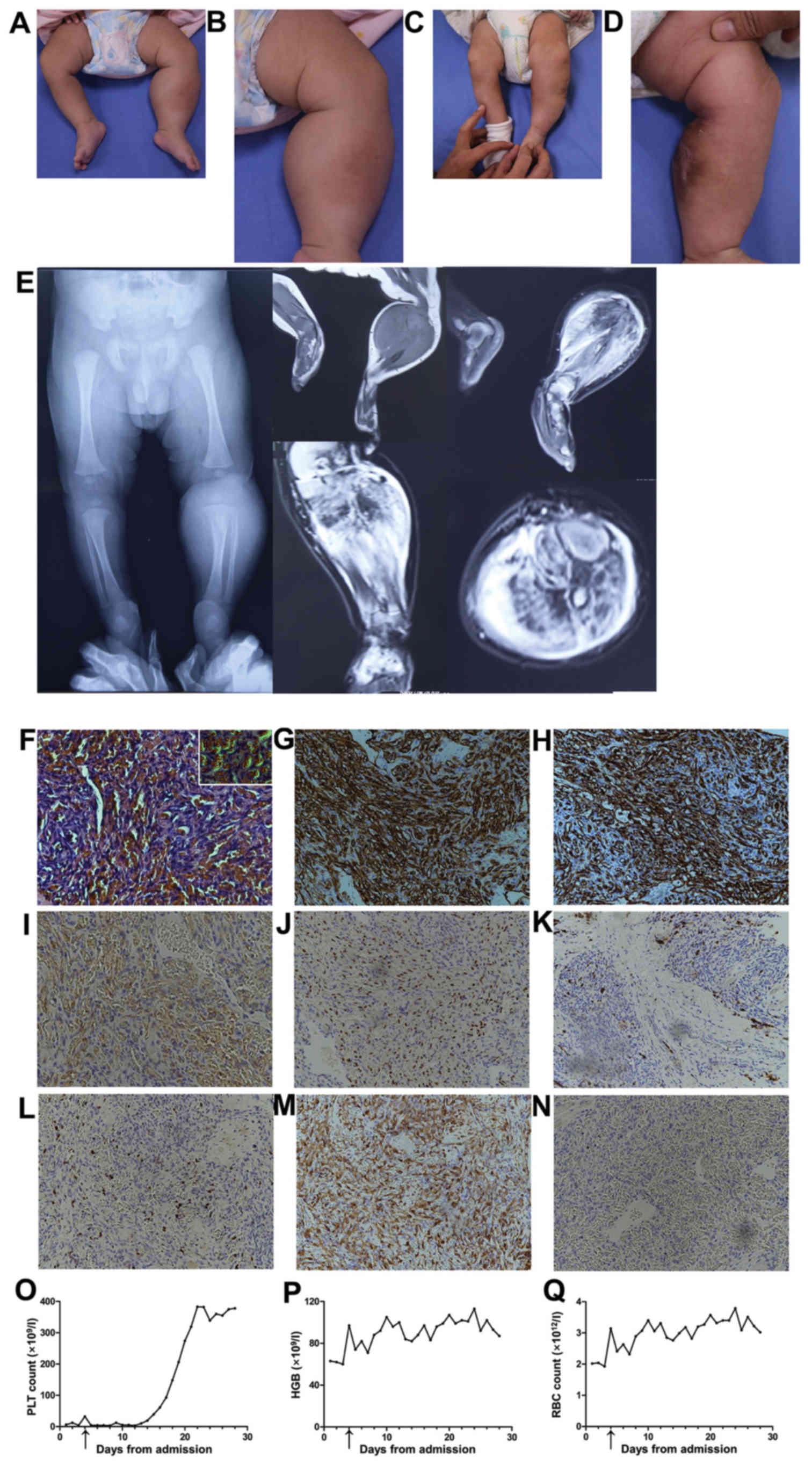 | Figure 3.Clinical, radiological, pathological
and laboratory evaluations of case 3. Pre-treatment image (A) of
the patient and (B) a close-up of the affected area. Images
obtained at 4 weeks following suture ligation treatment of (C) the
patient and (D) a close-up of the affected area. (E) Pre-treatment
radiological evaluations. Histopathological examinations including
(F) haematoxylin-eosin staining and immunohistochemical staining
for (G) cluster of differentiation (CD)34, (H) CD31, (I)
podoplanin, (J) prospero homeobox protein 1, (K) glucose
transporter 1, (L) proliferation marker protein Ki-67, (N) desmin
of the lesion and (M) smooth muscle actin. Original magnification
for (F-N), ×200; inset image in (F), ×400. (O-Q) Laboratory
evaluations: (O) PLT, (P) HGB and (Q) RBC counts. The arrow
indicates the time-point of suture ligation treatment. PLT,
platelet; HGB, haemoglobin; RBC, red blood cell. |
 | Table I.Demographics and clinical
presentation of patients with Kaposiform haemangioendotheliomas
associated with the Kasabach-Merritt phenomenon. |
Table I.
Demographics and clinical
presentation of patients with Kaposiform haemangioendotheliomas
associated with the Kasabach-Merritt phenomenon.
| Patient | Sex | Age, months | Location | Dimensions, cm | Appearance |
|---|
| 1 | M | 2 | Left lower
abdomen | 3×5 | Indurated
ecchymotic mass with local swelling |
| 2 | F | 5 | Left cluneal
region | 10×15 | Indurated
ecchymotic mass with local swelling |
| 3 | M | 1 | Left shin | 5×7 | Firm mass without
cutaneous changes, limiting ankle extension |
| Table II.Initial relevant haematological
investigations of patients with Kaposiform haemangioendotheliomas
associated with the Kasabach-Merritt phenomenon. |
Table II.
Initial relevant haematological
investigations of patients with Kaposiform haemangioendotheliomas
associated with the Kasabach-Merritt phenomenon.
| Patient | PLT, n
(×109/l) | HGB, g/l | RBC, n
(×1012 cells/l) | DD, µg/ml | FDP, µg/ml |
|---|
| 1 | 70 | 101 | 3.62 | 11.13 |
51.0 |
| 2 | 59 | 111 | 4.16 |
6.96 |
25.6 |
| 3 | 6 | 63 | 2.02 | 83.86 | 284.0 |
Treatment process
The patients underwent a stepwise synthetic serial
therapy consisting of percutaneous sclerotherapy and adjunctive
pharmacotherapy, accompanied by a suture ligation procedure.
Local suture ligation-assisted percutaneous
sclerotherapy was performed under general anaesthesia with a
standard aseptic technique. A suspension containing 10
mg/m2 (body surface area) bleomycin and 8 ml 0.9% NaCl
was injected into the tumour using a 10-ml syringe. Percutaneous
punctures were located at several points, including adjacent to,
and at the centre of, the lesion. Subsequently, a tight local
suture ligation was applied to limit the blood supply of the
lesion. A 9×34-mm circular needle and 3-0 absorbable sutures were
used. An interrupted suture method was applied to form a grid-like
pattern on the surface of local lesions. The spacing between
sutures was 2–3 cm and the suture depth was 2–3 cm. Every knot was
tight and tensile, and the lesions were covered with wound
dressings following the suture ligation procedure. A biopsy was
performed during the suture ligation procedure and specimens were
used for pathological diagnosis. The suture ligation procedure
limited the blood supply to the lesion and reduced intraoperative
blood loss during biopsy incision.
Pre- and post-adjunctive pharmacotherapy was
administered to maintain a relatively acceptable physiological
status during hospitalisation. Patients began treatment with
intravenous corticosteroids (methylprednisolone, 6 mg/kg/day) from
the first day of hospital admission throughout the treatment
procedure. Drug dosages were gradually reduced after KMP vanished
and PLT returned to normal (normal range of PLT:
100–300×109/l) for three days. Oral administration of
prednisone acetate was reduced from 2 to 1.5 mg/kg/day until drug
withdrawal.
Histological observation
All tumour tissue samples were fixed in 10% neutral
buffered formalin solution (24 h at room temperature) for
histopathological evaluation. Later, they were embedded in paraffin
and sectioned at a thickness of 4 µm were prepared from
formalin-fixed and paraffin-embedded blocks, deparaffinised in
xylene, rehydrated and microwaved for 10 min at 30% power in
citrate buffer, pH 6.0 (Poly Scientific, Bay Shore, NY, USA).
Endogenous peroxidase activity was blocked using 0.3% hydrogen
peroxide in 80% methanol for 5 min at room temperature. The
sections were either stained with haematoxylin-eosin (HE) (2 g/l
haematoxylin for 5 min followed by 0.5% eosin for 1–3 min, both at
room temperature) or incubated with primary antibodies, including
cluster of differentiation 34 (CD34), CD31, desmin (Des),
podoplanin (D2-40), prospero homeobox protein 1 (Prox-1), factor
VIII (FVIII), vascular endothelial growth factor receptor 3,
glucose transporter 1 (GLUT-1), proliferating cell nuclear antigen
(PCNA), proliferation marker protein Ki-67 (Ki-67) and smooth
muscle actin (SMA) overnight at 4°C subsequent to blocking with 3%
bovine serum albumin (Sigma-Aldrich; Merck KGaA, Darmstadt,
Germany). Secondary antibodies were applied to the slides for 1 h
at room temperature. The primary and secondary antibodies involved
in the present study are listed in Table III. Photographs were obtained with
an Eclipse Ti-E inverted microscope (Nikon Corporation, Tokyo,
Japan; magnification, ×200 and ×400) and a Nikon E600 camera.
| Table III.Antibodies used for immunochemical
analysis. |
Table III.
Antibodies used for immunochemical
analysis.
| Primary
antigen | Source (cat.
no.) | Clone | Dilution | Secondary antigen,
source, cat. no., dilution |
|---|
| CD34 | CSTa (3569S) | ICO0115 | 1:30 | Peroxidase
AffiniPure Donkey Anti-Mouse IgG (H+L), Jacksonb, 715-035-150, 1:500-1:5,000 |
| Des |
R&Dc
(AF3844) | DES | 5–15 µg/ml | AffiniPure Bovine
Anti-Goat IgG (H+L), Jacksonb, 805-035-180, 1:500-1:5,000 |
| D2-40 | Abcamd (ab77854) | D2-40 | 1:40 | Peroxidase
AffiniPure Donkey Anti-Mouse IgG (H+L), Jacksonb, 715-035-150, 1:500-1:5,000 |
| CD31 | CSTa (3528S) | PECAM-1′89C2 | 1/1,600 | Peroxidase
AffiniPure Donkey Anti-Mouse IgG (H+L), Jacksonb, 715-035-150, 1:500-1:5,000 |
| FVIII | Novuse (NB100-91761) | F8 | 1:50-1:200 | Peroxidase
AffiniPure Donkey Anti-Rabbit IgG (H+L), Jacksonb, 711-035-152, 1:500-1:5,000 |
| GLUT-1 | Novuse (NB110-39113) | SLC2A1 | 1:200 | Peroxidase
AffiniPure Donkey Anti-Rabbit IgG (H+L), Jacksonb, 711-035-152, 1:500-1:5,000 |
| Ki-67 | CSTa (9449S) | 8D5 | 1:400 | Peroxidase
AffiniPure Donkey Anti-Rabbit IgG (H+L), Jacksonb, 711-035-152, 1:500-1:5,000 |
| PCNA | CSTa (13110S) | D3H8P | 1:8,000 | Peroxidase
AffiniPure Donkey Anti-Rabbit IgG (H+L), Jacksonb, 711-035-152, 1:500-1:5000 |
| Prox-1 | CSTa (14963S) | D2J6J | 1:500 | Peroxidase
AffiniPure Donkey Anti-Rabbit IgG (H+L), Jacksonb, 711-035-152, 1:500-1:5,000 |
| SMA |
R&Dc
(MAB1420) | 1A4 | 8–25 µg/ml | Peroxidase
AffiniPure Donkey Anti-Mouse IgG (H+L), Jacksonb, 715-035-150, 1:500-1:5,000 |
| VEGFR-3 |
R&Dc
(AF349) | FLT4 | 5–15 µg/ml | Peroxidase
AffiniPure Donkey Anti- Goat IgG (H+L), Jacksonb, 805-035-180, 1:500-1:5,000 |
Results
Clinical outcomes
The proposed local suture ligation-assisted
percutaneous sclerotherapy strategy was effective in all patients,
with notable relief of clinical symptoms and improvement of
haematological indicators. Pre- and post-treatment clinical,
radiological, pathological and laboratory evaluations are presented
in Figs. 1–3.
Regarding clinical manifestations, the tumour mass
was reduced, local swelling was diminished, the tendencies for
ecchymosis and bleeding were weakened, and the overall health
status was largely improved in all patients. Furthermore, in one
patient (presented in Fig. 3) who had
suffered from limited range of motion in ankle joint due to KHE in
his left lower limb, no difference in the range of motion in
bilateral lower limbs was detected through post treatment clinical
examination. More importantly, none of the patients experienced
tissue defects or local dysfunction. The entire treatment process
was successful without any adverse effects. At the time of writing
the present study, the patients were undergoing regular follow-ups
(28-, 19- and 4-month follow-ups for cases 1, 2 and 3,
respectively). Following operating local suture ligation-assisted
percutaneous sclerotherapy there were eusemia and no signs of
recurrence in all cases.
Laboratory evaluations
Pre-treatment haematological data of the cases were
presented in Table II, which
indicated KMP through evaluation of PLT, haemoglobin, RBC, D-Dimer
and fibrin degradation products. According to laboratory and
haematological evaluations, immediate and notable improvements in
PLT, RBC and haemoglobin measurements were observed in all patients
on the first day after suture ligation treatment. In cases 1 and 2,
the PLT continued to increase until reaching normal thresholds and
showed similar plateau trends that remained stable within normal
range (normal range: 100–300×109/l) for 22 and 60 days,
respectively, according to haematological evaluations. However, in
case 3, although the relevant indicators remained at subnormal
levels several days after suture ligation, a marked and rapid
increase in PLT levels was observed between days 13 and 21, which
reached a plateau of ~300×109/l after the 21st day.
Histological findings
Histological analysis by HE staining showed that the
lesions were characterised by irregular sheets of spindle-shaped
endothelial cells and characteristic slit-like vascular channels,
confirming the diagnosis of KHE by pathology. In all cases,
immunohistochemical staining results were positive for vascular
markers (CD31, CD34 and FVIII) and proliferation markers (PCNA),
and a population of parietal cells was partially positive for
lymphatic markers D2-40 and Prox-1, ~5% positive for Ki-67
staining, and negative for GLUT-1 and Des. Muscular marker staining
of SMA revealed a negative result in case 1, and positive results
in cases 2 and 3 (Figs. 1–3).
Discussion
KHE/KMP is a type of haemangioma characterised by
consumption coagulopathy and potentially high mortality rates; it
is usually located in the extremities and retroperitoneum (10). In the present study, a stepwise
synthetic serial treatment for patients with KHE was explored,
particularly for those who failed to respond to, or tolerate,
conventional treatment. The clinical results showed that severe
KHE/KMP responded to the proposed treatment, local suture
ligation-assisted percutaneous sclerotherapy, in a limited series.
This therapy is particularly adaptable to lesions on the trunk and
extremities.
Previous studies have demonstrated that a definitive
diagnosis is essential in the management of KHE/KMP (16,27–30). KHE
presents during early childhood and manifests as violaceous
subcutaneous masses with ill-defined borders and a purpuric,
bruised appearance, located on the extremities and trunk in ~75% of
cases (27). KMP, a thrombocytopenic
coagulopathy often associated with more aggressive KHEs (28,29), is
one of the most dangerous and life-threatening clinical conditions,
with a 30–40% mortality rate due to uncontrollable haemorrhage
(16). The present cases showed
clinical features consistent with KMP located in the extremities
and trunk. Radiological evaluations provided supporting images of
the lesions. More importantly, pathological diagnosis is widely
accepted as the gold standard for KHE. Pathologically, the features
of KHE resemble a capillary haemangioma and Kaposi's sarcoma.
Infiltrating sheets composed of slit-like vascular channels and
irregularly shaped spindle endothelial cells are characteristic
features of KHEs (30). Regarding
pathological findings, all cases in the present study showed
classic features that confirmed the pathological diagnosis of
KHE.
In addition to histological features, expression
levels and changes in markers revealed by immunohistochemical
analysis are essential in assessing KHE and may provide potential
guidance for clinical treatment. According to Putra and Gupta
(31), as vascular lesions, KHE
lesions are immunoreactive to non-specific endothelial markers,
including CD31 and CD34, in addition to lymphatic marker
immunoreactivity with either Prox-1 or D2-40 within the neoplastic
spindled endothelial cells supports the diagnosis of KHE (32). Detailed and systematic evidence of
immunohistochemical staining was provided in the present study,
showing endothelial cells in the nodules to be positive for
vascular markers (CD31, CD34 and FVIII) and proliferation markers
(PCNA). In addition, a population of parietal cells was partially
positive for lymphatic markers D2-40 and Prox-1, ~5% positive for
Ki-67 staining, indicating a relatively low proliferation ability,
and negative for GLUT-1; these results facilitate a clear
differential diagnosis from infantile haemangioma. Such findings
are consistent with those of previous studies and support the
pathological diagnosis of KHE. Various therapies for KHE/KMP
treatment have long been considered and several methods have been
reported in the literature, including oral glucocorticoids,
vincristine chemotherapy, interferon therapy and surgical ablation
(33,34). However, clinical outcomes have not
improved due to limited efficacy, high recurrence possibilities,
high risks of adverse reactions and iatrogenic tissue defects
(7,35–38).
Previously, mesh suture treatment was observed to be effective in
the management of KMP (39); however,
a more specific association between mesh sutures and KHE was not
clearly reported, as only 1 patient in the series was
pathologically diagnosed with KHE. In the present study, the
reported cases were based on definite pathological KHE diagnoses
and were characterised by extremely low PLT levels, partial drug
sensitivities, larger tumour types compared with benign lesions,
high resection trauma and high potential mortality risks. A local
suture ligation procedure was paired with percutaneous
sclerotherapy to eliminate the tumour, and patient recovery was
achieved through physical and chemical mechanisms. Following this
stepwise synthetic serial treatment, which involved a minimally
invasive suture ligation procedure supplemented with appropriate
pre- and post-treatment medication, all cases presented notable
improvement in PLT levels. Indeed, haematological indices rapidly
increased and a plateau period indicated a favourable prognosis.
All patients recovered and remained stable throughout treatment.
Notably, a clear difference in the treatment response times was
observed between cases; cases 1 and 2 responded to our treatment 24
h after the suture ligation procedure, while case 3 required 7–8
days prior to presenting an upward trend in his PLT level. The
difference in response time may have been associated with the
lesion depth; the lesions were subcutaneous in cases 1 and 2,
whereas the lesion was within the muscular layer in case 3, which
was much deeper and the availability through percutaneous suture
ligation and sclerotherapy may be limited. However, further
investigation with a larger sample size is required to fully
elucidate the potential association.
Due to the complex mechanism involved, the true
aetiology and pathogenesis of KHE/KMP remain poorly understood.
Gruman et al (40) suggested
that the severity of thrombocytopaenia is associated with the
extent of local lesions. Pathological characterisation revealed
that local KHE lesions consist of irregular, infiltrating nodules
of fascicles of spindle-shaped endothelial cells and slit-like
vascular channels (41). Local
morphological sites of PLT consumption are likely characterised by
scattered epithelioid or glomeruloid islands featuring endothelium
associated with plump α-SMA-positive pericytes, stippled
haemosiderin and CD61-positive fibrin thrombi. According to a
previous study, such unique architectural features favouring
turbulent blood flow and platelet activation likely partially
contribute to the association with KMP (10). Such structural features may have been
damaged by mechanical pressure from the local suture ligation
procedure. Furthermore, PLT consumption may have been interrupted
and local PLTs may have been relieved through certain unknown
pathways. A second possible explanation for the clinical results is
that mechanical pressure may have cut off the supply of
nutrient-rich blood to tumours and lead to ischaemia and hypoxia in
tumour tissues; a microenvironment of ischaemia and hypoxia may
contribute to tumour elimination and disease relief. However, the
exact mechanism requires further elucidation. Additionally, only a
few studies have focused on the initiation and progression of KHE
at the genetic level. Egashira et al (42) performed exome sequencing using DNA
from a patient with KHE and identified germline missense single
nucleotide variants in the tumour protein p53 and adenomatous
polyposis coli genes, and tumour-specific somatic mutations in the
integrin subunit β2, interleukin 32 and death inducer-obliterator 1
genes. To provide effective experimental evidence for genetic
diagnosis and therapy in patients with KHE/KMP, additional in
vivo and in vitro studies are warranted.
The present study is limited by the following:
First, it is a retrospective report of a limited number of cases.
Larger sample sizes and a prospective study design are required to
perform a more precise assessment. Second, the exact mechanisms
underlying the physical reactions to local suture ligation-assisted
percutaneous sclerotherapy require investigation to obtain a deeper
understanding of KHE/KMP.
In conclusion, the outcomes of the present study
demonstrate that local suture ligation-assisted percutaneous
sclerotherapy is a safe and effective therapy for KHE/KMP, and that
it is minimally invasive, involves simple manipulation and results
in a clear treatment effect. Therefore, the discussed procedure can
be considered as a therapeutic option for treating KHE/KMP.
Acknowledgements
Not applicable.
Funding
The present study was funded by grants from the
National Natural Science Foundation of China (no. 81271681), the
State Key Laboratory of Molecular Engineering of Polymers Fudan
University (no. K2017-03) and the China Postdoctoral Science
Foundation (no. 2017M611585).
Availability of data and materials
The datasets used and/or analysed during the current
study are available from the corresponding author upon reasonable
request.
Authors' contributions
XDF designed the study and performed the
experiments. LXS detailed the study plan. XL and MZW performed the
experiments and wrote the manuscript. XTY and YFH analysed the
data.. All authors read and approved the final manuscript.
Ethics approval and consent to
participate
The present study was approved by the Scientific
Research Projects Approval Determination of Independent Ethics
Committee of Shanghai Ninth People's Hospital affiliated to
Shanghai Jiao Tong University, School of Medicine (approval no.
2017070). Informed consent to participate in the study was obtained
in all cases.
Patient consent for publication
The guardians of the patients provided written
informed consent for publication of any associated data and
accompanying images.
Competing interests
The authors declare that they have no competing
interests.
Glossary
Abbreviations
Abbreviations:
|
KHE
|
kaposiform haemangioendothelioma
|
|
KMP
|
Kasabach-Merritt phenomenon
|
|
PLT
|
platelet
|
|
RBC
|
red blood cell
|
|
CD
|
cluster of differentiation
|
|
FVIII
|
factor VIII
|
|
PCNA
|
proliferating cell nuclear antigen
|
|
SMA
|
smooth muscle actin
|
|
Prox-1
|
prospero homeobox protein 1
|
|
GLUT-1
|
glucose transporter 1
|
|
Ki-67
|
proliferation marker protein Ki-67
|
|
Des
|
desmin
|
References
|
1
|
Zukerberg LR, Nickoloff BJ and Weiss SW:
Kaposiform hemangioendothelioma of infancy and childhood. An
aggressive neoplasm associated with Kasabach-Merritt syndrome and
lymphangiomatosis. Am J Surg Pathol. 17:321–328. 1993. View Article : Google Scholar : PubMed/NCBI
|
|
2
|
Croteau SE, Liang MG, Kozakewich HP,
Alomari AI, Fishman SJ, Mulliken JB and Trenor CC: Kaposiform
hemangioendothelioma: Atypical features and risks of
Kasabach-Merritt phenomenon in 107 referrals. J Pediatr.
162:142–147. 2013. View Article : Google Scholar : PubMed/NCBI
|
|
3
|
Kasabach HH and Merritt KK: Capillary
hemangioma with extensive purpura. Am J Dis Child. 59:1063–1070.
1940. View Article : Google Scholar
|
|
4
|
Garcia-Monaco R, Giachetti A, Peralta O,
Napoli N, Lobos P, Gioseffi L and Mariani G: Kaposiform
hemangioendothelioma with Kasabach-Merritt phenomenon: Successful
treatment with embolization and vincristine in two newborns. J Vasc
Interv Radiol. 23:417–422. 2012. View Article : Google Scholar : PubMed/NCBI
|
|
5
|
Mahajan P, Margolin J and Iacobas I:
Kasabach-Merritt phenomenon: Classic presentation and management
options. Clin Med Insights Blood Disord. 10:1179545X176998492017.
View Article : Google Scholar : PubMed/NCBI
|
|
6
|
Chiu YE, Drolet BA, Blei F, Carcao M,
Fangusaro J, Kelly ME, Krol A, Lofgren S, Mancini AJ, Metry DW, et
al: Variable response to propranolol treatment of Kaposiform
hemangioendothelioma, tufted angioma, and Kasabach-Merritt
phenomenon. Pediatr Blood Cancer. 59:934–938. 2012. View Article : Google Scholar : PubMed/NCBI
|
|
7
|
Jiang RS and Hu R: Successful treatment of
Kasabach-Merritt syndrome arising from Kaposiform
hemangioendothelioma by systemic corticosteroid therapy and
surgery. Int J Clin Oncol. 17:512–516. 2012. View Article : Google Scholar : PubMed/NCBI
|
|
8
|
Beaubien ER, Ball NJ and Storwick GS:
Kaposiform hemangioendothelioma: A locally aggressive vascular
tumor. J Am Acad Dermatol. 38:799–802. 1998. View Article : Google Scholar : PubMed/NCBI
|
|
9
|
Liu X, Li J, Qu X, Yan W, Zhang L, Zhang
S, Yang C and Zheng J: Clinical outcomes for systemic
corticosteroids versus vincristine in treating Kaposiform
hemangioendothelioma and tufted angioma. Medicine. 95:e34312016.
View Article : Google Scholar : PubMed/NCBI
|
|
10
|
Lyons LL, North PE, Mac-Moune Lai F,
Stoler MH, Folpe AL and Weiss SW: Kaposiform hemangioendothelioma:
A study of 33 cases emphasizing its pathologic, immunophenotypic,
and biologic uniqueness from juvenile hemangioma. Am J Surg Pathol.
28:559–568. 2004. View Article : Google Scholar : PubMed/NCBI
|
|
11
|
Vivas-Colmenares GV, Ramirez-Villar GL,
Bernabeu-Wittel J, Matute de Cardenas JA and Fernandez-Pineda I:
The importance of early diagnosis and treatment of Kaposiform
hemangioendothelioma complicated by Kasabach-Merritt phenomenon.
Dermatol Pract Concept. 5:91–93. 2015. View Article : Google Scholar : PubMed/NCBI
|
|
12
|
van de Velde ME, Kaspers GL, Abbink FCH,
Wilhelm AJ, Ket JCF and van den Berg MH: Vincristine-induced
peripheral neuropathy in children with cancer: A systematic review.
Crit Rev Oncol Hematol. 114:114–130. 2017. View Article : Google Scholar : PubMed/NCBI
|
|
13
|
Mota JM, Scaranti M, Fonseca LG, Tolói DA,
de Camargo VP, Munhoz RR, Feher O and Hoff PM: Response to
paclitaxel in an adult patient with advanced Kaposiform
hemangioendothelioma. Case Rep Oncol. 9:481–487. 2016. View Article : Google Scholar : PubMed/NCBI
|
|
14
|
Alaqeel AM, Alfurayh NA, Alhedyani AA and
Alajlan SM: Sirolimus for treatment of Kaposiform
hemangioendothelioma associated with Kasabach-Merritt phenomenon.
JAAD Case Rep. 2:457–461. 2016. View Article : Google Scholar : PubMed/NCBI
|
|
15
|
Ji Y, Chen S, Xiang B, Li K, Xu Z, Yao W,
Lu G, Liu X, Xia C, Wang Q, et al: Sirolimus for the treatment of
progressive Kaposiform hemangioendothelioma: A multicenter
retrospective study. Int J Cancer. 141:848–855. 2017. View Article : Google Scholar : PubMed/NCBI
|
|
16
|
Daemen J, Wenaweser P, Tsuchida K, Abrecht
L, Vaina S, Morger C, Kukreja N, Jüni P, Sianos G, Hellige G, et
al: Early and late coronary stent thrombosis of sirolimus-eluting
and paclitaxel-eluting stents in routine clinical practice: Data
from a large two-institutional cohort study. Lancet. 369:667–678.
2007. View Article : Google Scholar : PubMed/NCBI
|
|
17
|
Reichel A, Hamm H, Wiegering V, Wiewrodt
B, Neubauer H, Ernestus K and Winkler B: Kaposiform
hemangioendothelioma with Kasabach-Merritt syndrome: Successful
treatment with sirolimus. J Dtsch Dermatol Ges. 15:329–331. 2017.
View Article : Google Scholar
|
|
18
|
Triana PJ, Dore M, Nuñez VC, Jimenez JG,
Miguel MF, Díaz MG, Ricardo JN, Andres A, Santamaria ML and
Lopez-Gutierrez JC: Pancreatic Kaposiform hemangioendothelioma not
responding to sirolimus. Eur J Pediatr Surg Rep. 5:e32–e35. 2017.
View Article : Google Scholar
|
|
19
|
Azma R, Alavi S, Khoddami M, Arzanian MT,
Nourmohammad A and Esteghamati S: Multifocal kaposiform
hemangioendothelioma of soft tissue with bilateral pulmonary
involvement in an adolescent. Korean J Pediatr. 57:500–504. 2014.
View Article : Google Scholar : PubMed/NCBI
|
|
20
|
Guo X, Gong Y and Dong C: Surgical
treatment of a huge kaposiform hemangioendothelioma in the chest
wall: A case study. SAGE Open Med Case Rep.
4:10.1177/2050313X16684742. 2016.PubMed/NCBI
|
|
21
|
Zahir ST, Benrazavi SS and Binesh F:
Kaposiform hemangioendothelioma: Report of a case unresponsive to
usual medical treatments. J Res Med Sci. 14:389–392.
2009.PubMed/NCBI
|
|
22
|
Leung M, Chao NS, Tang PM, Liu K and Chung
KL: Pancreatic kaposiform hemangioendothelioma presenting with
duodenal obstruction and kasabach-merritt phenomenon: A neonate
cured by Whipple operation. Eur J Pediatr Surg Rep. 2:7–9. 2014.
View Article : Google Scholar
|
|
23
|
Vashi P, Abboud E, Bier-Laning C and Gupta
D: Adult-onset kaposiform hemangioendothelioma of the tongue: Case
report and review of the literature. Curr Oncol. 23:e517–e520.
2016. View Article : Google Scholar : PubMed/NCBI
|
|
24
|
Vetter-Kauczok CS, Ströbel P, Bröcker EB
and Becker JC: Kaposiform hemangioendothelioma with distant
lymphangiomatosis without an association to
Kasabach-Merritt-Syndrome in a female adult! Vasc Health Risk
Manag. 4:263–266. 2008. View Article : Google Scholar : PubMed/NCBI
|
|
25
|
Kurian JJ, Kishore R, John TJ and Parmer
H: A rare case of kaposiform hemangioendothelioma presenting as
intussusception in a 4-month-old child without Kasabach-Merrit
syndrome: A case report. J Indian Assoc Pediatr Surg. 19:233–235.
2014. View Article : Google Scholar : PubMed/NCBI
|
|
26
|
Dong A, Zhang L, Wang Y, He T and Zuo C:
Abdominal kaposiform hemangioendothelioma associated with
lymphangiomatosis involving mesentery and ileum: A case report of
MRI, CT, and 18F-FDG PET/CT findings. Medicine (Baltimore).
95:e28062016. View Article : Google Scholar : PubMed/NCBI
|
|
27
|
Cinotti E and Rongioletti F: Kaposiform
hemangioendothelioma. In: Rare malignant skin tumors. Rongioletti
F, Margaritescu I and Smoller BR: Springer; New York, NY: pp.
161–164. 2015
|
|
28
|
Sarkar M, Mulliken JB, Kozakewich HP,
Robertson RL and Burrows PE: Thrombocytopenic coagulopathy
(Kasabach-Merritt phenomenon) is associated with Kaposiform
hemangioendothelioma and not with common infantile hemangioma.
Plast Reconstr Surg. 100:1377–1386. 1997. View Article : Google Scholar : PubMed/NCBI
|
|
29
|
Esterly NB: Kasabach-Merritt syndrome in
infants. J Am Acad Dermatol. 8:504–513. 1983. View Article : Google Scholar : PubMed/NCBI
|
|
30
|
O'Rafferty C, O'Regan GM, Irvine AD and
Smith OP: Recent advances in the pathobiology and management of
Kasabach-Merritt phenomenon. Br J Haematol. 171:38–51. 2015.
View Article : Google Scholar : PubMed/NCBI
|
|
31
|
Putra J and Gupta A: Kaposiform
haemangioendothelioma: A review with emphasis on histological
differential diagnosis. Pathology. 49:356–362. 2017. View Article : Google Scholar : PubMed/NCBI
|
|
32
|
Le HA, Jokinen CH, Rubin BP, Mihm MC,
Weiss SW, North PE and Dadras SS: Expression of prox1, lymphatic
endothelial nuclear transcription factor, in Kaposiform
hemangioendothelioma and tufted angioma. Am J Surg Pathol.
34:1563–1573. 2010.PubMed/NCBI
|
|
33
|
Szlachetka DM: Kasabach-Merritt syndrome:
A case review. Neonat Netw. 17:7–15. 1998.
|
|
34
|
Ryan C, Price V, John P, Mahant S,
Baruchel S, Brandão L, Blanchette V, Pope E and Weinstein M:
Kasabach-Merritt phenomenon: A single centre experience. Eur J
Haematol. 84:97–104. 2010. View Article : Google Scholar : PubMed/NCBI
|
|
35
|
Traivaree C, Lumkul R, Torcharus K,
Krutuecho T and Sriphaisal T: Outcome of Kasabach-Merritt
phenomenon: The role of vincristine as monotherapy: Report of a
case. J Med Assoc Thai. 95 Suppl 5:S181–S185. 2012.PubMed/NCBI
|
|
36
|
Dubois J, Hershon L, Carmant L, Bélanger
S, Leclerc JM and David M: Toxicity profile of interferon alfa-2b
in children: A prospective evaluation. J Pediatr. 135:782–785.
1999. View Article : Google Scholar : PubMed/NCBI
|
|
37
|
Murgia MG, Jordan S and Kahan BD: The side
effect profile of sirolimus: A phase I study in quiescent
cyclosporine-prednisone-treated renal transplant patients. Kidney
Int. 49:209–216. 1996. View Article : Google Scholar : PubMed/NCBI
|
|
38
|
Merkel S, Mogilevskaja N, Mengel M, Haller
H and Schwarz A: Side effects of sirolimus. Transplant Proc.
38:714–715. 2006. View Article : Google Scholar : PubMed/NCBI
|
|
39
|
Li K, Tai M, Qin Z and Ge C: Clinical
observations in mesh suture treatment for infants of
Kasabach-Merritt phenomenon. J Paediatr Child Health. 51:529–533.
2015. View Article : Google Scholar : PubMed/NCBI
|
|
40
|
Gruman A, Liang MG, Mulliken JB, Fishman
SJ, Burrows PE, Kozakewich HP, Blei F and Frieden IJ: Kaposiform
hemangioendothelioma without Kasabach-Merritt phenomenon. J Am Acad
Dermatol. 52:616–622. 2005. View Article : Google Scholar : PubMed/NCBI
|
|
41
|
Liu Q, Jiang L, Wu D, Kan Y, Fu F, Zhang
D, Gong Y, Wang Y, Dong C and Kong L: Clinicopathological features
of Kaposiform hemangioendothelioma. Int J Clin Exp Pathol.
8:13711–13718. 2015.PubMed/NCBI
|
|
42
|
Egashira S, Jinnin M, Harada M, Masuguchi
S, Fukushima S and Ihn H: Exome sequence analysis of Kaposiform
hemangioendothelioma: Identification of putative driver mutations.
An Bras Dermatol. 91:748–753. 2016. View Article : Google Scholar : PubMed/NCBI
|


