Introduction
The morbidity and mortality rates for colon cancer
(CC) rank third among all types of malignant tumor, worldwide
(1). CC incidence is increasing
annually, and occurring in increasingly younger populations
(2,3).
The early stages of CC are frequently asymptomatic, meaning that
invasion and metastasis have often already occurred when CC is
diagnosed, thus affecting how effectively the disease can be
clinically treated. The identification of CC-specific molecular
markers and strategies to inhibit tumor invasion has crucial
importance towards its early diagnosis, and potentially in the
development of gene-targeted therapy.
CCAAT/enhancer binding protein α (C/EBPα) is widely
expressed in human tissue samples (4), particularly in the liver, lung, adipose
tissue and placenta (5,6). C/EBPα is a transcription factor that has
been demonstrated not only to serve a role in inhibiting cell
proliferation and promoting cell differentiation (7,8), but also
in inhibiting the invasion and metastasis of tumor cells (9). Therefore, it demonstrates the typical
characteristics of a tumor suppressor gene.
The present study examined 48 CC tumor specimens and
determined their C/EBPα expression level, as well as the
association between its expression and clinicopathological
parameters. Then the effect of the induced overexpression of C/EBPα
on the invasiveness of the CC cell line, SW480, was investigated,
with the aim of providing a theoretical basis for novel molecular
markers and the gene-targeted therapy of CC.
Patients and methods
Clinical data
A total of 48 patients with CC admitted into the
Affiliated Nanhua Hospital (Hengyang, China) between October 2013
and November 2014 were selected, including 30 males and 18 females
aged 32 to 78 years, with a mean age of 54.2 years. A total of 22
cases exhibited a tumor diameter ≤5 cm, whereas the remaining 26
cases were >5 cm; 22 cases were stage T1 or 2, and 26 cases were
in stage T3 or 4, according to the National Comprehensive Cancer
Network Guidelines for Colon Cancer Tumor-Node-Metastasis (TNM)
staging system (10); 21 cases had no
lymph node metastasis, whereas 27 cases were lymph node
metastasis-positive. All patients were diagnosed as CC and had not
received chemotherapy prior to surgery. Tumor and adjacent normal
tissue samples (distance from the tumor margin, >5 cm) were
extracted from all patients, fixed in formalin, and embedded in
paraffin. The study was conducted in accordance with the
declaration of Helsinki, and was approved by the Ethics Committee
of the University of South China (Hengyang, China). Written
informed consent was obtained from all participants.
Immunohistochemistry
Representative formalin fixed, paraffin-embedded
tissue blocks were selected from each sample. Sections of 5-µm
thickness were cut, deparaffinized by xylene (twice) for 10 min and
rehydrated through a graded ethanol series (95, 85, 75 and 50%, for
5 min each). Antigen retrieval was performed by heating the slides
in citrate buffer (Beijing Solarbio Science & Technology Co.,
Ltd., Beijing, China) at 98°C for 30 min in a water bath.
Endogenous peroxidase activity was quenched for 10 min with a
peroxidase blocking reagent (Dako; Agilent Technologies, Inc.,
Santa Clara, CA, USA). The sample was incubated with primary
antibodies, including anti-C/EBPα (dilution, 1:200; cat. no. sc-61;
Santa Cruz Biotechnology, Inc., Dallas, TX, USA) at 4°C overnight,
and then were incubated with secondary antibodies anti-mouse IgG
(dilution, 1:200; cat. no. A21010; Amyjet Scientific, Wuhan, China)
for 30 min at 37°C. Antibody staining was visualized using the
ChemMate Envision detection system (Dako; Agilent Technologies,
Inc.). Sections were counterstained with Meyer's hematoxylin
solution at room temperature for 30–60 sec. Negative controls were
run simultaneously using a pre-immune serum without any antibody
diluted to 1:10 and 1:5 (Thermo Fisher Scientific, Inc., Waltham,
MA, USA). The C/EBPα IHC signals were scored on the following
scale, considering the proportion of cells stained as follows:
Score 0, no cells stained; score 1, weak or absent nuclear staining
in <5% of cells; score 2, nuclear staining in 5–25% of the
cells; score 3, nuclear staining in 26–50% of the cells; score 4,
nuclear staining in >50% of the cells. Two observers quantified
the staining results independently.
Plasmid construction
A pCDGFP-C/EBPα eukaryotic expression plasmid was
constructed. PrimeSTAR polymerase was used to amplify the C/EBPα
coding sequence from a human C/EBPα-containing cDNA library using
polymerase chain reaction (PCR) with the following primers:
Forward, CGCGGATCCGCGAGCCACCATGGAGTCGGCCGACT and reverse,
CCGGAATTCCGGCGCGCAGTTGCCCATG. Pre-denaturation at 95°C for 10 min,
denaturation at 95°C for 10 sec, 62°C for 30 sec and extension at
72°C for 32 sec for 40 cycles. The PCR products were then purified
by agarose gel electrophoresis followed by double-enzyme digestion
(BamHI and EcoRI); the pCDGFP vector was prepared
with the same double-enzyme digestion. T4 ligase was used to
connect the digested PCR products and the vector at 16°C for 12 h,
followed by plasmid transformation into TG1 competent E.
coli cells. The transformed cells were then incubated on
inverted ampicillin agar plates at 37°C until clones were visible
(~10 h). A sterilized toothpick was used to select several
monoclones and seed them into LB medium with ampicillin for
incubation with shaking (37°C, 250 RPM) until the broth appeared
cloudy (~10 h), when the bacteria were collected. The alkaline
lysis method was used to extract the plasmids. The extracted
plasmids were digested with BamHI and EcoRI and
separated with gel electrophoresis for verification; the plasmids
were then DNA sequenced for further confirmation.
Cell culture
The human CC cell line SW480 was purchased from
Biowit Technologies Ltd. (Shenzhen, China), and stored in liquid
nitrogen until the experiments. The cells were then cultured in 5%
FBS (cat. no. 10099158; Thermo Fisher Scientific, Inc.), 10% FBS,
and mycillin-containing (50 U/ml) Dulbecco's modified Eagle's
medium (DMEM; Merck KGaA, Darmstadt, Germany) at 37°C with 5%
CO2; the cells were observed and the culture medium
replaced on a daily basis. When cell confluence was >90%, they
were passaged and re-seeded to an appropriate density. Cells in the
logarithmic phase were used for transfection and cryopreserved.
Cell transfection
The liposomal transfection method was used.
Opti-MEM™ (Thermo Fisher Scientific, Inc.) was used to dilute
Lipofectamine 2000 (1:1; 15 µl) and pCDGFP-C/EBPα plasmids (5 µg;
500 ng/µl) with empty plasmid as the negative control; upon
standing for 5 min, they were agitated gently, left to stand for a
further 20 min, and added to SW480 cells in the logarithmic phase.
After 4–6 h of incubation, the culture medium was replaced with
fresh complete medium, followed by a further 24–72 h incubation;
the medium was changed once every 24 h. Then the C/EBPα expression
level was determined with reverse transcription-quantitative PCR
(RT-qPCR).
Wound-healing assay
The pCDGFP-C/EBPα-transfected and non-transfected
SW480 cells were used in a wound-healing assay. At 6 h after
transfection, the medium was changed, a pipette tip was used to
draw lines in the cell monolayer of culture dishes and PBS was used
to rinse the surface 2–3 times to remove debris. The culture dishes
were then incubated and the medium was replaced daily. The cell
migration was observed at 4 timepoints (0, 24, 48 and 72 h); 10
observation points were selected at each timepoint, and the
distance between one side of the wound to the farthest point was
measured and recorded. The mean distance was then used, and the
cell mobility was calculated using the following formula:
Mobility=(initial scratch width-scratch width at each time
point)/initial scratch width ×100%.
RNA isolation and RT-qPCR
The mRNA expression levels for C/EBPαwere examined
by RT-qPCR using RNA isolated from pCDGFP-C/EBPα-transfected and
non-transfected SW480 cells (TRIzol® kit; Thermo Fisher
Scientific, Inc.). Taq polymerase (Qiagen GmbH, Hilden, Germany)
was used to add bases to primers and a PCR kit (Thermo Fisher
Scientific, Inc.) was used for amplification in qPCR, and the
thermocycling protocol was as follows: Pre-denaturation at 95°C for
10 min, denaturation at 95°C for 10 sec and 62°C for 30 sec, and
extension at 72°C for 32 sec for 40 cycles. Using 2 µg RNA, cDNA
was synthesized using a Superscript III first-strand cDNA synthesis
kit (Invitrogen; Thermo Fisher Scientific, Inc.). qPCR was
subsequently performed using a StepOne™ Real-Time PCR machine
(Applied Biosystems; Thermo Fisher Scientific, Inc.). Reactions of
20 µl were performed using 100 ng/ml cDNA; the amplification of the
product was measured via SYBR-Green fluorescence relative to
endogenous cyclophilin, which was expressed in the comparative Cq
format (2−∆Cq) (11). The
amplification thermocycling profile was 94°C for 0.5 min, 58°C for
1 min, and 72°C for 1 min, for 28 cycles. Primers for C/EBPα
(forward, 5′-TCGCCATGCCGGGAGAACTCTAAC-3′ and reverse,
5′-CTGGAGGTGGCTGCTCATCGGGG-3′) were purchased from from GenScript
(Nanjing, China). β-actin was used as an internal control (forward,
5′-CCGACAGGATGCAGAAGGAG-3′ and reverse,
5′-GGCACGAAGGCTCATCATTC-3′).
Western blot analysis
Samples were centrifuged at 12,000 × g for 5 min at
4°C (Centrifuge 5804/5804 R; Eppendorf, Hamburg, Germany) and the
total protein content in the supernatant was determined using an
Enhanced BCA Protein Assay kit (Beyotime Institute of
Biotechnology, Haimen, China). A total of 30 µg protein from the
cell lysates was separated by 10% SDS-PAGE and transferred to a
polyvinylidene difluoride membrane. The membrane was subsequently
blocked with 5% skimmed milk at room temperature for 1 h, and
incubated at 4°C overnight with KLF5 (cat. no. bs-2020R), MMP-2
(cat. no. bs-4599R), MMP-9 (cat. no. bs-20619R) or ECD rabbit
polyclonal antibodies (dilution, 1:500; BIOSS, Beijing, China), or
a mouse anti-human anti-β-actin antibody (dilution, 1:1,000; cat.
no. sc-47778; Santa Cruz Biotechnology, Inc., Dallas, TX, USA).
Subsequent to washing with 0.01 M Tris-buffered saline containing
0.1% Tween-20, the membrane was incubated with horseradish
peroxidase-labeled goat-anti-rabbit (cat. no. E030120-01) or
anti-mouse (cat. no. E030110-01) IgG antibody (dilution, 1:1,000;
EarthOx Life Sciences, Millbrae, CA, USA) at room temperature for 1
h. A standard enhanced chemiluminescence reaction (Sangon Biotech
Co., Ltd., Shanghai, China) was performed, according to the
manufacturer's protocol.
Statistical analysis
SPSS statistical software 10.0 (SPSS, Inc., Chicago,
IL, USA) was used for all data analysis. Experimental data were
expressed as the mean ± standard deviation, and were analyzed with
a paired t test. The association of clinicopathological parameters
with expression status was analyzed with the χ2 test.
P<0.05 was considered to represent a statistically significant
difference.
Results
Expression of C/EBPα in CC tumor and
adjacent tissue samples
The CC tumor and adjacent tissue samples of from 48
patients with CC were analyzed with immunohistochemistry to assess
the expression of C/EBPα. Among the 48 normal tissue samples, 3
cases (6.25%) exhibited the negative or low expression of C/EBPα,
whereas the remaining 45 cases exhibited high C/EBPα expression;
among the 48 CC tumor samples, 33 cases (68.75%) exhibited the
negative or low expression of C/EBPα, whereas the remaining 15
cases exhibited high C/EBPα expression. Compared with the adjacent
tissue samples, the expression of C/EBPα in CC tissue was
significantly decreased (P<0.05).
Associations between C/EBPα expression
and CC clinicopathological parameters
Among the 48 cases, the C/EBPα expression was not
associated with such clinicopathological parameters as the sex and
age of the patients, whereas it was associated with the tumor size,
the TNM stage and the lymph node metastasis status (P<0.05;
Table I).
 | Table I.Relationships between C/EBPα
expression and the clinicopathological parameters of colon
cancer. |
Table I.
Relationships between C/EBPα
expression and the clinicopathological parameters of colon
cancer.
|
|
| Expression of C/EBPα,
n (% of all patients) |
|
|
|---|
|
|
|
|
|
|
|---|
| Clinicopathological
parameter | Total | Low | High | χ2 | P-value |
|---|
| Total | 48 | 33 | 15 |
|
|
| Sex (%) |
|
|
| 0.058 | 0.028 |
| Male | 30 | 21 (63.6) | 9 (60.0) |
|
|
|
Female | 18 | 12 (36.4) | 6 (40.0) |
|
|
| Age, years |
|
|
| 0.076 | 0.074 |
| ≤50 | 27 | 19 (57.6) | 8 (53.3) |
|
|
|
>50 | 21 | 14 (42.4) | 7 (46.7) |
|
|
| Tumor diameter,
cm |
|
|
| 4.663 | 0.015 |
| ≤5 | 21 | 11 (33.3) | 10 (66.7) |
|
|
|
>5 | 27 | 22 (66.7) | 5 (33.3) |
|
|
| Tumor-node-metastasis
stage |
|
|
| 10.381 | 0.043 |
| 1/2 | 19 | 8 (24.2) | 11 (73.3) |
|
|
| 3/4 | 29 | 25 (75.8) | 4 (26.7) |
|
|
| Lymph node
metastasis |
|
|
| 13.191 | 0.037 |
| Yes | 28 | 25 (75.8) | 3 (20.0) |
|
|
| No | 20 | 8 (24.2) | 12 (80.0) |
|
|
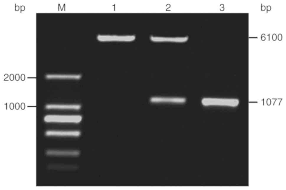
Identification of pCDGFP-C/EBPα
plasmid
The recombinant plasmid pCDGFP-C/EBPα was digested
using BamHI and EcoRI to produce two bright bands.
Comparison with the marker bands demonstrated that one band was the
vector, pCDGFP (6,100 bp), and the other was the C/EBPα fragment
(1,077 bp), indicating that the plasmid was successfully ligated.
The subsequent sequencing results further verified the construct.
The enzyme digestion results are displayed in Fig. 1. The expression level of C/EBPα in the
transfected cells was detected using RT-qPCR. The relative
expression level of C/EBPα was increased by >200-fold compared
with the control group (238:1). This result indicated that C/EBPα
was successfully expressed after transfection.
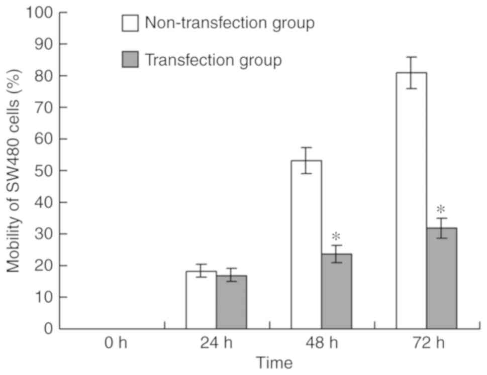
Impacts of overexpressed C/EBPα on the
mobility of SW480 cells
At 24 h in the wound-healing assay, the cell
mobility rate did not significantly differ between the
non-transfection and transfection groups (P>0.05),
whereas at 48 and 72 h, the cell mobility rates were significantly
different (P<0.05), indicating the overexpression of C/EBPα
significantly reduced the mobility of SW480 cells (Fig. 2).
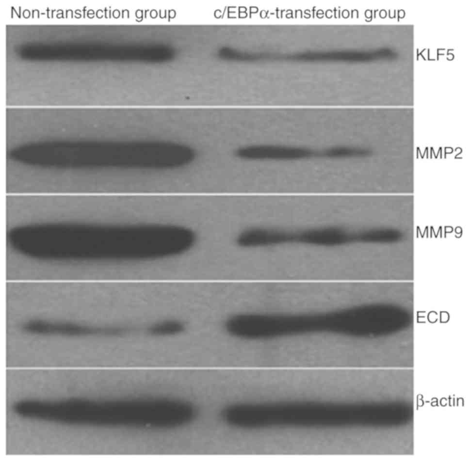
Impacts of overexpressed C/EBPα on
tumor invasion-associated proteins
At 48 h after plasmid transfection, western blotting
analysis was applied to determine the effect of C/EBPα
overexpression on the expression of tumor invasion-associated
proteins, including KLF5, MMP-2, MMP-9, and ECD. The results
revealed that, compared with the non-transfection group, the
protein expression levels of KLF5, MMP-2 and MMP-9 in the
C/EBPα-overexpression group were decreased, whereas the level of
ECD was increased. The results demonstrated that the overexpression
of C/EBPα reduced the expression of tumor invasion-promoting
proteins, thus potentially inhibiting tumor invasion (Fig. 3).
Discussion
Tumor invasion and metastasis are processes in which
cancer cells leave the primary lesion, invade surrounding tissue
and continue to proliferate, ultimately resulting in the formation
of new tumor lesions. This is the most critical biological property
of malignant tumors. The C/EBPs are a family of transcription
factors involved in the regulation of embryonic gut development in
rodents that have also been detected in various types of
malignancy; for example, Rask et al (12) identified that the expression of C/EBPβ
was increased in all the assessed CC tumor samples compared with
normal colon mucosa samples. Although the inter-patient variability
was large, it was identified that liver-enriched inhibitory protein
(LIP), the isoform of C/EBPβ associated with the inhibition of
transcription, was expressed at higher levels in Duke's stage B
tumors compared with stage A, whereas stage C tumors exhibited the
lowest LIP expression (12). However,
in the present study, it was identified that the expression of the
C/EBPα subtype in CC tissues was significantly reduced relative to
in the adjacent tissue. Thus, we hypothesize that C/EBPα may be a
tumor suppressor in CC, whereas C/EBPβ may increase CC
invasiveness. This result is similar to other research in solid
tumors; Yong et al (13)
concluded that C/EBPα is a tumor suppressor in lung cancer, and
that BMI1 is required for the oncogenic process downstream of
C/EBPα loss.
The present study identified that C/EBPα was
downregulated in CC, and was associated with tumor size, the TNM
stage and the lymph node metastasis status, suggesting that the low
expression of C/EBPα may affect the invasion and metastasis of CC.
When observing the effect of C/EBPα expression on the invasiveness
of SW480 CC cells, it was identified that the SW480 cells with
C/EBPα overexpression exhibited significantly decreased
invasiveness compared with untransfected cells. In addition, in
SW480 cells with overexpressed C/EBPα, the expression of KLF5,
MMP-2 and MMP-9 were reduced, whereas ECD was increased; this
result was consistent with the patient data analysis. Previous
studies have reported that the upregulation of KLF5, MMP-2 and
MMP-9, and the low expression of ECD, promote the invasiveness of
CC (14–18), which is consistent with the results of
the present study.
C/EBPα has been demonstrated to inhibit cell growth
via the direct repression of E2F-DP-mediated transcription
(19). C/EBPα has also been observed
to arrest cell proliferation through the direct inhibition of
cyclin-dependent kinases 2 and 4 (20,21).
However, the detailed mechanism for the C/EBPα-mediated inhibition
of invasion-associated protein expression remains unclear, and
should be considered in further research. In addition, the present
study did not compare the pairs of CC tumor and adjacent tissues
directly, so this is a limitation of the study that will be
addressed in further research.
In summary, C/EBPα expression was associated with
the occurrence and development of CC.
Acknowledgements
Not applicable.
Funding
The present study was supported by the Projects of
Science and Research Foundation of Educational Department of Hunan
(grant no. 13C831).
Availability of data and materials
The analyzed datasets generated during the study are
available from the corresponding author, on reasonable request.
Authors' contributions
XJZ and LFL designed the experiment. XZL, LFL and SW
reviewed the protocol. XJZ and LFL conducted the experiment. WL and
LJJ wrote the manuscript. WL and LJJ performed statistical
analysis. XZL and SW reviewed the manuscript.
Ethics approval and consent to
participate
The study was conducted in accordance with the
declaration of Helsinki, and was approved by the Ethics Committee
of the University of South China. Written informed consent was
obtained from all participants.
Patient consent for publication
Not applicable.
Competing interests
The authors declare that they have no competing
interests.
References
|
1
|
Siegel RL, Miller KD and Jemal A: Cancer
Statistics, 2016. CA Cancer J Clin. 66:7–30. 2016. View Article : Google Scholar : PubMed/NCBI
|
|
2
|
Shaukat A, Mongin SJ, Geisser MS, Lederle
FA, Bond JH, Mandel JS and Church TR: Long-term mortality after
screening for colorectal cancer. N Engl J Med. 369:1106–1114. 2013.
View Article : Google Scholar : PubMed/NCBI
|
|
3
|
Eichhorn JM, Alford SE, Sakurikar N and
Chambers TC: Molecular analysis of functional redundancy among
anti-apoptotic Bcl-2 proteins and its role in cancer cell survival.
Exp Cell Res. 322:415–424. 2014. View Article : Google Scholar : PubMed/NCBI
|
|
4
|
Cast A, Valanejad L, Wright M, Nguyen P,
Gupta A, Zhu L, Shin S and Timchenko N: C/EBPα-dependent
pre-neoplastic tumor foci are the origin of hepatocellular
carcinoma and aggressive pediatric liver cancer. Hepatology.
67:1857–1871. 2018. View Article : Google Scholar : PubMed/NCBI
|
|
5
|
Tan EH, Hooi SC, Laban M, Wong E, Ponniah
S, Wee A and Wang ND: CCAAT/enhancer binding protein alpha knock in
mice exhibit early liver glycogen storage and reduced
susceptibility to hepatocellular carcinoma. Cancer Res.
65:10330–10337. 2005. View Article : Google Scholar : PubMed/NCBI
|
|
6
|
Chen Y, Costa RM, Love NR, Soto X, Roth M,
Paredes R and Amaya E: C/EBPalpha initiates primitive myelopoiesis
in pluripotent embryonic cells. Blood. 114:40–48. 2009. View Article : Google Scholar : PubMed/NCBI
|
|
7
|
McFie PJ, Wang GL, Timchenko NA, Wilson
HL, Hu X and Roesler WJ: Identification of a co-repressor that
inhibits the transcriptional and growth-arrest activities of
CCAAT/enhancer binding protein alpha. J Biol Chem. 281:18069–18080.
2006. View Article : Google Scholar : PubMed/NCBI
|
|
8
|
Fukuchi Y, Shibata F, Ito M, Goto-Koshino
Y, Sotomaru Y, Ito M, Kitamura T and Nakajima H: Comprehensive
analysis of myeloid lineage conversion using mice expressing an
inducible form of C/EBP alpha. EMBO J. 25:3398–3410. 2006.
View Article : Google Scholar : PubMed/NCBI
|
|
9
|
Reddy SD, Pakala SB, Ohshiro K, Rayala SK
and Kumar R: MicroRNA-661, a c/EBPα target, inhibits metastatic
tumor antigen 1 and regulates its functions. Cancer Res.
69:5639–5642. 2009. View Article : Google Scholar : PubMed/NCBI
|
|
10
|
Benson AB III, Venook AP, Cederquist L,
Chan E, Chen YJ, Cooper HS, Deming D, Engstrom PF, Enzinger PC,
Fichera A, et al: Colon Cancer, Version 1.2017, NCCN clinical
practice guidelines in oncology. J Natl Compr Canc Netw.
15:370–398. 2017. View Article : Google Scholar : PubMed/NCBI
|
|
11
|
Mestdagh P, Feys T, Bernard N, Guenther S,
Chen C, Speleman F and Vandesompele J: High-throughput stem-loop
RT-qPCR miRNA expression profiling using minute amounts of input
RNA. Nucleic Acids Res. 36:e1432008. View Article : Google Scholar : PubMed/NCBI
|
|
12
|
Rask K, Thörn M, Pontén F, Kraaz W,
Sundfeldt K, Hedin L and Enerbäck S: Increased expression of the
transcription factors CCAAT-enhancer binding protein-beta
(C/EBBeta) and C/EBzeta (CHOP) correlate with invasiveness of human
colorectal cancer. Int J Cancer. 86:337–343. 2000. View Article : Google Scholar : PubMed/NCBI
|
|
13
|
Yong KJ, Basseres DS, Welner RS, Zhang WC,
Yang H, Yan B, Alberich-Jorda M, Zhang J, de Figueiredo-Pontes LL,
Battelli C, et al: Targeted BMI1 inhibition impairs tumor growth in
lung adenocarcinomas with low CEBPα expression. Sci Transl Med.
8:350ra1042016. View Article : Google Scholar : PubMed/NCBI
|
|
14
|
Basu N, Saha S, Khan I, Ramachandra SG and
Visweswariah SS: Intestinal cell proliferation and senescence are
regulated by receptor guanylyl cyclase C and p21. J Biol Chem.
289:581–593. 2014. View Article : Google Scholar : PubMed/NCBI
|
|
15
|
Zhen C, Chen L, Zhao Q, Liang B, Gu YX,
Bai ZF, Wang K, Xu X, Han QY, Fang DF, et al: Gankyin promote
breast cancer cell metastasis by regulating Racl activity.
Oncogene. 32:3452–3460. 2013. View Article : Google Scholar : PubMed/NCBI
|
|
16
|
Geng L, Sun B, Gao B, Wang Z, Quan C, Wei
F and Fang XD: MicroRNA-103 promotes colorectal cancer by targeting
tumor suppressor DICER and PTEN. Int J Mol Sci. 15:8458–8472. 2014.
View Article : Google Scholar : PubMed/NCBI
|
|
17
|
Chen J, Zheng B, Wang C, Chen Y, Du C,
Zhao G, Zhou Y and Shi Y: Prognostic role of microRNA-100 in
various carcinomas: Evidence from six studies. Tumour Biol.
35:3067–3071. 2014. View Article : Google Scholar : PubMed/NCBI
|
|
18
|
Liu Y, Sheng J, Dai D, Liu T and Qi F:
Smad4 acts as tumor suppressor by antagonizing lmphangiogenesis in
colorectal cancer. Pathol Res Pract. 211:286–292. 2015. View Article : Google Scholar : PubMed/NCBI
|
|
19
|
Zaragoza K, Bégay V, Schuetz A, Heinemann
U and Leutz A: Repression of transcriptional activity of C/EBPalpha
by E2F-dimerization partner complexes. Mol Cell Biol. 30:2293–2304.
2010. View Article : Google Scholar : PubMed/NCBI
|
|
20
|
Wang GL, Shi X, Salisbury E, Sun Y,
Albrecht JH, Smith RG and Timchenko NA: Cyclin D3 maintains
growth-inhibitory activity of C/EBPalpha by stabilizing
C/EBPalpha-cdk2 and C/EBPalpha-Brm complexes. Mol Cell Biol.
26:2570–2582. 2006. View Article : Google Scholar : PubMed/NCBI
|
|
21
|
Wang H, Iakova P, Wilde M, Welm A, Goode
T, Roesler WJ and Timchenko NA: C/EBPalpha arrests cell
proliferation through direct inhibition of Cdk2 and Cdk4. Mol Cell.
8:817–828. 2001. View Article : Google Scholar : PubMed/NCBI
|