Introduction
Rectal cancer is an increasing, and important
disease worldwide (1). For patients
with American Joint Committee on Cancer (AJCC) stage II or III
disease, neoadjuvant concurrent chemoradiotherapy (CCRT) followed
by radical proctectomy remains the preferred therapy as it has been
shown to provide better local control, greater anal preservation,
and lesser toxicity (2). However, up
to 10–20% of these patients will eventually develop local or distal
disease recurrence (3).
Rectal cancer post neoadjuvant CCRT or not are all
staged by using the AJCC tumor-node-metastasis (TNM) staging
system, and further adjuvant treatment is recommended. There is no
modification of staging for patients who receive neoadjuvant CCRT.
Prediction of prognosis using TNM staging is suboptimal, and
survival of patients with the same stage disease can vary markedly.
Therefore, additional prognostic biomarkers to aid in risk
stratification and identify patients who may benefit from more
intensive treatment may help to improve outcomes.
Epidermal growth factor receptor (EGFR) is a
transmembrane glycoprotein that belongs to the HER family of
receptor tyrosine kinases, and promotes multiple signaling cascades
for cellular survival, such as RAS/MAPK, PI3K/Akt, Stat, and Src
(4). Overexpression of cytoplasmic
EGFR (EGFR-C) is associated with tumor differentiation,
proliferation, and migration, and has been extensively studied as
the most important cancer molecular target in recent decades
(5,6).
Recently, studies have reported that EGFR family members can be
transported from the plasma membrane to the nucleus, and regulate
target genes involved in cell proliferation and angiogenesis
(7,8).
The prognostic significance of nuclear EGFR (EGFR-N) has been
demonstrated, and is highly associated with tumor progression and
worse overall survival (OS) (9,10). It has
also been shown to be associated with enhanced resistance to
systemic and radiation therapy (7).
Therefore, overexpression of EGFR-N is frequently a poor prognostic
factor. However, little is known about the relevance of EGFR-N in
rectal cancer patients treated with neoadjuvant CCRT.
The aim of this study was to evaluate the clinical
correlates of EGFR-C and EGFR-N expression in rectal cancer
patients after neoadjuvant CCRT, and also to compare the impact of
EGFR-N and EGFR-C expression on prognosis.
Materials and methods
Ethics statement
The present study was reviewed and approved by the
Institutional Review Board of Chi-Mei Medical Center in Taiwan
(Tainan, Taiwan; IRB, CMFHR10501-008). The requirement for informed
consent was waived as all identifying information was removed from
the dataset prior to analysis.
Patient demographic characteristics
and tumor specimens
This retrospective study was performed on
formalin-fixed paraffin-embedded (FFPE) tissue specimens from 172
newly diagnosed rectal adenocarcinoma patients treated at Chi-Mei
Medical Center between 1998 and 2004. The data was collected and
analyzed in February 2014. All patients received preoperative
5-fluorouracil-based chemotherapy concomitant with radiotherapy
(45–50 Gy), followed by radical proctectomy with total mesorectal
excision. The clinical stage was determined by endoscopic
ultrasound (EUS), abdominal computed tomography (CT), or magnetic
resonance imaging (MRI) findings. Adjuvant systemic chemotherapy
was given if the post-treatment (Post-Tx) tumor or nodal status was
beyond T3 or N1. All patients were regularly monitored after
diagnosis until death or last follow-up. Patients who had a
previous cancer history, distal metastasis at diagnosis, or were
unable to complete a full course of CCRT were excluded. The
histopathological parameters were reviewed by our pathologist (Dr.
Li) who was blind to the patients' clinical data.
The data extracted from the medical records included
the date of diagnosis, age, sex, clinical TNM stage, pathological
characteristics such as lymphovascular and perineural invasion,
chemotherapy regimen and timing, and cause of death. All patients
were re-staged and re-graded according to the 7th edition of the
AJCC staging system, and World Health Organization (WHO)
classification of Tumors of the Colon and Rectum. The primary
endpoints were 5-year disease-specific survival (DSS), local
recurrent-free survival (LRFS), and metastases-free survival (MeFS)
rates. Deaths due to cancer were defined as valid events, and
deaths secondary to other causes were censored.
Immunohistochemistry analysis
Immunohistochemistry of tissue microarray (TMA)
slides and whole sections were prepared as previously described
(11). Briefly, tissue sections from
Pre-Tx rectal tumor biopsies were deparaffinized and rehydrated for
EGFR immunostaining. Overexpression of EGFR-C was defined as an
intensity of 3+ EGFR reactivity seen in the cytoplasm (and/or
membrane) of tumor cells (12). To
define overexpression, the cutoff percentage of immunoreactive
tumor cells was 50% for EGFR-N in this analysis (13).
Statistical analysis
The results are presented as the mean ± standard
deviation. All statistical analyses and graphics were performed
using IBM SPSS statistical software for Windows, v14.0 (SPSS, Inc.,
Chicago, IL, USA). P<0.05 was considered to indicate a
statistically significant difference. Examination of the
associations of EGFR expression with clinicopathological features
was conducted using the chi-square test. The Kaplan-Meier method
was used to analyze the cumulative 5-year DSS, LRFS, and MeFS
rates, and rates were compared using the log-rank test to evaluate
prognostic differences between subgroups. The primary event was
defined as disease-specific mortality, and survival curves were
calculated from the date of diagnosis. Those parameters that
demonstrated prognostic significance in the univariate analysis
were input into the Cox multivariate regression analysis.
Results
Patient demographic and clinical characteristics are
summarized in Table I. A total of 172
rectal cancer patients who underwent neoadjuvant CCRT and radical
proctectomy with total mesorectal excision from January 1998 to
December 2008 were included. The median age was 63 years (range:
22–88 years). The percentages of patients with initial stage I, II,
and III disease were 41.9, 29.9, and 28.1% and all stage II, III
patients received adjuvant chemotherapy Fifteen (8.7%) patients
presented with lymphovascular invasion, and 5 (2.9%) with
perineural invasion.
 | Table I.Associations and comparisons between
Nuclear/Cytoplasmic epidermal growth factor receptor expression and
clinicopathological factors in 172 rectal cancer patients. |
Table I.
Associations and comparisons between
Nuclear/Cytoplasmic epidermal growth factor receptor expression and
clinicopathological factors in 172 rectal cancer patients.
|
|
|
| EGFR-N (n) |
| EGFR-C (n) |
|
|---|
|
|
|
|
|
|
|
|
|---|
| Parameter | Variable | No. of cases | Low Exp. | High Exp. | P-value | Low Exp. | High Exp. | P-value |
|---|
| Gender | Male | 108 | 84 | 24 | 0.0768 | 82 | 26 | 0.741 |
|
| Female | 64 | 51 | 13 |
| 50 | 14 |
|
| Age | <60 | 65 | 51 | 14 | 0.995 | 49 | 16 | 0.742 |
|
| ≥60 | 107 | 84 | 23 |
| 83 | 24 |
|
| Pre-Tx tumor status
(Pre-T) | T1-T2 | 81 | 70 | 11 | 0.017a | 69 | 12 | 0.013a |
|
| T3-T4 | 91 | 65 | 26 |
| 63 | 28 |
|
| Pre-Tx nodal status
(Pre-N) | N0 | 125 | 101 | 24 | 0.229 | 103 | 22 | 0.004b |
|
| N1-N2 | 47 | 34 | 13 |
| 29 | 18 |
|
| Post-Tx tumor
status (Post-T) | T1-T2 | 86 | 77 | 9 |
<0.001c | 71 | 15 | 0.071 |
|
| T3-T4 | 86 | 58 | 28 |
| 65 | 25 |
|
| Post-Tx nodal
status (Post-N) | N0 | 123 | 99 | 24 | 0.312 | 99 | 24 | 0.066 |
|
| N1-N2 | 49 | 36 | 13 |
| 33 | 16 |
|
| Lymphovascular
invasion | Absent | 157 | 123 | 34 | 0.881 | 124 | 33 | 0.025a |
|
| Present | 15 | 12 | 3 |
| 8 | 7 |
|
| Perineural
invasion | Absent | 167 | 131 | 36 | 0.933 | 130 | 37 | 0.048a |
|
| Present | 5 | 4 | 1 |
| 2 | 3 |
|
| Cytoplasmic EGFR
expression | Low Exp. | 132 | 105 | 27 | 0.540 | n/a | n/a | n/a |
|
| High Exp. | 40 | 30 | 10 |
| n/a | n/a |
|
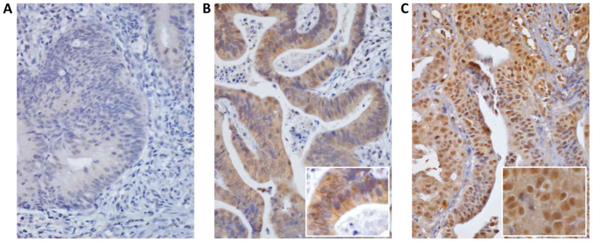
The results of EGFR immunostaining to investigate
EGFR expression in the cytoplasm and the nucleus of rectal tumor
tissues are shown in Fig. 1. Of the
patients, 20.9% had EGFR-N overexpression, 23.3% EGFR-C
overexpression and 5.2% have both high EGFR-N and high EGFR-C
expression. EGFR-N overexpression was significantly related to
advanced pre-Tx tumor T-stage (T3, 4; P=0.017) and post-Tx tumor
T-stage (T3, 4; P<0.001). EGFR-C overexpression was
significantly related to pre-Tx tumor T-stage (T3, 4; P=0.013),
post-Tx tumor T-stage (T3, 4; P=0.004), lymphovascular invasion
(P=0.025), and perineural invasion (P=0.048).
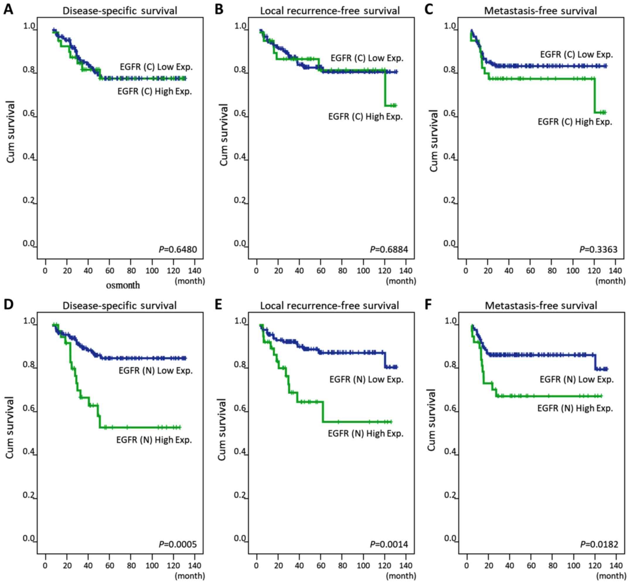
Univariate and multivariate survival
analyses for factors predicting survival
In univariate analyses (Table II), post-Tx tumor status and nuclear
EGFR expression were significantly correlated with poorer 5-year
DSS, LRFS, and MeFS rate (all, P<0.05).
| Table II.Univariate log-rank analysis for
important clinicopathological variables and epidermal growth factor
receptor expression. |
Table II.
Univariate log-rank analysis for
important clinicopathological variables and epidermal growth factor
receptor expression.
|
|
|
| DSS | LRFS | MeFS |
|---|
|
|
|
|
|
|
|
|---|
| Parameter | Variable | No. of cases | No. of events | P-value | No. of events | P-value | No. of events | P-value |
| Gender | Male | 108 | 20 | 0.6027 | 5 | 0.3096 | 14 | 0.1047 |
|
| Female | 64 | 11 |
| 17 |
| 15 |
|
| Age | <60 | 65 | 13 | 0.3259 | 7 | 0.7575 | 17 | 0.0052b |
|
| ≥60 | 107 | 18 |
| 15 |
| 12 |
|
| Pre-Tx tumor status
(Pre-T) | T1-T2 | 81 | 10 | 0.0484a | 7 | 0.0836 | 10 | 0.1288 |
|
| T3-T4 | 91 | 21 |
| 15 |
| 19 |
|
| Pre-Tx nodal status
(Pre-N) | N0 | 125 | 19 | 0.0059b | 12 | 0.0025b | 18 | 0.0866 |
|
| N1-N2 | 47 | 21 |
| 10 |
| 11 |
|
| Post-Tx tumor
status (Post-T) | T1-T2 | 86 | 12 | 0.0006c | 5 | 0.0056b | 8 | 0.0123a |
|
| T3-T4 | 86 | 34 |
| 17 |
| 21 |
|
| Post-Tx nodal
status (Post-N) | N0 | 123 | 31 | 0.3442 | 15 | 0.6267 | 20 | 0.8403 |
|
| N1-N2 | 49 | 15 |
| 7 |
| 9 |
|
| Lymphovascular
invasion | Absent | 157 | 38 | 0.0107a | 17 | 0.0023b | 26 | 0.7236 |
|
| Present | 15 | 8 |
| 5 |
| 3 |
|
| Perineural
invasion | Absent | 167 | 43 | 0.0297a | 20 | 0.0083b | 28 | 0.8157 |
|
| Present | 5 | 3 |
| 2 |
| 1 |
|
| Cytoplasmic EGFR
expression | Low Exp. | 132 | 33 | 0.6480 | 16 | 0.6884 | 20 | 0.3363 |
|
| High Exp. | 40 | 13 |
| 6 |
| 9 |
|
| Nuclear EGFR
expression | Low Exp. | 135 | 28 | 0.0005c | 12 | 0.0014b | 18 | 0.0182a |
|
| High Exp. | 37 | 18 |
| 10 |
| 11 |
|
Pre-Tx nodal status, lymphovascular invasion, and
perineural invasion were negatively associated with 5-year DSS and
LRFS (all, P<0.05). Pre-Tx tumor status was significantly
associated with poorer 5-year DSS only (P=0.0484). Multivariate
analyses results are shown in Table
III. EGFR-N, but not EGFR-C, was an independent prognostic
factor for 5-year DSS (hazard ratio [HR]=2.755, 95% confidence
interval [CI]: 1.313–5.783) and LRFS (HR=3.026, 95% CI:
1.224–7.281), but not for MeFS (HR=1.853, 95% CI: 0.852–4.032).
Pre-Tx nodal status and post-Tx tumor status were independent
prognostic factors for 5-year DSS only (HR=2.640, 95% CI:
1.317–5.290; HR=2.210, 95% CI: 1.084–4.503, respectively).
| Table III.Multivariate analysis. |
Table III.
Multivariate analysis.
|
|
| DSS | LRFS | MeFS |
|---|
|
|
|
|
|
|
|---|
| Parameter | Category | HR | 95% CI | P-value | HR | 95% CI | P-value | HR | 95% CI | P-value |
|---|
| EGFR-N | Low Exp. | 1 | – | 0.0061b | 1 | – | 0.0164a | 1 | – | 0.1200 |
|
| High Exp. | 2.418 | 1.287–4.543 | | 3.026 | 1.224–7.281 |
| 1.853 | 0.852–4.032 |
|
| Pre-Tx nodal status
(Pre-N) | N0 | 1 | – | 0.0062b | 1 | – | 0.0948 | – | – | – |
|
| N1-N2 | 2.640 | 1.317–5.290 | | 2.308 | 0.865–6.157 |
| – | – | – |
| Post-Tx tumor
status (Post-T) | T1-T2 | 1 | – | 0.0291a | 1 | – | 0.1217 | 1 | – | 0.0587 |
|
| T3-T4 | 2.210 | 1.084–4.503 | | 2.305 | 0.800–6.637 |
| 2.257 | 0.971–5.247 |
|
| Age, years | ≥60 | 1 | – | 0.0203a | – | – | – | 1 | – | 0.0112a |
|
| <60 | 0.205 | 1.183–3.765 | | – | – |
| 2.608 | 1.243–5.469 |
|
| Lymphovascular
invasion | Absent | 1 | – | 0.0651 | 1 | – | 0.0683 | – | – | – |
|
| Present | 2.213 | 0.951–5.149 | | 2.780 | 0.926–8.345 |
| – | – |
|
| Pre-Tx tumor status
(Pre-T) | T1-T2 | 1 |
| 0.8048 | – | – | – | – | – | – |
|
| T3-T4 | 1.091 | 0.460–1.824 | | – | – |
| – | – |
|
| Perineural
invasion | Absent | 1 |
| 0.8397 | 1 | – | 0.4012 | – | – | – |
|
| Present | 1.145 | 0.236–3.249 | | 1.999 | 0.397–10.075 |
| – | – |
|
EGFR-N overexpression as an
independent prognostic factor
Overexpression of EGFR-N, but not EGFR-C, was
significantly correlated with poor DSS, LRFS, and MeFS in
univariate analyses (all, P<0.05) (Table II; Fig.
2). Higher expression of EGFR-N remained an independent
predictor of worse DSS and LRFS in multivariate analyses, as shown
in Table III (HR=2.755, 95% CI:
1.3137–5.783; HR=3.026, 95% CI: 1.224–7.281, respectively).
Discussion
There have been great advances in the treatment of
rectal cancer in recent decades, and neoadjuvant CCRT followed by
radical proctectomy has become primary treatment. However, the
long-term survival of rectal cancer patients has remained stagnant.
Therefore, it is necessary to find effective predictive biomarkers
to stratify high-risk patients for more intensive therapy. In this
study, we demonstrated that high expression of EGFR-N, but not
EGFR-C, was correlated with aggressive tumor behavior and poor
outcome. These results suggest that EGFR-N may be useful for
outcome prediction in rectal cancer patients after CCRT, and may
possibly be a potential therapeutic target.
Molecular methods usually provide reliable
information for cancer patients. However, studies that have
evaluated prognostic biological markers for rectal cancer have
yielded inconclusive results (14,15). A
variety of studies have suggested that overexpression of EGFR was
positively correlated with aggressive tumor behavior and poor
prognosis in colorectal cancer (12,16,17). But,
there was considerable discrepancy in the frequency and
distribution of EGFR expression in previous immunohistochemical
studies (18,19). Many of these studies provided
speculative information on the association of protein expression
and clinicopathological features. Furthermore, in clinical trials
evaluating the efficacy of cetuximab, treatment response was not
related to the level of EGFR expression; many patients with
colorectal cancer expressing EGFR failed to respond to treatment,
and those with EGFR negative tumors responded to therapy. A
possible reason that EGFR levels are a poor predictor of response
to anti-EGFR therapies is disparity between the form or epitope of
EGFR detected by immunohistochemistry and the one targeted by
anti-EGFR monoclonal antibodies (7).
Recent evidence has suggested that EGFR can
translocate to the nucleus (7). After
activation, the nuclear translocation of EGFR has been shown to be
dependent on phosphorylation events by many intracellular kinases
such as SRC family and AKT (9). The
nuclear transport of EGFR is guided by COPI-mediated vesicular
trafficking from the Golgi to the endoplasmic reticulum (ER)
(20). After being shuttled into the
ER membrane, EGFR and importin β1 interface in the nuclear pore
complex, and shuttle EGFR from the outer to the inner nuclear
membrane. Once in the nucleus, the function of EGFR is widely
different from its plasma membrane bound counterpart. EGFR-N is
involved in many cellular signaling pathways such as cell
proliferation, tumor behavior, DNA synthesis, and DNA repair
(21–24). EGFR-N also plays a functional role in
the response to molecular therapeutic agents, and has been linked
to poor prognosis in a variety of malignancies (9,10,25). Hadzisejdic et al (10), reported that higher EGFR-N staining
was associated with shorter OS in breast cancer patients.
Overexpression of EGFR-N carried a 3.4-times greater mortality risk
as compared to EGFR-N negative patients (HR=3.402; P=0.0026).
Hoshino et al (25), also
demonstrated that EGFR-N was associated with an increase in the
malignant potential of esophageal squamous cell carcinoma that may
affect the prognosis these patients. These intriguing findings
emphasize the relevance of evaluating both EGFR-C and EGFR-N
expression in colorectal cancer in order to provide additional
independent prognostic information.
Our results showed that EGFR-N overexpression was
significantly related to advanced pre-Tx tumor T-stage (T3, 4;
P=0.017) and post-Tx tumor T-stage (T3, 4; P<0.001). In
multivariate survival analysis, EGFR-N expression was also
correlated with worse 5-year DSS and LRFS. These findings are
different than those of prior studies where EGFR-C was associated
with aggressive tumor behavior and poor outcomes (17,19).
Similar to our study, Doger et al (16) evaluated 60 colon tumor specimens and
reported that EGFR-C expression did not predict lymph nodes
metastasis or survival. Moreover, EGFR-N overexpression was
significantly associated with worse 5-year DSS (HR=2.755, P=0.007)
and LRFS (HR=3.026, P=0.0164), but not MeFS (HR=1.85, P=0.12). The
possible reasons for these findings may be that nuclear
translocation of EGFR decreases cell survival, and is associated
with enhanced resistance to radiation and chemotherapy (7). EGFR-N is correlated with increased
DNA-dependent protein kinase (DNA-PK) activity, which plays an
essential role in DNA damage repair and radioresistance (26). Thus, the overexpression of EGFR-N in
rectal cancers could contribute to the identification of patients
who have an increased risk of local recurrences and poor outcomes
after neoadjuvant CCRT.
The present study has some limitations. First, the
number of patients was relatively small, and the findings should be
verified by larger-scale studies. Second, our study cohort included
some early-stage rectal cancer patients because of the intention of
organ preservation via neoadjuvant CCRT. Further studies focused on
locally advanced rectal cancer should be performed. Finally, our
results are applicable to only patients with non-metastatic rectal
cancer who undergo CCRT.
In summary, the present study revealed that
overexpression of EGFR-N, but not EGFR-C, may help identify and
stratify high-risk patients after neoadjuvant CCRT. The strong
inverse correlation with DFS and LFRS suggested that EGFR-N may be
a potential prognostic biomarker and promising therapeutic
target.
Acknowledgements
Not applicable.
Funding
The study was supported from the Health and Welfare
surcharge of tobacco products (MOHW107-TDU-B-212-114020, WanFang
Hospital, Chi-Mei Medical Center, and Hualien Tzu-Chi Hospital
Joing Cancer Center Grant-Focus on Colon Cancer Research).
Availability of data and materials
The datasets used and/or analyzed during the present
study are available from the corresponding author on reasonable
request.
Authors' contributions
CY, LL, YL, YT, CYL, MS, CFL and MT conceived and
designed the experiments. CY, LL and CFL participated in the design
of the study and performed the statistical analysis. CY, LL, CFL
and MT drafted the manuscript. All authors read and approved the
final manuscript.
Ethics approval and consent to
participate
The present study was reviewed and approved by the
Institutional Review Board of Chi-Mei Medical Center in Taiwan
(Tainan, Taiwan; IRB, CMFHR10501-008). The requirement for informed
consent was waived as all identifying information was removed from
the dataset prior to analysis.
Patient consent for publication
Not applicable.
Competing interests
The authors declare that they have no competing
interests.
Glossary
Abbreviations
Abbreviations:
|
CCRT
|
concurrent chemoradiotherapy
|
|
AJCC
|
American Joint Committee on Cancer
|
|
TNM
|
tumor-node-metastasis
|
|
EGFR
|
epidermal growth factor receptor
|
|
EGFR-C
|
cytoplasmic EGFR
|
|
EGFR-N
|
nuclear EGFR
|
|
Pre-Tx
|
pre-treatment
|
|
Post-Tx
|
post-treatment
|
|
DSS
|
disease-specific survival
|
|
LRFS
|
local-recurrence-free survival
|
|
MeFS
|
metastases-free survival
|
|
HR
|
hazard ratio
|
|
CI
|
confidence interval
|
References
|
1
|
Siegel RL, Miller KD and Jemal A: Cancer
statistics, 2016. CA Cancer J Clin. 66:7–30. 2016. View Article : Google Scholar : PubMed/NCBI
|
|
2
|
Sauer R, Becker H, Hohenberger W, Rödel C,
Wittekind C, Fietkau R, Martus P, Tschmelitsch J, Hager E, Hess CF,
et al: Preoperative versus postoperative chemoradiotherapy for
rectal cancer. N Engl J Med. 351:1731–1740. 2004. View Article : Google Scholar : PubMed/NCBI
|
|
3
|
van den Brink M, Stiggelbout AM, van den
Hout WB, Kievit J, Klein Kranenbarg E, Marijnen CA, Nagtegaal ID,
Rutten HJ, Wiggers T and van de Velde CJ: Clinical nature and
prognosis of locally recurrent rectal cancer after total mesorectal
excision with or without preoperative radiotherapy. J Clin Oncol.
22:3958–3964. 2004. View Article : Google Scholar : PubMed/NCBI
|
|
4
|
Yarden Y and Sliwkowski MX: Untangling the
ErbB signalling network. Nat Rev Mol Cell Biol. 2:127–137. 2001.
View Article : Google Scholar : PubMed/NCBI
|
|
5
|
Navolanic PM, Steelman LS and McCubrey JA:
EGFR family signaling and its association with breast cancer
development and resistance to chemotherapy (Review). Int J Oncol.
22:237–252. 2003.PubMed/NCBI
|
|
6
|
Zhang YL, Yuan JQ, Wang KF, Fu XH, Han XR,
Threapleton D, Yang ZY, Mao C and Tang JL: The prevalence of EGFR
mutation in patients with non-small cell lung cancer: A systematic
review and meta-analysis. Oncotarget. 7:78985–78993.
2016.PubMed/NCBI
|
|
7
|
Brand TM, Iida M, Luthar N, Starr MM,
Huppert EJ and Wheeler DL: Nuclear EGFR as a molecular target in
cancer. Radiother Oncol. 108:370–377. 2013. View Article : Google Scholar : PubMed/NCBI
|
|
8
|
Lo HW, Ali-Seyed M, Wu Y, Bartholomeusz G,
Hsu SC and Hung MC: Nuclear-cytoplasmic transport of EGFR involves
receptor endocytosis, importin beta1 and CRM1. J Cell Biochem.
98:1570–1583. 2006. View Article : Google Scholar : PubMed/NCBI
|
|
9
|
Traynor AM, Weigel TL, Oettel KR, Yang DT,
Zhang C, Kim K, Salgia R, Iida M, Brand TM, Hoang T, et al: Nuclear
EGFR protein expression predicts poor survival in early stage
non-small cell lung cancer. Lung Cancer. 81:138–141. 2013.
View Article : Google Scholar : PubMed/NCBI
|
|
10
|
Hadzisejdić I, Mustać E, Jonjić N,
Petković M and Grahovac B: Nuclear EGFR in ductal invasive breast
cancer: Correlation with cyclin-D1 and prognosis. Mod Pathol.
23:392–403. 2010. View Article : Google Scholar : PubMed/NCBI
|
|
11
|
Yoo PS, Sullivan CA, Kiang S, Gao W, Uchio
EM, Chung GG and Cha CH: Tissue microarray analysis of 560 patients
with colorectal adenocarcinoma: High expression of HuR predicts
poor survival. Ann Surg Oncol. 16:200–207. 2009. View Article : Google Scholar : PubMed/NCBI
|
|
12
|
Richter I, Dvořák J, Urbanec M, Bluml A,
Čermáková E, Bartoš J and Petera J: The prognostic significance of
tumor epidermal growth factor receptor (EGFR) expression change
after neoadjuvant chemoradiation in patients with rectal
adenocarcinoma. Contemp Oncol (Pozn). 19:48–53. 2015.PubMed/NCBI
|
|
13
|
Li CF, Fang FM, Wang JM, Tzeng CC, Tai HC,
Wei YC, Li SH, Lee YT, Wang YH, Yu SC, et al: EGFR nuclear import
in gallbladder carcinoma: Nuclear phosphorylated EGFR upregulates
iNOS expression and confers independent prognostic impact. Ann Surg
Oncol. 19:443–454. 2012. View Article : Google Scholar : PubMed/NCBI
|
|
14
|
Molinari C, Matteucci F, Caroli P and
Passardi A: Biomarkers and molecular imaging as predictors of
response to neoadjuvant chemoradiotherapy in patients with locally
advanced rectal cancer. Clin Colorectal Cancer. 14:227–238. 2015.
View Article : Google Scholar : PubMed/NCBI
|
|
15
|
Smith FM, Reynolds JV, Miller N, Stephens
RB and Kennedy MJ: Pathological and molecular predictors of the
response of rectal cancer to neoadjuvant radiochemotherapy. Eur J
Surg Oncol. 32:55–64. 2006. View Article : Google Scholar : PubMed/NCBI
|
|
16
|
Doger FK, Meteoglu I, Tuncyurek P, Okyay P
and Cevikel H: Does the EGFR and VEGF expression predict the
prognosis in colon cancer? Eur Surg Res. 38:540–544. 2006.
View Article : Google Scholar : PubMed/NCBI
|
|
17
|
Rego RL, Foster NR, Smyrk TC, Le M,
O'Connell MJ, Sargent DJ, Windschitl H and Sinicrope FA: Prognostic
effect of activated EGFR expression in human colon carcinomas:
Comparison with EGFR status. Br J Cancer. 102:165–172. 2010.
View Article : Google Scholar : PubMed/NCBI
|
|
18
|
Garouniatis A, Zizi-Sermpetzoglou A, Rizos
S, Kostakis A, Nikiteas N and Papavassiliou AG: FAK, CD44v6, c-Met
and EGFR in colorectal cancer parameters: Tumour progression,
metastasis, patient survival and receptor crosstalk. Int J
Colorectal Dis. 28:9–18. 2013. View Article : Google Scholar : PubMed/NCBI
|
|
19
|
Spano JP, Lagorce C, Atlan D, Milano G,
Domont J, Benamouzig R, Attar A, Benichou J, Martin A, Morere JF,
et al: Impact of EGFR expression on colorectal cancer patient
prognosis and survival. Ann Oncol. 16:102–108. 2005. View Article : Google Scholar : PubMed/NCBI
|
|
20
|
Wang YN, Wang H, Yamaguchi H, Lee HJ, Lee
HH and Hung MC: COPI-mediated retrograde trafficking from the Golgi
to the ER regulates EGFR nuclear transport. Biochem Biophys Res
Commun. 399:498–504. 2010. View Article : Google Scholar : PubMed/NCBI
|
|
21
|
Lin SY, Makino K, Xia W, Matin A, Wen Y,
Kwong KY, Bourguignon L and Hung MC: Nuclear localization of EGF
receptor and its potential new role as a transcription factor. Nat
Cell Biol. 3:802–808. 2001. View Article : Google Scholar : PubMed/NCBI
|
|
22
|
Lo HW, Hsu SC, Ali-Seyed M, Gunduz M, Xia
W, Wei Y, Bartholomeusz G, Shih JY and Hung MC: Nuclear interaction
of EGFR and STAT3 in the activation of the iNOS/NO pathway. Cancer
Cell. 7:575–589. 2005. View Article : Google Scholar : PubMed/NCBI
|
|
23
|
Wang SC, Nakajima Y, Yu YL, Xia W, Chen
CT, Yang CC, McIntush EW, Li LY, Hawke DH, Kobayashi R and Hung MC:
Tyrosine phosphorylation controls PCNA function through protein
stability. Nat Cell Biol. 8:1359–1368. 2006. View Article : Google Scholar : PubMed/NCBI
|
|
24
|
Dittmann K, Mayer C, Fehrenbacher B,
Schaller M, Raju U, Milas L, Chen DJ, Kehlbach R and Rodemann HP:
Radiation-induced epidermal growth factor receptor nuclear import
is linked to activation of DNA-dependent protein kinase. J Biol
Chem. 280:31182–31189. 2005. View Article : Google Scholar : PubMed/NCBI
|
|
25
|
Hoshino M, Fukui H, Ono Y, Sekikawa A,
Ichikawa K, Tomita S, Imai Y, Imura J, Hiraishi H and Fujimori T:
Nuclear expression of phosphorylated EGFR is associated with poor
prognosis of patients with esophageal squamous cell carcinoma.
Pathobiology. 74:15–21. 2007. View Article : Google Scholar : PubMed/NCBI
|
|
26
|
Hsu SC, Miller SA, Wang Y and Hung MC:
Nuclear EGFR is required for cisplatin resistance and DNA repair.
Am J Transl Res. 1:249–258. 2009.PubMed/NCBI
|