Introduction
Endometrial carcinoma is the most common invasive
neoplasm of the female genital tract in the Western world, with a
rising incidence. Furthermore, endometrial carcinoma is a
significant contributor to gynecological mortality and the fourth
most common cancer in women after breast, colon and lung cancer.
Endometrial carcinoma primarily affects perimenopausal and
postmenopausal women at a median age of diagnosis of 60 years old.
Likely risk factors for this disease include diabetes, thyroid
disease, hypertension, postmenopausal status, nulliparity,
increased obesity, polycystic ovarian syndrome, early menarche and
late menopause, radiation exposure, long-term use of unopposed
exogenous estrogenic stimulation, a personal history of endometrial
hyperplasia or breast cancer, and a family history of endometrial
cancer (1–7).
Endometrial carcinoma is classified into two
clinicopathological types (type I and type II). Type I endometrial
carcinoma is the most common subtype, accounting for >80% of
endometrial tumors, and typically has a favorable prognosis. They
are usually low-grade, well-differentiated endometrioid
adenocarcinomas. These tumors are pathogenetically linked to an
excess of unopposed estrogen, arise from endometrial hyperplasia
and have hormone-receptor positivity. However, type II endometrial
carcinoma is a less common type of serous or clear cell
adenocarcinoma, accounting for only ~10% of endometrial tumors.
They are poorly differentiated, estrogen-independent tumors, which
are associated with atrophic endometrium and have poorer outcomes
(8,9). Endometrial carcinoma is believed to
arise from a variety of genetic alterations involving signaling
pathways, activation of proto-oncogenes and inactivation of tumor
suppressor genes. The development and progression of each group of
endometrial carcinoma follows distinct molecular mechanisms of
oncogenesis, reflecting the presence of type-specific genetic
alterations. Although there are well-established surgical, radio-
and chemotherapeutic treatments, the identification and
characterization of biomarkers is necessary for improving the
understanding of molecular pathways of the disease and for the
development of specific novel molecular targeted therapies, with
the aim to achieve greater specificity in tumor progression and
metastatic processes, and to accurately evaluate the prognosis,
particularly for recurrent and unfavorable disease course (3,5,10,11).
Phosphatase and tensin homolog (PTEN) was identified
in 1997, and is a tumor suppressor gene located on chromosome 10
(10q23) that suppresses cell proliferation and differentiation and
is involved in the insulin signaling pathway. The protein encoded
by this gene is a 55-kDa protein composed of 403 amino acids, which
has protein tyrosine phosphatase activities. PTEN protein
negatively regulates the phosphatidylinositol 3-kinase (PI3K)
signaling pathway. A downstream effector that emanates from PI3K is
the Akt protein, which is a serine-threonine kinase. Therefore,
PTEN protein can act through the Akt signaling pathway (12–14).
PTEN protein under normal physiological conditions has an
antagonistic effect on intracellular signaling pathways induced by
integrin or growth factors. Furthermore, PTEN protein inhibits
intracellular signaling, cell proliferation, cell migration and
cellular adhesion formation. PTEN protein can also induce apoptosis
in damaged cells (15,16). Notably, PTEN protein lowers the
levels of phosphatidylinositol-3,4,5-triphosphate (PIP3) in cells
and down regulates cell proliferation by dephosphorylating the
3-position of PIP3, a second messenger of PI3K (14,16–18). In
addition, PTEN is a proapoptotic molecule. Overexpression of
wild-type PTEN is associated with increased expression of p27,
which leads to suppression of cell growth through arrest of the
cell cycle in G1. Previous findings indicated that
wild-type PTEN restricts murine double minute 2 (mdm2) to the
cytoplasm and promotes p53 function (19,20).
However, lack of functional PTEN protein contributes to
tumorigenesis by preventing apoptosis and increasing growth and
proliferative activity. In addition, loss of PTEN protein function
leads to increased activity of mammalian target of rapamycin (mTOR)
kinase, which is major downstream effector of Akt. Activation of
the mTOR signaling pathway modulates angiogenesis, protein
translation, growth and survival signals in neoplastic cells
(21,22). PTEN loss occurs through inactivation
of the two alleles of PTEN via mutations or deletions, promoter
hypermethylation, loss of heterozygosity without mutation, aberrant
expression of regulatory microRNA and protein degradation (18,23,24). The
majority of mutations of the PTEN gene in tumors are localized in
the phosphatase domain, which influences phosphatase activity
(16). Decreased expression of PTEN
gene has been indicated in various types of human cancer, including
glioblastoma, melanoma, prostate cancer, breast cancer, lung
cancer, ovary cancer and endometrial cancer (25). Furthermore, previous studies have
revealed that PTEN expression is decreased in endometrial
hyperplasia and in endometrial carcinoma compared to proliferative
endometrium (14,26,27).
Proapoptotic gene p53 is a tumor suppressor gene,
which is located in 17p13.1 and expresses a nuclear 53-kDa
phosphoprotein called p53. The p53 protein is a transcription
factor that induces the expression of genes necessary for cell
cycle arrest at the G1 checkpoint and promotes the
repair of damaged DNA. Additionally, the p53 protein initiates
apoptosis (programmed cell death) in case of failed DNA repair
(17). The p53 content of cells is
maintained at low levels as the protein mdm2 binds with wild-type
p53 protein and inhibits p53 transcriptional activity. The protein
mdm2 acts as a negative regulator of p53. This p53-mdm2 feedback
loop is vital for cell-cycle regulation (28). Mutant forms of p53 are stable and
accumulate to high levels intracellularly due to inability of the
p53 mutant protein to optimally transactivate its negative
regulator, mdm2 (28). Mdm2 also
serves an oncogene role independent of p53. Notably, mdm2
overexpression leads to excessive cell proliferation and promotes
tumor formation (29). Inactivation
of p53 protein provides the neoplastic cells with a higher capacity
for division and proliferation, and therefore contributes to
malignant change and tumor formation (17,30).
Inactivation of p53 protein may occur through mutation of the p53
gene, allelic loss, expansion of its negative regulators or complex
formation with other nuclear proteins that are involved in
p53-mediated signaling (28).
Mutations in the p53 gene can induce changes of the protein
conformation and may alter the tumor suppressive function (31). It has been indicated that the
PI3K-Akt signaling pathway can be deregulated by inactivation of
PTEN or activation of p53, resulting in malignant transformation
(32). Notably, wild-type p53 is
rapidly degraded and is rarely detectable with
immunohistochemistry. Mutant p53 proteins are not degraded and
accumulate in the nucleus. The immunohistochemical expression of
p53 in the majority of endometrial carcinoma cases results from p53
alterations or functional changes. Furthermore, complete absence of
p53 protein can be result from some missense mutations (33–35). In
addition, overexpression of p53 protein has been associated with
endometrioid carcinoma without gene alterations. Previous findings
have indicated that the overexpression of p53 protein is associated
with the formation of highly stable protein complexes by the
binding of p53 to other overexpressed nuclear proteins, for example
mdm-2 protein (36–38). In non-endometrioid endometrial
carcinoma, p53 gene mutation and the loss of p53 function are the
more common genetic alterations (39–41).
Notably, mutational analysis is the gold standard examination for
determining p53 status (35).
The purpose of the present study was to investigate
the distribution of tumor suppressor genes p53 and PTEN in primary
endometrial carcinoma specimens acquired from Greek patients. In
addition, the associations of p53 and PTEN as separate factors with
well-established clinicopathological prognostic factors, including
patient age, histologic type, clinical stage, histologic grade,
depth of myometrial invasion, lymph-vascular space invasion,
presence of tumor necrosis and fallopian tube and/or ovarian
invasion, were analyzed in order to understand the mechanism of
endometrial carcinogenesis and clarify their prognostic
significance. This was performed because results in the literature
regarding this matter are contradictory (42). Also, the aim of the present study was
to analyze the combination of p53 and PTEN expression with
well-established clinicopathological prognostic factors and
evaluate their prognostic significance by examining their potential
interactions in endometrial carcinoma, as such evidence in the
literature is poor.
Materials and methods
Patients
A total of 99 women with primary endometrial
carcinoma and who underwent surgery were randomly selected and
analyzed retrospectively. The mean age of the patients was 64 years
old (range, 42–90 years old). The standard primary treatment for
patients with endometrial carcinoma and localized disease was
surgery, which consisted of total abdominal hysterectomy and
salpingo-oophorectomy. Adjuvant radiation therapy was
postoperatively administered in patients with ≥50% invasion of the
myometrium, a histologic grade of 3 or a nonendometrioid histologic
type. None of the patients examined had received irradiation,
hormonal therapy or chemotherapy prior to surgery. Clinical staging
for all patients was performed with computerized tomography
scanning and magnetic resonance imaging. Patients with metastases
in the pelvic or paraaortic lymph nodes were excluded from the
study (FIGO stages IIIc and IVb). In all patients with endometrial
carcinoma, the following histopathologic parameters were
determined: Histologic type and grade, depth of myometrial
invasion, lymphovascular space invasion, fallopian tube and/or
ovarian invasion and presence of tumor necrosis. Histologic grades
(tumor differentiation) of endometrial carcinomas were based on the
ratio of glandular or papillary structures vs. solid tumor growth
(grade 1, <5% solid tumor; grade 2, 6–50% solid; and grade 3,
>50% solid). The depth of myometrial invasion was defined as the
percentage of the myometrium invaded by the carcinoma.
Lymphovascular invasion was considered to be present when cancerous
cells were within or attached to the wall of a capillary-like
space.
Histopathologic analysis
For histological examination, endometrial carcinoma
specimens were routinely fixed with formalin, embedded in paraffin,
sliced into thin sections and stained with hematoxylin and eosin.
Four-micrometers-thick sections included sufficient quantities of
neoplasm mass. The sections were mounted on silane-coated glass
slides.
Immunohistochemical analysis for p53
and PTEN
The following primary antibodies were used for
analysis: Mouse monoclonal anti-p53 antibody (clone DO-7; Thermo
Fisher Scientific Inc., Waltham, MA, USA) and monoclonal PTEN
(clone MMAC; Novocastra, Newcastle, UK). Immunohistochemical
staining was performed on tissue sections deparaffinized in xylene,
using the standard avidin-biotin-peroxidase complex method with an
automated immunostainer (Benchmark XT; Ventana Medical System,
Inc., Tuscon, AZ, USA). Sections were incubated for 45 min at room
temperature with a diluted solution of primary antibodies (1:200
for p53 and 1:100 for PTEN). Visualization was performed using a
DAKO EnVision immunostainer. The final stage involved dehydration
and coverage of the tile.
Evaluation of
immunohistochemistry
A total of 100 cells were counted in 10 random
fields (with ×400 objectives) and the percentage of positive cells
was calculated. The semi-quantitative immunoreaction scoring system
was evaluated based on the percentage of positive cells added to
the stain intensity.
Regarding stain intensity, negative staining was
defined as 0, weakly positive was defined as 1, moderately positive
as 2 and strongly positive as 3. The scores of immunopositive
positive cells were defined as follows: <5% positive cells was
defined as 0 (negative); 5–25% immunopositive positive cells as 1
(low); 25–75% immunopositive cells as 2 (moderate); and >75%
immunopositive positive cells as 3 (high). The sum of the stain
intensity and positive cell scores was the result for each section.
It was determined as -(0), + (1, 2), ++ (3, 4), and +++ (5, 6).
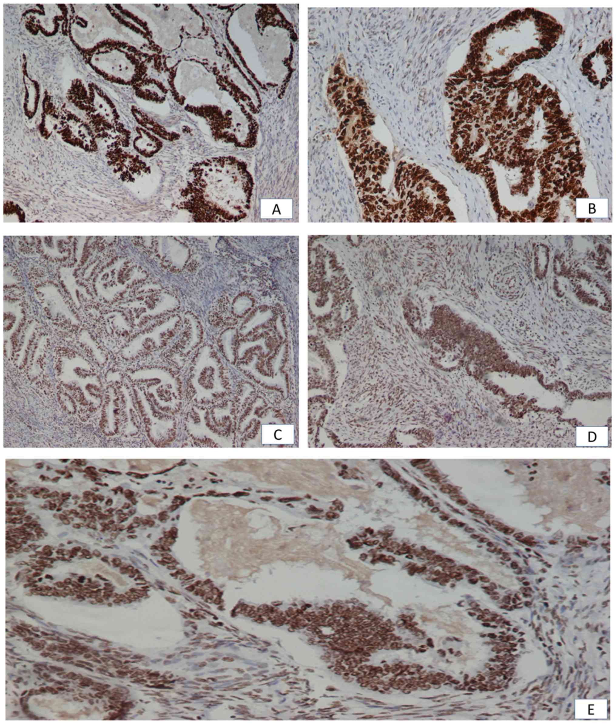
Fig. 1A and B indicate the positive
immunohistochemical expression of p53 in the nucleus. Fig. 1C-E indicate the positive
immunohistochemical expression of PTEN in the nucleus.
Statistical analysis
Categorical variables were presented as absolute (n)
and relative (%) frequencies, while continuous variables were
presented as median (min, max). Associations between categorical
variables were assessed using exact Pearson's χ2 test.
For continuous variables, differences in the median between two
groups were assessed using the Mann-Whitney U test and differences
between three groups were assessed with the Kruskal-Wallis test.
Correlations between continuous variables were assessed with
Spearman's rho (ρ). Statistical significance was set at a
two-tailed P-value of <0.05. Data were analyzed using SPSS
software, version 23.0 (IBM Corporation, Armonk, NY, USA).
Results
Assessment of histologic types indicated that 86
(86.9%) cases of endometrial carcinoma were endometrioid and 13
(13.1%) cases were non-endometrioid. Assessment of histologic
grades revealed that 20 (20.2%) cases were in grade 1, 49 (49.5%)
cases were in grade 2 and 30 (30.3%) cases were in grade 3.
According to tumor depth assessment, 34 (34.3%) cases had <50%
myometrial invasion and 65 (65.7%) cases had >50%. Disease
clinical stage classification revealed that 68 (68.7%) cases were
in stage I, 15 (15.2%) cases were in stage II and 5 (5.1%) cases
were in stage III. Lymph-vascular space invasion was identified in
14 (14.1%) cases, while fallopian tube and ovarian invasion was
revealed in 19 (19.1%) cases. Tumor necrosis was detected in 7
(7.1%) cases.
Table I indicates the
characteristics of the 99 patients with endometrial carcinoma,
whereas Table II indicates the
clinicopathological parameters of the patients according to the
histologic subtypes.
 | Table I.Clinicopathological characteristics
of endometrial adenocarcinomas according to histological
subtypes. |
Table I.
Clinicopathological characteristics
of endometrial adenocarcinomas according to histological
subtypes.
| Clinicopathological
parameters | Endometrioid
adenocarcinomas (n=86) cases, n (%) | Clear cell and
papillary serous adenocarcinomas (n=13) cases, n (%) |
|---|
| Age (years) |
|
<60 | 23 (26.7) | 0 (0.0) |
|
>60 | 63 (73.3) | 13 (100.0) |
| Clinical stage |
| I | 62 (72.1) | 6 (46.2) |
| II | 10 (11.6) | 5 (38.5) |
|
III | 4 (4.7) | 1 (7.7) |
| IV | 0 (0.0) | 0 (0.0) |
| Histological
differentiation |
| G1 | 20 (23.3) | 0 (0.0) |
| G2 | 47 (54.7) | 2 (15.4) |
| G3 | 19 (22.1) | 11 (84.6) |
| Myometrial
invasion |
|
<1/2 | 32 (37.2) | 2 (15.4) |
|
≥1/2 | 54 (62.8) | 11 (84.6) |
| Lymph-vascular
space invasion |
|
Positive | 10 (11.6) | 4 (30.8) |
|
Negative | 44 (51.2) | 7 (53.8) |
| Fallopian tube
and/or ovarian invasion |
|
Positive | 12 (14.0) | 7 (53.8) |
|
Negative | 25 (29.1) | 2 (15.4) |
| Tumoral
necrosis |
|
Yes | 5 (5.8) | 2 (15.4) |
| No | 43 (50.0) | 9 (69.2) |
| Table II.Characteristics of the 99 endometrial
adenocarcinoma patients. |
Table II.
Characteristics of the 99 endometrial
adenocarcinoma patients.
| Clinicopathological
parameters | No. of patients
(%) |
|---|
| Age (years) |
|
<60 | 23 (23.2) |
|
≥60 | 76 (76.8) |
| Clinical stage |
| I | 68 (68.7) |
| II | 15 (15.2) |
|
III | 5 (5.1) |
| Histological
differentiation |
| G1 | 20 (20.2) |
| G2 | 49 (49.5) |
| G3 | 30 (30.3) |
| Myometrial
invasion |
|
<1/2 | 34 (34.3) |
|
≥1/2 | 65 (65.7) |
| Lymph-vascular
space invasion |
|
Positive | 14 (14.1) |
|
Negative | 51 (51.5) |
| Fallopian tube and
ovarian invasion |
|
Positive | 19 (19.2) |
|
Negative | 27 (27.3) |
| Tumoral
necrosis |
|
Yes | 7 (7.1) |
| No | 52 (52.5) |
p53 immunohistochemistry
Scores of p53 immunohistochemical expression were
not significantly associated with the mean age of the patients
(P=0.131), histologic types (P=0.349), clinical stages (P=0.100),
histologic grades (P=0.165), depth of myometrial invasion (P=0.323)
or the presence of tumor necrosis (P=0.313). However, there was a
significant association between lymph-vascular space invasion and
scores of immunohistochemical p53 expression (P=0.007). In the
presence of lymph-vascular space invasion, immunopositivity for p53
was detected in 25–75% of cells in 10 (90.9%) cases and in >75%
of cells in 1 (9.1%) case. In the absence of lymph-vascular space
invasion, 5–25% immunopositive cells were identified in 17 (33.3%)
cases, 25–75% in 22 (43.1%) cases and >75% in 1 (2.0%) case.
Patients with lymph-vascular space invasion had a larger percentage
of immunopositivity for p53 compared with patients without
lymph-vascular space invasion.
The intensity of p53 expression was not
significantly associated with the mean age of patients (P=0.489),
histologic grades (P=0.539), histologic types (P=0.191), depth of
myometrial invasion (P=0.696), clinical stage (P=0.253),
lymph-vascular space invasion (P=0.185), the presence of tumor
necrosis (P=0.411) or fallopian tube invasion (P=0.321).
Table III reveals
the sum of stain intensity and scores of p53-immunopositive cells
and the association of this with the clinicopathological
characteristics. There was a significant association between the
sum of stain intensity and scores of p53-immunopositive cells and
the age of the patients (P=0.037), histologic subtypes (P=0.008),
histologic grades (P=0.002) and fallopian tube and/or ovarian
invasion (P=0.014). In addition, results implied the association
between the sum of stain intensity and scores of p53-immunopositive
cells with clinical stage (P=0.089).
| Table III.Correlations between
clinicopathological characteristics and sum of stain intensity and
scores of p53 expression. |
Table III.
Correlations between
clinicopathological characteristics and sum of stain intensity and
scores of p53 expression.
|
|
| IHC results of p53,
N (%) |
|
|---|
|
|
|
|
|
|---|
|
Characteristics | Cases, n (%) | 0 | + | ++ | +++ | P-value |
|---|
| Age (years) |
|
<60 | 23 (23.2) | 0 (0.0) | 4 (33.3) | 16 (38.1) | 3 (10.3) | 0.037 |
|
≥60 | 76 (76.8) | 0 (0.0) | 8 (66.7) | 26 (61.9) | 26 (89.7) |
|
| Histological
type |
|
Endometrioid | 86 (86.9) | 0 (0.0) | 12 (100.0) | 40 (95.2) | 21 (72.4) | 0.008 |
| Clear
cell and papillary serous | 13 (13.1) | 0 (0.0) | 0 (0.0) | 2 (4.8) | 8 (27.6) |
|
| Clinical stage |
| I | 68 (68.7) | 0 (0.0) | 8 (66.7) | 34 (81.0) | 17 (58.6) | 0.089 |
| II | 15 (15.2) | 0 (0.0) | 1 (8.3) | 2 (4.8) | 6 (20.7) |
|
|
III | 5 (5.1) | 0 (0.0) | 0 (0.0) | 1 (2.4) | 3 (10.3) |
|
| Histological
differentiation |
| G1 | 20 (20.2) | 0 (0.0) | 3 (25.0) | 7 (16.7) | 7 (24.1) | 0.002 |
| G2 | 49 (49.5) | 0 (0.0) | 8 (66.7) | 26 (61.9) | 6 (20.7) |
|
| G3 | 30 (30.3) | 0 (0.0) | 1 (8.3) | 9 (21.4) | 16 (55.2) |
|
| Myometrial
invasion |
|
<1/2 | 34 (34.3) | 0 (0.0) | 5 (41.7) | 16 (38.1) | 9 (31.0) | 0.778 |
|
≥1/2 | 65 (65.7) | 0 (0.0) | 7 (58.3) | 26 (61.9) | 20 (69.0) |
|
| Lymph-vascular
space invasion |
|
Positive | 14 (14.1) | 0 (0.0) | 0 (0.0) | 6 (14.3) | 5 (17.2) | 0.101 |
|
Negative | 51 (51.5) | 0 (0.0) | 10 (83.3) | 22 (52.4) | 9 (31.0) |
|
| Fallopian tube
and/or ovarian invasion |
|
Positive | 19 (19.2) | 0 (0.0) | 1 (8.3) | 4 (9.5) | 8 (27.6) | 0.014 |
|
Negative | 27 (27.3) | 0 (0.0) | 7 (58.3) | 15 (357) | 4 (13.8) |
|
| Tumoral
necrosis |
|
Yes | 7 (7.1) | 0 (0.0) | 1 (8.3) | 2 (4.8) | 3 (10.3) | 0.524 |
| No | 52 (52.5) | 0 (0.0) | 9 (75.0) | 22 (52.4) | 10 (34.5) |
|
PTEN immunohistochemistry
The scores of immunohistochemical expression of PTEN
were not significantly associated with the mean age of the patients
(P=0.844), histologic grade (P=0.352), lymph-vascular space
invasion (P=0.451) or the presence of tumor necrosis (P=1.000).
There was a negative statistical significance between the scores of
PTEN immunohistochemical expression and the depth of myometrial
invasion (P=0.002; ρ=−0.377). Among the 28 cases that demonstrated
positive immunostaining for PTEN in 5–25% of cells, 6 (21.4%) cases
had a depth of myometrial invasion less than half the thickness of
the myometrium, 1 (3.6%) case had a depth of myometrial invasion
equal to half the thickness of the myometrium, 7 (25.0%) cases had
a depth of myometrial invasion equal to two thirds of the thickness
of the myometrium, 7 (25.0%) cases had a depth of myometrial
invasion equal to three quarters of the thickness of the myometrium
and 7 (25.0%) cases had a depth of myometrial invasion equal to the
entire thickness of the myometrium. Regarding the 27 cases that
exhibited positive immunostaining for PTEN in 25–75% of cells, 6
(22.2%) cases had a depth of myometrial invasion less than half the
thickness of the myometrium, 10 (37.0%) cases had a depth of
myometrial invasion equal to half the thickness of the myometrium,
1 (3.7%) case had a depth of myometrial invasion equal to two
thirds of the thickness of the myometrium, 4 (14.8%) cases had a
depth of myometrial invasion equal to three quarters of the
thickness of the myometrium, 2 (7.4%) cases had a depth equal to
the superficial lining of the myometrium and 4 (14.8%) cases had a
depth of myometrial invasion equal to the entire thickness of the
myometrium. Among the 13 cases that demonstrated positive
immunostaining for PTEN in >75% of cells, 4 (30.8%) cases had a
depth of myometrial invasion less than half the thickness of the
myometrium, 3 (23.1%) cases had a depth of myometrial invasion
equal to half the thickness of the myometrium, 1 (7.7%) case had a
depth of myometrial invasion equal to three quarters of the
thickness of the myometrium, 4 (30.8%) cases had a depth equal to
the superficial lining of the myometrium and 1 (7.7%) case had a
depth of myometrial invasion equal to the entire thickness of the
myometrium.
Notably, there was a significant correlation between
the scores of immunohistochemical PTEN expression and the clinical
stage (P=0.019). Among those classified as clinical stage I, 18
(26.5%) cases exhibited 5–25% PTEN-immunopositive cells, 22 (32.4%)
cases exhibited 25–75% PTEN-immunopositive cells and 13 (19.1%)
cases exhibited >75% PTEN-immunopositive cells. In clinical
stage II, immunopositivity for PTEN was detected in 5–25% of cells
in 6 (40.0%) cases, whereas there were no cases with
immunopositivity for PTEN in 25–75% or in >75% of cells.
Finally, in clinical stage III, 2 (40.0%) cases had 5–25%
PTEN-immunopositive cells and another 2 (40.0%) cases exhibited
25–75% PTEN-immunopositive cells.
The intensity of PTEN expression was not
significantly associated with the mean age of patients (P=0.387),
histologic type of the tumor (P=0.630), depth of myometrial
invasion (P=0.124), clinical stage (P=0.621), lymph-vascular space
invasion (P=0.442), presence of tumor necrosis (P=1.000) or the
presence of fallopian tube invasion (P=0.524). Furthermore, the
results suggested that there was no significant association was
observed between the intensity of PTEN staining and histologic
grade (P=0.071). Strong positive PTEN expression was observed in 4
(20.0%) cases of histologic grade G1, in 21 (42.9%) cases of grade
G2 and in 5 (16.7%) cases of histologic grade G3. The corresponding
frequencies for moderate PTEN expression were 9 (45.0%), 17 (34.7%)
and 14 (46.7%), respectively.
Table IV indicates
the sum of stain intensity and scores of PTEN-immunopositive cells
and the association of this with the clinicopathological
characteristics. There was no correlation between the sum of stain
intensity and scores of PTEN-immunopositive cells and the age of
the patients (P=0.371), histologic subtype (P=1.000), histologic
grade (P=0.439), myometrial invasion (P=0.308), clinical stage
(P=0.259), ovarian or fallopian tube invasion (P=0.752) or the
presence of tumor necrosis (P=1.000).
| Table IV.Correlations between
clinicopathological characteristics and sum of stain intensity and
scores of PTEN expression. |
Table IV.
Correlations between
clinicopathological characteristics and sum of stain intensity and
scores of PTEN expression.
|
|
|
Immunohistochemistry results of PTEN
(N) |
|
|---|
|
|
|
|
|
|---|
|
Characteristics | Cases (N) | 0 | + | ++ | +++ | P-value |
|---|
| Age (years) |
|
<60 | 19 | 0 (0.0) | 1 (10.0) | 12 (33.3) | 6 (27.3) | 0.371 |
|
≥60 | 49 | 0 (0.0) | 9 (90.0) | 24 (66.7) | 16 (72.7) |
|
| Histological
type |
|
Endometrioid | 64 | 0 (0.0) | 9 (90.0) | 34 (94.4) | 22 (5.5) | 1.000 |
| Clear
cell and papillary serous | 4 | 0 (0.0) | 1 (10.0) | 2 (5.6) | 1 (4.5) |
|
| Clinical stage |
| I | 53 | 0 (0.0) | 8 (80.0) | 24 (66.7) | 21 (95.5) | 0.259 |
| II | 6 | 0 (0.0) | 1 (10.0) | 5 (13.9) | 0 (0.0) |
|
|
III | 4 | 0 (0.0) | 0 (0.0) | 3 (8.3) | 1 (4.5) |
|
| Histological
differentiation |
| G1 | 13 | 0 (0.0) | 1 (10.0) | 8 (22.2) | 4 (18.2) | 0.439 |
| G2 | 36 | 0 (0.0) | 4 (40.0) | 18 (50.0) | 14 (63.6) |
|
| G3 | 19 | 0 (0.0) | 5 (50.0) | 10 (27.8) | 4 (18.2) |
|
| Myometrial
invasion |
|
<1/2 | 22 | 0 (0.0) | 3 (30.0) | 9 (25.0) | 10 (45.5) | 0.308 |
|
≥1/2 | 46 | 0 (0.0) | 7 (70.0) | 27 (75.0) | 12 (54.5) |
|
| Lymph-vascular
space invasion |
|
Positive | 11 | 0 (0.0) | 3 (30.0) | 6 (16.7) | 2 (9.1) | 0.292 |
|
Negative | 24 | 0 (0.0) | 4 (40.0) | 19 (52.8) | 1 (4.5) |
|
| Fallopian tube and
ovarian invasion |
|
Positive | 8 | 0 (0.0) | 1 (10.0) | 7 (19.4) | 0 (0.0) | 0.752 |
|
Negative | 18 | 0 (0.0) | 4 (40.0) | 13 (36.1) | 1 (4.5) |
|
| Tumoral
necrosis |
|
Yes | 5 | 0 (0.0) | 1 (10.0) | 4 (11.1) | 0 (0.0) | 1.000 |
| No | 24 | 0 (0.0) | 4 (40.0) | 19 (52.8) | 1 (4.5) |
|
Concomitant expression of p53 and PTEN
and the association with clinicopathogical parameters
According to the scores of immunopositive
endometrial carcinoma cells, p53 expression was identified in 73
(85%) cases and PTEN expression was indicated in 64 (74%) cases.
According to the intensity of immunopositive cells, p53 and PTEN
expression was indicated in 74 (86%) and 66 (77%) cases,
respectively. According to the sum of stain intensity and scores of
positive cells, endometrial carcinoma samples had a lower
proportion of PTEN-positive results (77.1%) compared with
p53-postive results (89.2%). Notably, 17% of patients exhibited
PTEN(−)/p53(+) expression, whereas 4.8% of patients exhibited
PTEN(+)/p53(−). In addition, p53 and PTEN concomitant sum
expression was identified in 45% of patients with endometrial
adenocarcinoma.
According to the proportion (score) of
immunopositive cells, there was a coexistence of p53 and PTEN
expression in 53.2% (33/62) of cases (group A) compared with 46.8%
(29/62) of cases, in which there was an absence of p53 and PTEN
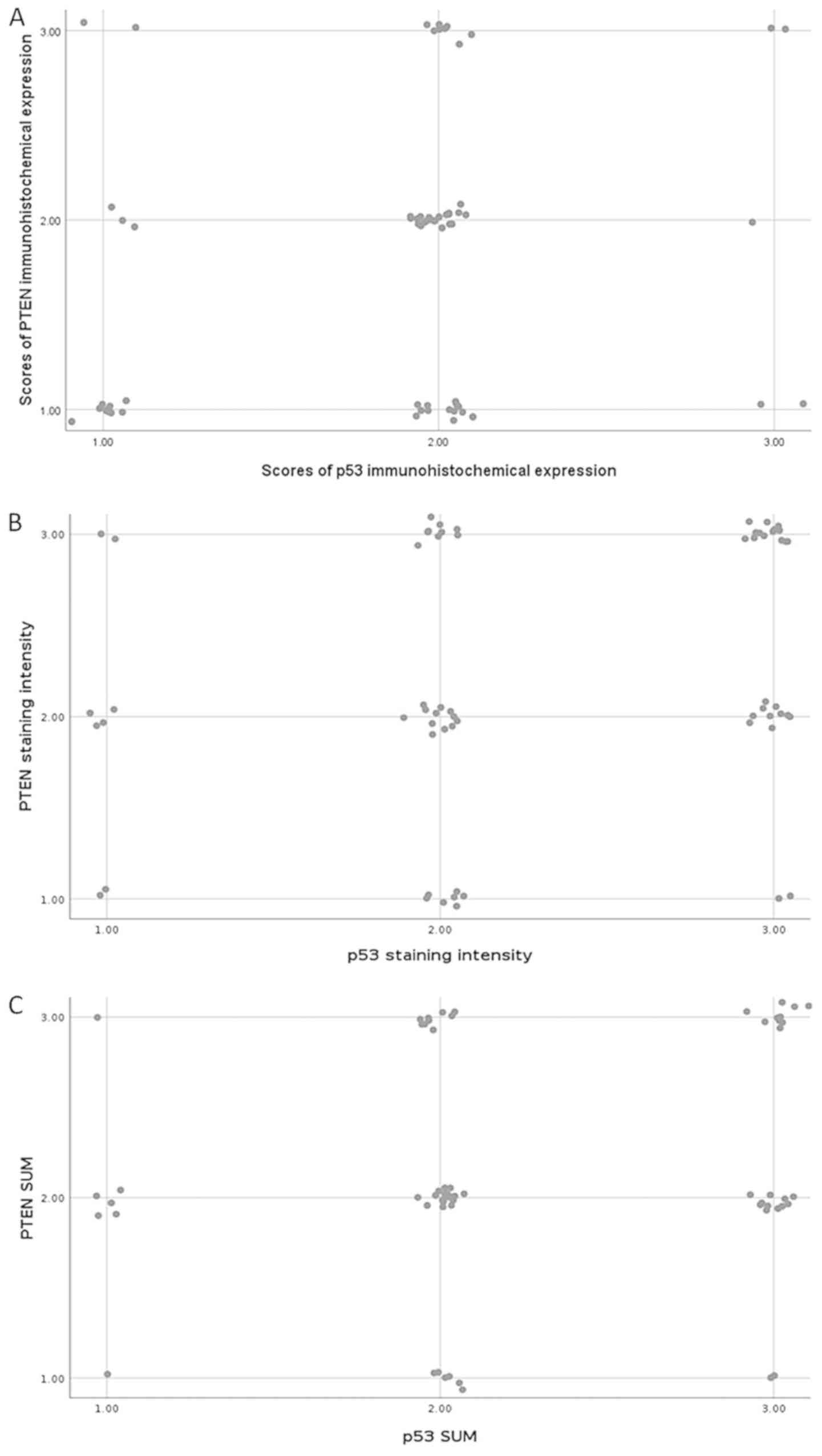
co-expression (group B). Spearman's coefficient for co-expression
of p53 and PTEN was ρ=0.248 (P=0.052), which was marginal for
statistical significance. This correlation was indicated in the
scatterplot (Fig. 2A). Low
concomitant staining was identified in 16.1% of patients, moderate
concomitant staining was identified in 33.9% of patients and high
concomitant staining was identified in 3.2% of patients.
Additionally, 40.0% of patients with high scores of p53 expression
also had high scores of PTEN expression (2/5 patients), whereas
15.4% of patients with high PTEN scores exhibited high scores of
p53 (2/13 patients).
According to the staining intensity, weak
concomitant staining was indicated in 3.2% of patients, moderate
concomitant staining was indicated in 19.0% of patients and strong
concomitant staining was indicated in 23.8%. A total of 44.1% of
patients with strong levels of p53 expression also exhibited strong
PTEN expression (15/34 patients), whereas 50.0% of patients with
strong PTEN levels exhibited strong levels of p53 expression (15/30
patients). There was a significantly positive correlation between
the intensity of PTEN and p53 staining. Spearman's coefficient for
the staining intensity of p53 and PTEN co-expression was ρ=0.282
(P=0.025; Fig. 2B). This suggests
that strong PTEN staining was associated with strong p53 staining
and vice versa.
According to the sum of stain intensity and scores
of positive cells, + concomitant staining was indicated in 1.6% of
patients, ++ was indicated in 27.4% and +++ was indicated in 16.1%
of patients. Notably, 34.5% of patients with +++ p53 staining also
had +++ PTEN staining (10/29 patients), whereas 45.5% of patients
with +++ PTEN staining levels exhibited +++ p53 staining (10/22
patients). Furthermore, it was demonstrated that the sum of stain
intensity and scores of p53-immunopositive cells significantly
correlated with PTEN expression (ρ=0.256; P=0.044; Fig. 2C).
According to the proportion (scores) of
immunopositive cells, the age of patients was significantly
different between the two groups; 33 cases with the coexistence of
p53 and PTEN (group A) and the remaining 29 cases without the
coexistence of p53 and PTEN (group B; P=0.002).
The scores of immunopositive cells between group A
and group B were not significantly associated with the histologic
type of the tumor (P=0.595), histologic grade (P=0.259), depth of
myometrial invasion (P=0.224), lymph-vascular space invasion
(P=0.253), presence of tumor necrosis (P=0.340) or fallopian tube
invasion (P=1.000).
To further study the co-expression of p53 and PTEN,
patients were divided into three groups that were defined as
follows: Patients with low p53 and PTEN expression scores; patients
with moderate expression scores of either p53 or PTEN; and patients
with high expression scores of p53 and PTEN. Table V summarizes the distribution of the
co-expression of p53 and PTEN in endometrial carcinomas according
to scores of immunopositive cells in correlation with
clinicopathological characteristics. Notably, there was a
correlation between the scores of p53 and PTEN co-expression and
the age of the patients (P=0.008) and histologic grade (P=0.028).
The findings also suggested a correlation between the scores of p53
and PTEN co-expression and lymphovascular invasion (P=0.084).
Table VI indicates the distribution
of p53 and PTEN co-expression in endometrial carcinomas according
to the stain intensity in correlation with clinicopathological
characteristics. Furthermore, Table
VII demonstrates p53 and PTEN co-expression in endometrial
carcinomas according to the sum of stain intensity and
immunoexpression scores.
| Table V.Co-expression of p53 and PTEN in
endometrial carcinomas according to scores of immunopositive cells
in relation to clinopathological parameters. |
Table V.
Co-expression of p53 and PTEN in
endometrial carcinomas according to scores of immunopositive cells
in relation to clinopathological parameters.
|
Characteristics | Patients with p53
and PTEN low scores expression cases, n (%) | Patients with
either p53 or PTEN moderate scores expression cases, n (%) | Patients with p53
and PTEN high scores expression cases, n (%) | P-value |
|---|
| Age (years) |
|
<60 | 7 (70.0) | 15 (24.6) | 0 (0.0) | 0.008 |
|
≥60 | 3 (30.0) | 46 (75.4) | 2 (100.0) |
|
| Histological
type |
|
Endometrioid | 10 (100.0) | 53 (86.9) | 1 (50.0) | 0.106 |
| Clear
cell and papillary serous | 0
(0.0) | 8 (13.1) | 1 (50.0) |
|
| Clinical stage |
| I | 9 (90.0) | 44 (72.1) | 2 (100.0) | 0.876 |
| II | 1 (10.0) | 4 (6.6) | 0 (0.0) |
|
|
III | 0 (0.0) | 5 (8.2) | 0 (0.0) |
|
| Histological
differentiation |
| G1 | 2 (20.0) | 14 (23.0) | 0 (0.0) | 0.028 |
| G2 | 8 (80.0) | 27 (44.3) | 0 (0.0) |
|
| G3 | 0 (0.0) | 20 (32.8) | 2 (100.0) |
|
| Myometrial
invasion |
|
<1/2 | 3 (30.0) | 22 (36.1) | 0 (0.0) | 0.651 |
|
≥1/2 | 7 (70.0) | 39 (63.9) | 2 (100.0) |
|
| Lymph-vascular
space invasion |
|
Yes | 0 (0.0) | 11 (18.0) | 0 (0.0) | 0.084 |
| No | 9 (90.0) | 23 (37.7) | 0 (0.0) |
|
| Fallopian tube
and/or ovarian invasion |
|
Yes | 1 (10.0) | 8 (13.1) | 0 (0.0) | 0.642 |
| No | 5 (50.0) | 17 (27.9) | 0 (0.0) |
|
| Tumoral
necrosis |
|
Yes | 1 (10.0) | 4 (6.6) | 0 (0.0) | 1.000 |
| No | 8 (80.0) | 24 (39.3) | 0 (0.0) |
|
| Table VI.Co-expression of p53 and PTEN in
endometrial carcinomas according to stain intensity of
immunopositive cells in relation to clinopathological
parameters. |
Table VI.
Co-expression of p53 and PTEN in
endometrial carcinomas according to stain intensity of
immunopositive cells in relation to clinopathological
parameters.
|
Characteristics | Patients with p53
and PTEN weak positive expression cases, n (%) | Patients with
either p53 or PTEN moderate positive expression cases, n (%) | Patients with p53
and PTEN strong positive expression cases, n (%) | P-value |
|---|
| Age (years) |
|
<60 | 1 (50.0) | 16 (31.4) | 2 (13.3) | 0.261 |
|
≥60 | 1 (50.0) | 35 (68.6) | 13 (86.7) |
|
| Histological
type |
|
Endometrioid | 2 (100) | 48 (94.1) | 14 (93.3) | 1.000 |
| Clear
cell and papillary serous | 0 (0.0) | 3 (5.9) | 1 (6.7) |
|
| Clinical stage |
| I | 2 (100.0) | 39 (76.5) | 14 (93.3) | 0.685 |
| II | 0 (0.0) | 5 (9.8) | 1 (6.7) |
|
|
III | 0 (0.0) | 3 (5.9) | 0 (0.0) |
|
| Histological
differentiation |
| G1 | 1 (50.0) | 9 (17.6) | 4 (26.7) | 0.801 |
| G2 | 1 (50.0) | 28 (54.9) | 7 (46.6) |
|
| G3 | 0 (0.0) | 14 (27.5) | 4 (26.7) |
|
| Myometrial
invasion |
|
<1/2 | 1 (50.0) | 16 (31.4) | 7 (46.7) | 0.513 |
|
≥1/2 | 1 (50.0) | 35 (68.6) | 8 (53.3) |
|
| Lymph-vascular
space invasion |
|
Yes | 0 (0.0) | 8 (15.7) | 1 (6.7) | 1.000 |
| No | 1 (50.0) | 27 (52.9) | 2 (13.3) |
|
| Fallopian tube
and/or ovarian invasion |
|
Yes | 0 (0.0) | 7 (13.7) | 1 (6.7) | 1.000 |
| No | 1 (50.0) | 18 (35.3) | 1 (6.7) |
|
| Tumoral
necrosis |
|
Yes | 0 (0.0) | 4 (7.8) | 1 (6.7) | 0.488 |
| No | 1 (50.0) | 26 (51.0) | 2 (13.3) |
|
| Table VII.Co-expression of p53 and PTEN in
endometrial carcinomas according to sum of stain intensity and
scores of immunopositive cells in relation to clinopathological
parameters. |
Table VII.
Co-expression of p53 and PTEN in
endometrial carcinomas according to sum of stain intensity and
scores of immunopositive cells in relation to clinopathological
parameters.
|
Characteristics | Patients with p53
and PTEN + expression cases, n (%) | Patients with
either p53 or PTEN + + expression cases, n (%) | Patients with p53
and PTEN + + + expression cases, n (%) | P-value |
|---|
| Age (years) |
|
<60 | 1 (100.0) | 20 (32.8) | 1 (10.0) | 0.122 |
|
≥60 | 0 (0.0) | 41 (67.2) | 9 (90.0) |
|
| Histological
type |
|
Endometrioid | 1 (100.0) | 57 (93.4) | 9 (90.0) | 1.000 |
| Clear
cell and papillary serous | 0 (0.0) | 4 (6.6) | 1 (10.0) |
|
| Clinical stage |
| I | 1 (100.0) | 46 (75.4) | 10 (100.0) | 0.548 |
| II | 0 (0.0) | 6 (9.8) | 0 (0.0) |
|
|
III | 0 (0.0) | 3 (4.9) | 0 (0.0) |
|
| Histological
differentiation |
| G1 | 0 (0.0) | 11 (18.0) | 4 (40.0) | 0.594 |
| G2 | 1 (100.0) | 34 (55.7) | 4 (40.0) |
|
| G3 | 0 (0.0) | 16 (26.2) | 2 (20.0) |
|
| Myometrial
invasion |
|
<1/2 | 1 (100.0) | 20 (32.8) | 5 (50.0) | 0.271 |
|
≥1/2 | 0 (0.0) | 41 (67.2) | 5 (50.0) |
|
| Lymph-vascular
space invasion |
|
Yes | 0 (0.0) | 10 (16.4) | 0 (0.0) | 0.762 |
| No | 1 (100.0) | 31 (50.8) | 0 (0.0) |
|
| Fallopian tube
and/or ovarian invasion |
|
Yes | 0 (0.0) | 9 (14.8) | 0 (0.0) | 1.000 |
| No | 1 (100.0) | 21 (34.4) | 0 (0.0) |
|
| Tumoral
necrosis |
|
Yes | 0 (0.0) | 5 (8.2) | 0 (0.0) | 1.000 |
| No | 1 (100.0) | 31 (50.8) | 0 (0.0) |
|
Discussion
The overall rate of p53 and PTEN positivity in the
present study was 89 and 77%, respectively, according to sum of
stain intensity and scores of immunopositive cells. In the study,
the intensity of p53 and PTEN staining was positively correlated
(ρ=0.282; P=0.025). Furthermore, the sum of stain intensity and
immunohistochemical scores of p53 was positively correlated with
PTEN expression (ρ=0.256; P=0.044). The findings indicate an
intrinsic association between the overexpression of the two major
tumors suppressor genes, p53 and PTEN. This supports the previous
suggestions that p53 induces PTEN expression and PTEN reduces
p53-induced degradation (20).
Notably, p53 and PTEN concomitant expression was demonstrated in
45% of patients with endometrial adenocarcinoma, and was considered
a common event.
Previous findings have indicated that p53
alterations seem to occur at early and late phases of endometrial
carcinogenesis (43,44). Early involvement of p53 alterations
in endometrial carcinogenesis has been suggested because p53 has
been indicated to be expressed in endometrial glands adjacent to
endometrial carcinoma and it is associated with endometrial
hyperplasia (30). In the present
study, no correlation was indicated with the sum of stain intensity
and scores of p53-immunopositive cells and clinical stage
(P=0.089), depth of myometrial invasion (P=0.778) or lymph-vascular
space invasion (P=0.101). Therefore, the findings support the
hypothesis that p53 alterations occur at early and late phases of
the endometrial carcinoma progression. In the literature, it has
been demonstrated that overexpression of p53 in endometrioid
adenocarcinomas of the uterus were significantly higher in serous
papillary (in 75–90% of cases) compared with endometrioid
endometrial carcinomas (in 10–35% of cases) (45–70). In
patients with endometrial carcinoma, overexpression of p53 has been
indicated to be a significantly negative prognostic factor and
associated with poor differentiation, advanced stage, increased
myometrial invasion, positive lymph node involvement and distant
metastases (71–81). In the present study, there was a
significant association between the scores of immunohistochemical
p53 expression and lymph-vascular invasion (P=0.007), suggesting
that a larger percentage of p53-immunopositive cells in endometrial
carcinoma may be involved in the metastatic process of the disease.
In addition, the sum of stain intensity and scores of p53
expression were significantly correlated with patient age
(P=0.037), histologic type (P=0.008), histologic grade (P=0.002)
and fallopian and/or ovarian invasion (P=0.014). The present
findings indicate that p53 protein expression serves an important
role in the differentiation and extension process of endometrial
neoplastic cells in older patients. Daniilidou et al
(70) revealed p53 expression, as a
separate factor, was correlated with stage but not with histologic
grade of endometriod endometrial adenocarcinoma; positive p53
expression correlated with stage IIIC, while the absence of p53
expression was connected with stages IB and IC. A key difference
between the present study and the study by Daniilidou et al
(70) was that all endometrial
carcinomas (including endometrioid, clear cell and serous papillary
adenocarcinomas) were examined as a whole in relation to the
clinicopathological factors in the present study, whereas
Daniilidou et al (70)
separately studied the clinicopathological and immunohistochemical
properties for endometrioid and serous papillary adenocarcinomas.
The different results probably reflect the different pathways of
carcinogenesis of type I and II endometrial carcinoma. In the
literature, a reduced 5-year survival has been demonstrated
(71,75,80).
However, there is controversy regarding the independent prognostic
value of p53 expression using multivariate analysis. In particular,
there are studies that have indicated p53 expression as an
independent prognostic factor compared with FIGO stage, tumor grade
and myometrial invasion (71,75,79,82),
whereas other studies have failed to demonstrate such independent
prognostic value of p53 expression (42,76,81,83). As
a result, there are reservations about the routine use of this
marker in clinical practice. For this reason, it is very important
to examine how the expression of p53 potentially interacts with
other tumor suppressor genes, and the prognostic significance of
their concomitant expression in endometrial carcinoma.
In endometrial carcinoma, particularly in type I,
mutations of PTEN have been described to occur in 25–83% of cases;
however, mutations of PTEN have also been described to occur in
endometrial hyperplasia (~55%) (13,15,84–88). In
a study by Lacey et al (26),
loss of PTEN expression in biopsies of endometrial hyperplasia was
not associated with subsequent risk of endometrial carcinoma.
Accordingly, inactivation of PTEN may be considered a crucial
factor for early endometrial carcinogenesis. PTEN gene mutations
have been revealed in more advanced stages of endometrial carcinoma
(15). Loss of heterozygosity at
chromosome 10q23 occurs in ~40% of endometrial carcinomas (89,90). It
has been indicated that loss of PTEN expression was associated with
endometrioid histology, and inversely associated with the presence
of lymphovascular space invasion (91). Risinger et al (84) indicated that PTEN mutations were
associated with low-grade and low-stage endometrial carcinomas,
whereas Konopka et al (15)
revealed a significant correlation between PTEN gene mutations and
histologic grade of endometrial carcinomas, suggesting that defects
in PTEN gene are associated with increased malignancy due to the
loss of the ability of endometrial cells to differentiate. Other
studies have indicated no correlation between PTEN expression and
standard prognostic factors (14,39,92–94). In
the present study, the immunohistochemical scores of PTEN
expression were negatively associated with myometrial invasion
(P=0.002; ρ=−0.377). The lower levels of positive PTEN
immunostaining scores were associated with deeper myometrial
invasion and vice versa. Furthermore, an association was identified
between clinical stages and the immunohistochemical scores of PTEN
expression (P=0.019). Patients at clinical stage I had higher
positive immunostaining scores, whereas patients at clinical stage
II had lower scores. The findings support the hypothesis that lower
PTEN expression in endometrial carcinoma occurs in later stages of
endometrial carcinogenesis. However, when the sum of stain
intensity and scores of PTEN expression were examined, no
significant correlations between the age of patients, histologic
type, clinical stage, histologic differentiation, myometrial
invasion, lymph-vascular space invasion, fallopian and/or ovarian
invasion or tumor necrosis were indicated. Daniilidou et al
(70) indicated an association
between PTEN expression and histologic grade of endometrioid
endometrial adenocarcinoma. Notably, the negative expression of
PTEN correlated with grade 3, whereas positive PTEN expression
correlated with grades I and II (70). In addition, their study revealed an
association between PTEN expression and stage of endometrioid
endometrial adenocarcinomas (negative expression of PTEN correlated
with stages IC and IIC, while positive PTEN expression with stage
IB). The findings in the literature regarding the loss PTEN protein
expression and clinical outcome in endometrial carcinomas are
inconsistent. Some studies have reported more favorable survival
(14,28,29,91,95,96),
while other studies have indicated less favorable prognosis
(19,90,97,98).
Terakawa et al (97)
suggested that overexpression of PTEN is a significant prognostic
indicator of improved overall survival for patients with advanced
endometrial carcinoma who undergo postoperative chemotherapy, as
PTEN was able to increase the chemosensitivity of neoplastic
cells.
In the literature, it is apparent that concomitant
genetic alterations may have a prognostic value in endometrial
carcinoma. It has been indicated that concomitant PI3K-Akt and p53
alterations were associated with poor prognosis (99). In addition, simultaneous activations
of p53 and microsatellite instability were strong genetic
prognostic factors for disease-free survival (100). Furthermore, Uegaki et al
(101) demonstrated that
PTEN-positive and phosphorylated-AKT-negative expression is a
predictor of survival for patients with advanced endometrial
carcinoma. In the present study, an association of the p53 and PTEN
co-expression with well-established clinicopathogical factors in
patients with endometrial carcinoma was indicated, which opposed
the findings of Daniilidou et al (70), in which there was no such
correlation. The levels of concomitant p53 and PTEN expression,
according to the scores of immunopositive cells, were correlated
with the age of patients (P=0.008) and histologic differentiation
(P=0.028) in the present study. These results suggested that p53
and PTEN co-expression may serve a role in the development of
high-grade endometrial carcinoma in older patients. The present
findings also suggest the involvement of different molecular
pathways in the development of low-grade and high-grade endometrial
carcinoma. The findings also suggested a correlation with
lymphovascular invasion (P=0.084), whereas no correlation was
identified between the co-expression of p53 and PTEN in endometrial
carcinoma (according to the stain intensity or the sum of stain
intensity and immunoexpression scores) or clinicopathological
characteristics. Therefore, the present study indicated that
concomitant p53 and PTEN expression may contribute to the
characterization of tumor behavior in endometrial carcinoma.
Because the findings of the present study indicated the expression
of p53 was positively associated with the levels of PTEN expression
in endometrial carcinoma, it was suggested that further molecular
studies to estimate and determine the impact of the co-expression
of these molecular factors on patient survival of the disease are
required.
To conclude, the present results suggest a strong
correlation between the expression of p53 and PTEN in endometrial
adenocarcinoma, indicating an intrinsic association between the
expression of these tumor suppressor genes. In addition, according
to the scores of immunopositive cells, which were correlated with
the age of patients and the histologic differentiation, concomitant
p53 and PTEN expression may contribute to the characterization of
tumor behavior in endometrial carcinoma. The findings suggest that
combination of p53 and PTEN expression may serve a role in the
development of high-grade endometrial carcinoma in older patients.
Furthermore, the results imply the involvement of different
molecular pathways between the progression of low-grade and
high-grade endometrial carcinoma.
Acknowledgements
The present study was part of a thesis for a Doctor
of Philosophy (PhD) in Obstetrics and Gynecology, Medical School,
Kapodistrian University of Athens, Greece for Mr. Aggelis
Stavropoulos.
Funding
Not applicable.
Availability of data and materials
The datasets used and/or analyzed during this study
are available from the corresponding author on reasonable
request.
Authors' contributions
All authors were responsible for the conception and
design of the present study. TV and AT were responsible for the
provision of the study materials. TV, AT, VKV and FNV were
responsible for the collection and assembly of the data. AS, MV,
TV, VKV, AT, FNV, AN, NK and ACL performed the data analysis and
interpretation. AS, MV, TV, VKV, AT, FNV, AN, NK and ACL
contributed in writing the manuscript. AS, MV, TV, VKV, AT, FNV,
AN, NK and ACL read and gave the final approval of the
manuscript.
Ethics approval and consent to
participate
The study was approved by the Ethics Committee of
Medical School of Kapodistrian University of Athens, Greece. The
patient included in the case provided consent for her data to be
used in this publication.
Patient consent for publication
All the patients included in this study at the time
of data collection provided consent for their data to be used in
this publication.
Competing interests
The authors declare that they have no competing
interests.
References
|
1
|
Liu FS: Molecular carcinogenesis of
endometrial cancer. Taiwan J Obstet Gynecol. 46:26–32. 2007.
View Article : Google Scholar : PubMed/NCBI
|
|
2
|
Sasnauskienė A, Jonušienė V,
Krikštaponienė A, Butkytė S, Dabkevičienė D, Kanopienė D,
Kazbarienė B and Didžiapetrienė J: NOTCH1, NOTCH3, NOTCH4, and JAG2
protein levels in human endometrial cancer. Medicina (Kaunas).
50:14–18. 2014. View Article : Google Scholar : PubMed/NCBI
|
|
3
|
Elbasateeny SS, Salem AA, Abdelsalam WA
and Salem R: Immunohistochemical expression of cancer stem cell
related markers CD44 and CD133 in endometrial cancer. Pathol Res
Pract. 212:10–16. 2016. View Article : Google Scholar : PubMed/NCBI
|
|
4
|
Li Y, Zhang X, Ge J, Liu X, Xu S, Zhu Z,
Fang G, Liu J, Zhang H and Sun X: Can Nup88 expression be
associated with atypical endometrial hyperplasia and endometrial
cancer? A preliminary study. Pathol Res Pract. 212:274–278. 2016.
View Article : Google Scholar : PubMed/NCBI
|
|
5
|
Agopianz M, Forgez P, Casse JM, Lacomme S,
Charra-Brunaud C, Clerc-Urmès I, Morel O, Bonnet C, Guéant JL,
Vignaud JM, et al: Expression of neurotensin receptor 1 in
endometrial adenocarcinoma is correlated with histological grade
and clinical outcome. Virchows Arch. 471:521–530. 2017. View Article : Google Scholar : PubMed/NCBI
|
|
6
|
Khabaz MN, Abdelrahman AS, Butt NS,
Al-Maghrabi B and Al-Maghrabi J: Cyclin D1 is significantly
associated with stage of tumor and predicts poor survival in
endometrial carcinoma patients. Ann Diagn Pathol. 30:47–51. 2017.
View Article : Google Scholar : PubMed/NCBI
|
|
7
|
Mittal P, Klingler-Hoffmann M, Arentz G,
Winderbaum L, Kaur G, Anderson L, Scurry J, Leung Y, Stewart CJ,
Carter J, et al: Annexin A2 and alpha actinin 4 expression
correlates with metastatic potential of primary endometrial cancer.
Biochim Biophys Acta Proteins Proteom. 1865:846–857. 2017.
View Article : Google Scholar : PubMed/NCBI
|
|
8
|
Qiu M, Bao W, Wang J, Yang T, He X, Liao Y
and Wan X: FOXA1 promotes tumor cell proliferation through AR
involving the Notch pathway in endometrial cancer. BMC Cancer.
14:782014. View Article : Google Scholar : PubMed/NCBI
|
|
9
|
Gu X, Liu Q, Yang N, Shen JF, Zhang XG,
Cao F and Ding HZ: Clinicopathological significance of increased
ZIC1 expression in human endometrial cancer. J Huazhong Univ Sci
Technolog Med Sci. 35:898–903. 2015. View Article : Google Scholar : PubMed/NCBI
|
|
10
|
Bansal N, Yendluri V and Wenham RM: The
molecular biology of endometrial cancers and the implications for
pathogenesis, classification, and targeted therapies. Cancer
Control. 16:8–13. 2009. View Article : Google Scholar : PubMed/NCBI
|
|
11
|
Dohi S, Ohno S, Ohno Y, Kyo S, Soma G,
Sugiyama H and Inoue M: WT1 expression correlates with angiogenesis
in endometrial cancer tissue. Anticancer Res. 30:3187–3192.
2010.PubMed/NCBI
|
|
12
|
Stambolic V, Suzuki A, de la Pompa JL,
Brothers GM, Mirtsos C, Sasaki T, Ruland J, Penninger JM,
Siderovski DP and Mak TW: Negative regulation of PKB/Akt-dependent
cell survival by the tumor suppressor PTEN. Cell. 95:29–39. 1998.
View Article : Google Scholar : PubMed/NCBI
|
|
13
|
Scully MM, Palacios-Helgeson LK, Wah LS
and Jackson TA: Rapid estrogen signaling negatively regulates PTEN
activity through phosphorylation in endometrial cancer cells. Horm
Cancer. 5:218–231. 2014. View Article : Google Scholar : PubMed/NCBI
|
|
14
|
Erkanli S, Kayaselcuk F, Kuscu E, Bagis T,
Bolat F, Haberal A and Demirhan B: Expression of survivin, PTEN and
p27 in normal, hyperplastic, and carcinomatous endometrium. Int J
Gynecol Cancer. 16:1412–1418. 2006. View Article : Google Scholar : PubMed/NCBI
|
|
15
|
Konopka B, Paszko Z, Janiec-Jankowska A
and Goluda M: Assessment of the quality and frequency of mutations
occurrence in PTEN gene in endometrial carcinomas and hyperplasias.
Cancer Lett. 178:43–51. 2002. View Article : Google Scholar : PubMed/NCBI
|
|
16
|
Kimura F, Watanabe J, Hata H, Fujisawa T,
Kamata Y, Nishimura Y, Jobo T and Kuramoto H: PTEN
immunohistochemical expression is suppressed in G1 endometrioid
adenocarcinoma of the uterine corpus. J Cancer Res Clin Oncol.
130:161–168. 2004. View Article : Google Scholar : PubMed/NCBI
|
|
17
|
Machwinnie N and Monaghan H: The use of
P53, PTEN, and C-erbB-2 to differentiate uterine serous papillary
carcinoma from endometrioid endometrial carcinoma. Int J Gynecol
Cancer. 14:938–946. 2004. View Article : Google Scholar : PubMed/NCBI
|
|
18
|
Kapucuoglu N, Aktepe F, Kaya H, Bircan S,
Karahan N and Ciriş M: Immunohistochemical expression of PTEN in
normal, hyperplastic and malignant endometrium and its correlation
with hormone receptors, bcl-2, bax, and apoptotic index. Pathol Res
Pract. 203:153–162. 2007. View Article : Google Scholar : PubMed/NCBI
|
|
19
|
Kanamori Y, Kigawa J, Itamochi H, Sultana
H, Suzuki M, Ohwada M, Kamura T, Sugiyama T, Kikuchi Y, Kita T, et
al: PTEN expression is associated with prognosis for patients with
advanced endometrial carcinoma undergoing postoperative
chemotherapy. Int J Cancer. 100:686–689. 2002. View Article : Google Scholar : PubMed/NCBI
|
|
20
|
Mayo LD, Dixon JE, Durden DL, Tonks NK and
Donner DB: PTEN protects p53 from Mdm2 and sensitizes cancer cells
to chemotherapy. J Biol Chem. 277:5484–5489. 2002. View Article : Google Scholar : PubMed/NCBI
|
|
21
|
Garg K, Broaddus RR, Soslow RA, Urbauer
DL, Levine DA and Djordjevic B: Pathologic scoring of PTEN
immunohistochemistry in endometrial carcinoma is highly
reproducible. Int J Gynecol Pathol. 31:48–56. 2012. View Article : Google Scholar : PubMed/NCBI
|
|
22
|
Jeczen R, Skomra D, Cybulski M,
Scheider-Stock R, Szewczuk W, Roessner A, Rechberger T and Semczuk
A: P53/MDM2 overexpression in metastatic endometrial cancer:
correlation with clinicopathological features and patient outcome.
Clin Exp Metastasis. 24:503–511. 2007. View Article : Google Scholar : PubMed/NCBI
|
|
23
|
Pallares J, Bussaglia E, Martínez-Guitarte
JL, Dolcet X, Llobet D, Rue M, Sanchez-Verde L, Palacios J, Prat J
and Matias-Guiu X: Immunohistochemical analysis of PTEN in
endometrial carcinoma: A tissue microarray study with a comparison
of four commercial antibodies in correlation with molecular
abnormalities. Mod Pathol. 18:719–727. 2005. View Article : Google Scholar : PubMed/NCBI
|
|
24
|
Westin SN, Ju Z, Broaddus RR, Krakstad C,
Li J, Pal N, Lu KH, Coleman RL, Hennessy BT, Klempner SJ, et al:
PTEN loss is a context-dependent outcome determinant in obese and
non-obese endometrioid endometrial cancer patients. Mol Oncol.
9:1694–1703. 2015. View Article : Google Scholar : PubMed/NCBI
|
|
25
|
Chen J, Li S, Yang Z, Lu G and Hu H:
Correlation between NDRG1 and PTEN expression in endometrial
carcinoma. Cancer Sci. 99:706–710. 2008. View Article : Google Scholar : PubMed/NCBI
|
|
26
|
Lacey JV Jr, Mutter GL, Ronnett BM, Ioffe
OB, Duggan MA, Rush BB, Glass AG, Richesson DA, Chatterjee N,
Langholz B and Sherman ME: PTEN expression in endometrial biopsies
as a marker of progression to endometrial carcinoma. Cancer Res.
68:6014–6020. 2008. View Article : Google Scholar : PubMed/NCBI
|
|
27
|
Merritt MA and Cramer DW: Molecular
pathogenesis of endometrial and ovarian cancer. Cancer Biomark.
9:287–305. 2010. View Article : Google Scholar : PubMed/NCBI
|
|
28
|
Fadare O and Parksh V: p53 aberrations in
low grade endometrioid carcinoma of the endometrium with nodal
metastases: Possible insights on pathogenesis discerned from
immunohistochemistry. Diagn Pathol. 12:812017. View Article : Google Scholar : PubMed/NCBI
|
|
29
|
Jiang Z, Xu W, Dan G, Liu Y and Xiong J:
P53 and murine double minute 2 (MDM2) expression changes and
significance in different types of endometrial lesions. Med Sci
Monit. 22:4786–4793. 2016. View Article : Google Scholar : PubMed/NCBI
|
|
30
|
Kounelis S, Kapranos N, Kouri E, Coppola
D, Papadaki H and Jones MW: Immunohistochemical profile of
endometrial adenocarcinoma: A study of 61 cases and review of the
literature. Mod Pathol. 13:379–388. 2000. View Article : Google Scholar : PubMed/NCBI
|
|
31
|
Mazurek A, Kuć P, Mazurek-Wadołkowska E
and Laudański T: A role of thymidine phosphorylase and P53 tissue
protein expression in biology of endometrial cancer. Neoplasma.
55:261–265. 2008.PubMed/NCBI
|
|
32
|
Oda K, Okada J, Timmerman L,
Rodriguez-Viciana P, Stokoe D, Shoji K, Taketani Y, Kuramoto H,
Knight ZA, Shokat KM and McCormick F: PIK3CA cooperates with other
phosphatidylinositol 3′-kinase pathway mutations to effect
oncogenic transformation. Cancer Res. 68:8127–8136. 2008.
View Article : Google Scholar : PubMed/NCBI
|
|
33
|
Li SF, Shiozawa T, Nakayama K, Nikaido T
and Fujii S: Stepwise abnormality of sex steroid hormone receptors,
tumor suppressor gene products (p53 and Rb), and cyclin E in
uterine endometrioid carcinoma. Cancer. 77:321–329. 1996.
View Article : Google Scholar : PubMed/NCBI
|
|
34
|
Ozkara SK and Corakci A: Significantly
decreased P27 expression in endometrial carcinoma compared to
complex hyperplasia with atypia (correlation with p53 expression).
Pathol Oncol Res. 10:89–97. 2004. View Article : Google Scholar : PubMed/NCBI
|
|
35
|
Edmondson RJ, Crosbie EJ, Nickkho-Amiry M,
Kaufmann A, Stelloo E, Nijman HW, Leary A, Auguste A, Mileshkin L,
Pollock P, et al: Markers of the p53 pathway further refine
molecular profiling in high-risk endometrial cancer: A TransPORTEC
initiative. Gynecol Oncol. 146:327–333. 2017. View Article : Google Scholar : PubMed/NCBI
|
|
36
|
Suzuki C, Matsumoto T, Sonoue H, Arakawa
A, Furugen Y and Kinoshita K: Prognostic significance of the
infiltrative pattern invasion in endometrioid adenocarcinoma of the
endometrium. Pathol Int. 53:495–500. 2003. View Article : Google Scholar : PubMed/NCBI
|
|
37
|
Ambros RA, Ross JS, Kallakury BV,
Malfetano J, Kim Y, Hwang J, Breese K and Figge J: p53 gene status
in endometrial carcinomas showing diffuse positivity for p53
protein by immunohistochemical analysis. Mod Pathol. 8:441–445.
1995.PubMed/NCBI
|
|
38
|
Ambros RA, Sheehan CE, Kallakury BV, Ross
JS, Malfetano J, Paunovich E and Figge J: MDM2 and p53 protein
expression in the histologic subtypes of endometrial carcinoma. Mod
Pathol. 9:1165–1169. 1996.PubMed/NCBI
|
|
39
|
Tashiro H, Blazes MS, Wu R, Cho KR, Bose
S, Wang SI, Li J, Parsons R and Ellenson LH: Mutations in PTEN are
frequent in endometrial carcinoma but rare in other common
gynecological malignancies. Cancer Res. 57:3935–3940.
1997.PubMed/NCBI
|
|
40
|
Sakuragi N, Hirai A, Tada M, Yamada H,
Yamamoto R, Fujimoto S and Moriuchi T: Dominant-negative mutation
of p53 tumor suppressor gene in endometrial carcinoma. Gynecol
Oncol. 83:485–490. 2001. View Article : Google Scholar : PubMed/NCBI
|
|
41
|
Appel ML, Edelweiss MI, Fleck J, Rivero
LF, Rivoire WA, Mônego HI and Dos Reis R: P53 and BCL-2 as
prognostic markers in endometrial carcinoma. Pathol Oncol Res.
14:23–30. 2008. View Article : Google Scholar : PubMed/NCBI
|
|
42
|
Athanassiadou P, Athanassiades P, Grapsa
D, Gonidi M, Athanassiadou AM, Stamati PN and Patsouris E: The
prognostic value of PTEN, p53, and beta-catenin in endometrial
carcinoma: A prospective immunocytochemical study. Int J Gynecol
Cancer. 17:697–704. 2007. View Article : Google Scholar : PubMed/NCBI
|
|
43
|
Ellenson LH: Early molecular changes in
endometrial cancer. Int J Gynecol Cancer. 15:399–400. 2005.
View Article : Google Scholar
|
|
44
|
Lax SF: Molecular genetic pathways in
various types of endometrial carcinoma: From a phenotypical to a
molecular-based classification. Virchows Arch. 444:213–223. 2004.
View Article : Google Scholar : PubMed/NCBI
|
|
45
|
Darvishian F, Hummer AJ, Thaler HT,
Bhargava R, Linkov I, Asher M and Soslow RA: Serous endometrial
cancers that mimic endometrioid adenocarcinomas: A
clinicopathologic and immunohistochemical study of a group of
problematic cases. Am J Surg Pathol. 28:1568–1578. 2004. View Article : Google Scholar : PubMed/NCBI
|
|
46
|
Cancer Genome Atlas Research Network, ;
Kandoth C, Schultz N, Cherniack AD, Akbani R, Liu Y, Shen H,
Robertson AG, Pashtan I, Shen R, et al: Integrated genomic
characterization of endometrial carcinoma. Nature. 497:67–73. 2013.
View Article : Google Scholar : PubMed/NCBI
|
|
47
|
Fadare O and Zheng W: Insights into
endometrial serous carcinogenesis and progression. Int J Clin Exp
Pathol. 2:411–432. 2009.PubMed/NCBI
|
|
48
|
Fadare O, Gwin K, Desouki MM, Crispens MA,
Jones HW III, Khabele D, Liang SX, Zheng W, Mohammed K, Hecht JL
and Parkash V: The clinicopathologic significance of p53 and
BAF-250a (ARID1A) expression in clear cell carcinoma of the
endometrium. Mod Pathol. 26:1101–1110. 2013. View Article : Google Scholar : PubMed/NCBI
|
|
49
|
Ramalingam P, Masand RP, Eucher ED and
Malpica A: Undifferentiated carcinoma of the endometrium: An
expanded immunohistochemical analysis including PAX-8 and
basal-like carcinoma surrogate markers. Int J Gynecol Pathol.
35:410–418. 2016. View Article : Google Scholar : PubMed/NCBI
|
|
50
|
Hoang LN, Lee YS, Karnezis AN,
Tessier-Cloutier B, Almandani N, Coatham M, Gilks CB, Soslow RA,
Stewart CJ, Köbel M and Lee CH: Immunophenotypic features of
dedifferentiated endometrial carcinoma-insights from
BRG1/INI1-deficient tumours. Histopathology. 69:560–569. 2016.
View Article : Google Scholar : PubMed/NCBI
|
|
51
|
Lopez-Garcia MA and Palacios J: Pathologic
and molecular features of uterine carcinosarcomas. Semin Diagn
Pathol. 27:274–286. 2010. View Article : Google Scholar : PubMed/NCBI
|
|
52
|
Cherniack AD, Shen H, Walter V, Stewart C,
Murray BA, Bowlby R, Hu X, Ling S, Soslow RA, Broaddus RR, et al:
Integrated molecular characterization of uterine carcinosarcoma.
Cancer Cell. 31:411–423. 2017. View Article : Google Scholar : PubMed/NCBI
|
|
53
|
Alvarez T, Miller E, Duska L and Oliva E:
Molecular profile of grade 3 endometrioid endometrial carcinoma: Is
it a type I or type II endometrial carcinoma? Am J Surg Pathol.
36:753–761. 2012. View Article : Google Scholar : PubMed/NCBI
|
|
54
|
Sherman ME, Bur ME and Kurman RJ: p53 in
endometrial cancer and its putative precursors: Evidence for
diverse pathways of tumorigenesis. Hum Pathol. 26:1268–1274. 1995.
View Article : Google Scholar : PubMed/NCBI
|
|
55
|
Moll MU, Chalas E, Auguste M, Meaney D and
Chumas J: Uterine papillary serous carcinoma evolves via a
p53-driven pathway. Hum Pathol. 27:1295–1300. 1996. View Article : Google Scholar : PubMed/NCBI
|
|
56
|
Zheng W, Cao P, Zheng M, Kramer EE and
Godwin TA: p53 overexpression and bcl-2 persistence in endometrial
carcinoma: Comparison of papillary serous and endometrioid
subtypes. Gynecol Oncol. 61:167–174. 1996. View Article : Google Scholar : PubMed/NCBI
|
|
57
|
Bur ME, Perlman C, Edelmann BS, Fey E and
Rose PG: p53 expression in neoplasms of the uterine corpus. Am J
Clin Pathol. 98:81–87. 1992. View Article : Google Scholar : PubMed/NCBI
|
|
58
|
Inoue M, Fujita M, Enomoto T, Morimoto H,
Monden T, Shinano T and Tanizawa O: Immunohistochemical analysis of
p53 in gynecologic tumors. Am J Clin Pathol. 102:665–670. 1994.
View Article : Google Scholar : PubMed/NCBI
|
|
59
|
Prat J, Oliva E, Lerma E, Vaquero M and
Matías-Guiu X: Uterine papillary serous adenocarcinoma. A 10-case
study of p53 and c-erbB-2 expression and DNA content. Cancer.
74:1778–1783. 1994. View Article : Google Scholar : PubMed/NCBI
|
|
60
|
Reinartz JS, George E, Lindgren BR and
Niehans GA: Expression of p53, transforming growth factor alpha,
epidermal growth factor receptor, and c-erbB-2 in endometrial
carcinoma and correlation with survival and known predictors of
survival. Hum Pathol. 25:1075–1083. 1994. View Article : Google Scholar : PubMed/NCBI
|
|
61
|
Khalifa MA, Mannel RS, Haraway SD, Walker
J and Min KW: Expression of EGFR, HER-2/neu, P53, and PCNA in
endometrioid, serous papillary, and clear cell endometrial
adenocarcinomas. Gynecol Oncol. 53:84–92. 1994. View Article : Google Scholar : PubMed/NCBI
|
|
62
|
King SA, Adas AA, LiVolsi VA, Takahashi H,
Behbakht K, McGovern P, Benjamin I, Rubin SC and Boyd J: Expression
and mutation analysis of the p53 gene in uterine papillary serous
carcinoma. Cancer. 75:2700–2705. 1995. View Article : Google Scholar : PubMed/NCBI
|
|
63
|
Tashiro H, Isacson C, Levine R, Kurman RJ,
Cho KR and Hedrick L: p53 gene mutations are common in uterine
serous carcinoma and occur early in their pathogenesis. Am J
Pathol. 150:177–185. 1997.PubMed/NCBI
|
|
64
|
Soslow RA, Shen PU, Chung MH and Isacson
C: Distinctive p53 and mdm2 immunohistochemical expression profiles
suggest different pathogenetic pathways in poorly differentiated
endometrial carcinoma. Int J Gynecol Pathol. 17:129–134. 1998.
View Article : Google Scholar : PubMed/NCBI
|
|
65
|
Garcia-Dios DA, Lambrechts D, Coenegrachts
L, Vandenput I, Capoen A, Webb PM, Ferguson K; ANECS, Akslen LA,
Claes B, et al: High-throughput interrogation of PIK3CA, PTEN,
KRAS, FBXW7 and TP53 mutations in primary endometrial carcinoma.
Gynecol Oncol. 128:327–334. 2013. View Article : Google Scholar : PubMed/NCBI
|
|
66
|
Seeger A, Kölbl H, Petry IB, Gebhard S,
Battista MJ, Böhm D and Steiner E: p53 is correlated with low BMI
negative progesterone receptor status and recurring disease in
patients with endometrial cancer. Gynecol Oncol. 125:200–207. 2012.
View Article : Google Scholar : PubMed/NCBI
|
|
67
|
Urabe R, Hachisuga T, Kurita T, Kagami S,
Kawagoe T, Matsuura Y and Shimajiri S: Prognostic significance of
overexpression of p53 in uterine endometrioid adenocarcinomas with
an analysis of nuclear grade. J Obstet Gynaecol Res. 40:812–819.
2014. View Article : Google Scholar : PubMed/NCBI
|
|
68
|
Lee EJ, Kim TJ, Kim DS, Choi CH, Lee JW,
Lee JH, Bae DS and Kim BG: p53 alteration independently predicts
poor outcomes in patients with endometrial cancer: A
clinicopathologic study of 131 cases and literature review. Gynecol
Oncol. 116:533–538. 2010. View Article : Google Scholar : PubMed/NCBI
|
|
69
|
Kurnit KC, Kim GN, Fellman BM, Urbauer DL,
Mills GB, Zhang W and Broaddus RR: CTNNB1 (beta-catenin) mutation
identifies low grade, early stage endometrial cancer patients at
increased risk of recurrence. Mod Pathol. 30:1032–1041. 2017.
View Article : Google Scholar : PubMed/NCBI
|
|
70
|
Daniilidou K, Frangou-Plemenou M,
Grammatikakis J, Grigoriou O, Vitoratos N and Kondi-Pafiti A:
Prognostic significance and diagnostic value of PTEN and p53
expression in endometrial carcinoma. A retrospective
clinicopathological and immunohistochemical study. J BUON.
18:195–201. 2013.PubMed/NCBI
|
|
71
|
Ohkouchi T, Sakuragi N, Watari H, Nomura
E, Todo Y, Yamada H and Fujimoto S: Prognostic significance of
Bcl-2, p53 overexpression, and lymph node metastasis in surgically
staged endometrial carcinoma. Am J Obstet Gynecol. 187:353–359.
2002. View Article : Google Scholar : PubMed/NCBI
|
|
72
|
Erdem O, Erdem M, Dursun A, Akyol G and
Erdem A: Angiogenesis, p53, and bcl-2 expression as prognostic
indicators in endometrial cancer: comparison with traditional
clinicopathologic variables. Int J Gynecol Pathol. 22:254–260.
2003. View Article : Google Scholar : PubMed/NCBI
|
|
73
|
Hirschowitz L, Ganesan R and McCluggage
WG: WT1, p53 and hormone receptor expression in uterine serous
carcinoma. Histopathology. 55:478–482. 2009. View Article : Google Scholar : PubMed/NCBI
|
|
74
|
Mariani A, Sebo TJ, Webb MJ, Riehle D,
Katzmann JA, Keeney GL, Roche PC, Lesnick TG and Podratz KC:
Molecular and histopathologic predictors of distant failure in
endometrial cancer. Cancer Detect Prev. 27:434–441. 2003.
View Article : Google Scholar : PubMed/NCBI
|
|
75
|
Ito K, Watanabe K, Nasim S, Sasano H, Sato
S, Yajima A, Silverberg SG and Garrett CT: Prognostic significance
of p53 overexpression in endometrial cancer. Cancer Res.
54:4667–4670. 1994.PubMed/NCBI
|
|
76
|
Alkushi A, Lim P, Coldman A, Huntsman D,
Miller D and Gilks CB: Interpretation of p53 immunoreactivity in
endometrial carcinoma: Establishing a clinically relevant cut-off
level. Int J Gynecol Pathol. 23:129–137. 2004. View Article : Google Scholar : PubMed/NCBI
|
|
77
|
Hamel NW, Sebo TJ, Wilson TO, Keeney GL,
Roche PC, Suman VJ, Hu TC and Podratz KC: Prognostic value of p53
and proliferating cell nuclear antigen expression in endometrial
carcinoma. Gynecol Oncol. 62:192–198. 1996. View Article : Google Scholar : PubMed/NCBI
|
|
78
|
Bancher-Todesca D, Gitsch G, Williams KE,
Kohlberger P, Neunteufel W, Obermair A, Heinze G, Breitenecker G
and Hacker NF: p53 protein overexpression: A strong prognostic
factor in uterine papillary serous carcinoma. Gynecol Oncol.
71:59–63. 1998. View Article : Google Scholar : PubMed/NCBI
|
|
79
|
Geisler JP, Geisler HE, Wiemann MC, Zhou
Z, Miller GA and Crabtree W: p53 expression as a prognostic
indicator of 5-year survival in endometrial cancer. Gynecol Oncol.
74:468–471. 1999. View Article : Google Scholar : PubMed/NCBI
|
|
80
|
Oreskovic S, Babic D, Kalafatic D, Barisic
D and Beketic-Oreskovic L: A significance of immunohistochemical
determination of steroid receptors, cell proliferation factor Ki-67
and protein p53 in endometrial carcinoma. Gynecol Oncol. 93:34–40.
2004. View Article : Google Scholar : PubMed/NCBI
|
|
81
|
Osmanağaoğlu MA, Kadioğlu S, Osmanağaoğlu
S, Bozkaya H, Reis A and Tekelioğlu Y: The relationship between
mutant p53 gene, DNA contents and conventional clinicopathological
prognostic variables in cases with endometrial carcinoma. Eur J
Gynaecol Oncol. 26:64–70. 2005.PubMed/NCBI
|
|
82
|
Soong R, Knowles S, Williams KE, Hammond
IG, Wysocki SJ and Iacopetta BJ: Overexpression of p53 protein is
an independent prognostic indicator in human endometrial carcinoma.
Br J Cancer. 74:562–567. 1996. View Article : Google Scholar : PubMed/NCBI
|
|
83
|
Jeon YT, Kang S, Kang DH, Yoo KY, Park IA,
Bang YJ, Kim JW, Park NH, Kang SB, Lee HP and Song YS:
Cyclooxygenase-2 and p53 expression in endometrial cancer. Cancer
Epidemiol Biomarkers Prev. 13:1538–1542. 2004.PubMed/NCBI
|
|
84
|
Risinger JI, Hayes K, Maxwell GL, Carney
ME, Dodge RK, Barrett JC and Berchuck A: PTEN Mutation in
endometrial cancers is associated with favorable clinical and
pathologic characteristics. Clin Cancer Res. 4:3005–3010.
1998.PubMed/NCBI
|
|
85
|
Maxwell GL, Risinger JI, Hayes KA, Alvarez
AA, Dodge RK, Barrett JC and Berchuck A: Racial disparity in the
frequency of PTEN mutations, but not microsatellite instability, in
advanced endometrial cancers. Clin Cancer Res. 6:2999–3005.
2000.PubMed/NCBI
|
|
86
|
Levine RL, Cargile CB, Blazes MS, van Rees
B, Kurman RJ and Ellenson LH: PTEN mutations and microsatellite
instability in complex atypical hyperplasia, a precursor lesion to
uterine endometrioid carcinoma. Cancer Res. 58:3254–3258.
1998.PubMed/NCBI
|
|
87
|
Yoshinaga K, Sasano H, Furukawa T,
Yamakawa H, Yuki M, Sato S, Yajima A and Horii A: The PTEN, BAX,
and IGFIIR genes are mutated in endometrial atypical hyperplasia.
Jpn J Cancer Res. 89:985–990. 1998. View Article : Google Scholar : PubMed/NCBI
|
|
88
|
Mutter GL, Lin MC, Fitzgerald JT, Kum JB,
Baak JP, Lees JA, Weng LP and Eng C: Altered PTEN expression as a
diagnostic marker for the earliest endometrial precancers. J Natl
Cancer Inst. 92:924–930. 2000. View Article : Google Scholar : PubMed/NCBI
|
|
89
|
Peiffer SL, Herzog TJ, Tribune DJ, Mutch
DG, Gersell DJ and Goodfellow PJ: Allelic loss of sequences from
the long arm of chromosome 10 and replication errors in endometrial
cancers. Cancer Res. 55:1922–1926. 1995.PubMed/NCBI
|
|
90
|
Nagase S, Sato S, Tezuka F, Wada Y, Yajima
A and Horii A: Deletion mapping on chromosome 10q25-q26 in human
endometrial cancer. Br J Cancer. 74:1979–1983. 1996. View Article : Google Scholar : PubMed/NCBI
|
|
91
|
Akiyama-Abe A, Minaguchi T, Nakamura Y,
Michikami H, Shikama A, Nakao S, Sakurai M, Ochi H, Onuki M,
Matsumoto K, et al: Loss of PTEN expression is an independent
predictor of favourable survival in endometrial carcinomas. Br J
Cancer. 109:1703–1710. 2013. View Article : Google Scholar : PubMed/NCBI
|
|
92
|
Bussaglia E, del Rio E, Matias-Guiu X and
Prat J: PTEN mutations in endometrial carcinomas: A molecular and
clinicopathologic analysis of 38 cases. Hum Pathol. 31:312–317.
2000. View Article : Google Scholar : PubMed/NCBI
|
|
93
|
Kanamori Y, Kigawa J, Itamochi H, Shimada
M, Takahashi M, Kamazawa S, Sato S, Akeshima R and Terakawa N:
Correlation between loss of PTEN expression and Akt phosphorylation
in endometrial carcinoma. Clin Cancer Res. 7:892–895.
2001.PubMed/NCBI
|
|
94
|
Minaguchi T, Yoshikawa H, Oda K, Ishino T,
Yasugi T, Onda T, Nakagawa S, Matsumoto K, Kawana K and Taketani Y:
PTEN mutation located only outside exons 5, 6, and 7 is an
independent predictor of favorable survival in endometrial
carcinomas. Clin Cancer Res. 7:2636–2642. 2001.PubMed/NCBI
|
|
95
|
Dellas A, Jundt G, Sartorius G, Schneider
M and Moch H: Combined PTEN and p27kip1 protein expression patterns
are associated with obesity and prognosis in endometrial
carcinomas. Clin Cancer Res. 15:2456–2462. 2009. View Article : Google Scholar : PubMed/NCBI
|
|
96
|
Mackay HJ, Gallinger S, Tsao MS, McLachlin
CM, Tu D, Keiser K, Eisenhauer EA and Oza AM: Prognostic value of
microsatellite instability (MSI) and PTEN expression in women with
endometrial cancer: Results from studies of the NCIC Clinical
Trials Group (NCIC CTG). Eur J Cancer. 46:1365–1373. 2010.
View Article : Google Scholar : PubMed/NCBI
|
|
97
|
Terakawa N, Kanomori Y and Yoshida S: Loss
of PTEN expression followed by Akt phosphorylation is poor
prognostic factor for patients with endometrial cancer. Endocr
Relat Cancer. 10:203–208. 2003. View Article : Google Scholar : PubMed/NCBI
|
|
98
|
Salvesen HB, Stefansson I, Kalvenes MB,
Das S and Akslen LA: Loss of PTEN expression is associated with
metastatic disease in patients with endometrial carcinoma. Cancer.
94:2185–2191. 2002. View Article : Google Scholar : PubMed/NCBI
|
|
99
|
Catasus L, Gallardo A, Cuatrecasas M and
Prat J: Concomitant PI3K-AKT and p53 alterations in endometrial
carcinomas are associated with poor prognosis. Mod Pathol.
22:522–529. 2009. View Article : Google Scholar : PubMed/NCBI
|
|
100
|
Nout RA, Bosse T, Creutzberg CL,
Jürgenliemk-Schulz IM, Jobsen JJ, Lutgens LC, van der Steen-Banasik
EM, van Eijk R, Ter Haar NT and Smit VT: Improved risk assessment
of endometrial cancer by combined analysis of MSI, PI3K-AKT,
Wnt/β-catenin and P53 pathway activation. Gynecol Oncol.
126:466–473. 2012. View Article : Google Scholar : PubMed/NCBI
|
|
101
|
Uegaki K, Kanamori Y, Kigawa J, Kawaguchi
W, Kaneko R, Naniwa J, Takahashi M, Shimada M, Oishi T, Itamochi H
and Terakawa N: PTEN-positive and phosphorylated-Akt-negative
expression is a predictor of survival for patients with advanced
endometrial carcinoma. Oncol Rep. 14:389–392. 2005.PubMed/NCBI
|