Introduction
Prostate cancer (PCa) is a common malignancy in male
patients. Statistics have shown that (1) PCa is second only to lung cancer in
middle-aged and elderly male malignancies. PCa patients in the
United States have surpassed lung cancer patients and it has become
a malignancy with the highest incidence (2). Although the incidence of PCa in
developing countries is not as high as in developed countries in
Europe and America, it has shown an upward trend in recent years
and has gradually developed towards a younger trend (3). Data have shown that there were 1.1
million PCa patients in the world in 2012, of whom 307,000
individuals succumbed to the disease (4). At present, PCa patients are mainly
elderly people, and studies have shown that the incidence of PCa in
patients over the age of 60 has increased significantly (5). However, the risk factors for PCa are
not completely clear except for age, race and heredity.
The prostate is an important androgen-dependent
organ, and the target and signaling pathways mediated by androgen
and its receptors play a key role in the growth and development of
the prostate (6). Currently, the
main treatment method for PCa is to inhibit the secretion of
androgens in patients by surgery or drugs, thereby reducing
androgen level (7). However, most
PCa patients undergoing castration therapy will turn into
castration-resistant PCa, which further deteriorates the patients'
conditions and imposes a great burden on their families and
themselves (8).
MicroRNAs (miRs) are non-coding RNAs approximately
10–22 nt in length and more than 1000 are known to exist in the
human genome (9). By binding to the
3′UTR end of the downstream target gene messenger RNA, the target
gene is degraded or inhibited during the translation, which has a
regulatory effect on the cell growth and differentiation of the
body (10). Findings have shown that
miRs that participate in the occurrence and development of a
variety of tumors have an important regulatory effect (11). miR-200c is a member of miR-200 and it
plays a key role in the growth of various tumors (12). For example, miR-200c is highly
expressed in the serum of ovarian cancer patients and is expected
to become a biomarker for the diagnosis of ovarian cancer.
However, reports on miR-200c are rare in PCa.
Therefore, this study evaluated the effect of miR-200c on the
proliferation, invasion and apoptosis of PCa LNCaP cells using
RT-qPCR. The results showed that, the low expression of miR-200c is
beneficial in the inhibition of the proliferation and invasion of
LNCaP cells in vitro and to promote apoptosis, which may be
a potential target for prostate cancer biotherapy.
Materials and methods
Main experimental reagents,
instruments and cells
PCa cell line LNCaP and normal prostate cell line
RWPE-1 were purchased from the Shanghai Institute of Life Sciences.
RPMI-1640 culture solution, fetal bovine serum (FBS), trypsin and
penicillin-streptomycin double antibody were obtained from Gibco
Co. (Grand Island, NY, USA). miR-200c primer sequences, miR-200c
mimics (mimics), miR-NC control vector and miR-inhibitor
(inhibitor) were all designed and synthesized by Shanghai Jema
Corporation (Shanghai, China). RNA extraction TRIzol reagent and
transfection kit Lipofectamine™ 2000 were purchased from
Invitrogen. Thermo Fisher Scientific, Inc. (Waltham, MA, USA);
Annexin V-FITC and cell counting kit-8 (CCK-8) kit were obtained
from Shanghai Biyuntian Institute of Biotechnology (Shanghai,
China). Transwell chamber was purchased from Corning Corporation
(Corning, NY, USA); SYBR-Green PCR Master Mix kit was from Applied
Biosystems (Foster City, CA, USA); the microplate reader SpectraMax
M5 was from Shanghai Meigu Molecule (Shanghai, China); ABI 7900 PCR
amplification instrument was purchased from Applied Biosystems; and
flow cytometry CytoFLEX LX was from Beckman Coulter, Inc. (Brea,
CA, USA).
Cell culture and transfection
LNCaP and RWPE-1 cells were cultured in RPMI-1640
medium (10% FBS, 1% penicillin-streptomycin double antibody) and in
a thermostatic incubator at 37°C and 5% CO2 to observe
cell growth. When the cells adhered to the wall and their fusion
reached 80–90%, they were collected, washed with PBS, digested with
0.25% trypsin and added to RPMI-1640 culture solution (10% FBS)
after digestion to culture. The LNCaP cells at logarithmic growth
phase were grouped and transfected. The experiments were divided
into 3 groups. miR-NC (NC group) transfected empty plasmids,
miR-200c-mimics (simulation group) were transferred into the
simulation sequence, and miR-200c-inhibitor (inhibition group) into
the inhibition sequence. The cells were transfected according to
the Lipofectamine™ 2000 manufacturer's kit instruction and
collected at 48 h after transfection for subsequent experiments.
The study was approved by the Ethics Committee of Shengzhou
People's Hospital (Shengzhou, China).
CCK-8 detection of cell proliferation
ability
The CCK-8 kit was used for detection of
proliferation of each group of cells after transfection. The main
steps were as follows: Cells in each group at 48 h after
transfection were collected and inoculated in a 96-well plate for
24 h. Cells (5×10³) were inoculated in each well. The
day the cells adhered to the wall was recorded as the first day,
and CCK-8 solution (20 µl/well) was added at 1st, 2nd, 3rd, 4th and
5th days, respectively. After the addition of the reagent, the
cells were cultured in an incubator (37°C, 5% CO2) for 4
h. The OD values were measured using a SpectraMax M5 microplate
reader at 450 nm for detection of cell proliferation, and the
growth curve was plotted. The experiment was repeated three
times.
Transwell chamber detection of cell
invasion ability
Transwell chamber was used for detection of invasion
ability of the cells in each group after transfection. The cells at
48 h after transfection were collected and inoculated in a 24-well
plate. The cell density was adjusted to 5×10⁴ cells/well
(200 µl serum-free culture solution) and added to the upper
chamber, and 400 µl of the culture solution containing 10% FBS
PRMI-1640 to the lower chamber, and incubated at 37°C and 5%
CO2 for 12 h. After the incubation, non-migrated cells
on the membrane were removed with a cotton swab and then washed
with PBS. The migrated cells at the bottom of the membrane were
fixed with 4% paraformaldehyde solution for 10 min, washed with PBS
and stained with 0.5% crystal violet for 10 min. With 95% ethanol
as the solvent, it was gently agitated at room temperature for 6 h.
The migrated cells were then quantified under light microscopy
(Olympus Corporation, Tokyo, Japan), the cell invasion of five
fields was calculated, and the average value was obtained for
comparison. The experiment was repeated three times.
Flow cytometry detection of cell
apoptosis
The Annexin V- FITC apoptosis detection kit was used
for the detection of apoptosis of the cells in each group after
transfection. The cells in each group were transfected for 48 h to
make a single cell suspension and washed with PBS, and centrifuged
at 3,000 × g for 8 min at 4°C to remove the supernatant,
resuspended again and incubated at room temperature. It was fixed
with an equal volume of ethanol (75%) for 20 min and washed with
PBS. Five microliters of Annexin V-FITC and 10 ml of PI were added
for incubation for 60 min at 4°C in the dark. CytoFLEX LX flow
cytometry was used for detection of cell apoptosis. The experiment
was repeated three times.
miR-200c expression in cells
Total RNA was extracted from untransfected LNCaP and
RWPE-1 cells and each group of cells at 48 h after transfection
using TRIzol reagent. A UV spectrophotometer (Bio-Rad Laboratories,
Inc., Hercules, CA, USA) was used for the detection of RNA
concentration and total RNA was transcribed into cDNA using the kit
as 2 µl 10 mM dNTP, 0.5 µl RNase inhibitor, 0.5 µl miR reverse
primer, 0.5 µl U6 reverse primer, 4 µl 5X buffer, 0.5 µl M-MLV
(Promega Corporation, Madison, WI, USA). Transcription method was
performed according to the manufacturer's kit. The transcribed cDNA
was used for PCR amplification and its system was configured
according to the manufacturer's instructions. PCR reaction
conditions were: Pre-denaturation at 95°C for 10 min, denaturation
at 95°C for 30 sec, annealing at 60°C for 30 sec and elongation at
74°C for 30 sec for a total of 40 cycles. The real-time
fluorescence quantitative PCR detection was performed using ABI
7900 real-time PCR amplification instrument. With U6 as the
internal reference, the relative expression of each group was
calculated using 2−ΔCq and three reactions were
performed for each sample (13).
Table I shows the primer sequences
used in the present study.
 | Table I.Primer sequences. |
Table I.
Primer sequences.
| Gene | Upstream primers | Downstream
primers |
|---|
| miR-200c |
5′-GGATAATACTGCCGGGT-3′ |
5′-GTGCGTGTCGTGGAGTC-3′ |
| U6 |
5′-CTCGCTTCGGCAGCACA-3′ |
5′-AACGCTTCACGAATTTGCGT-3′ |
Statistical analysis
In this study, SPSS 20.0 software package (Shanghai
Kaibei) was used for statistical analysis of the collected data.
GraphPad Prism 7 software was used to plot all the pictures of this
experiment. The measurement data are expressed as mean ± standard
deviation (mean ± SD), the Students' t-test was used for analysis
of comparison between the two groups, and variance for analysis of
comparison among multiple groups with LSD test. P<0.05,
indicates a statistical difference.
Results
miR-200c expression in each group of
cells
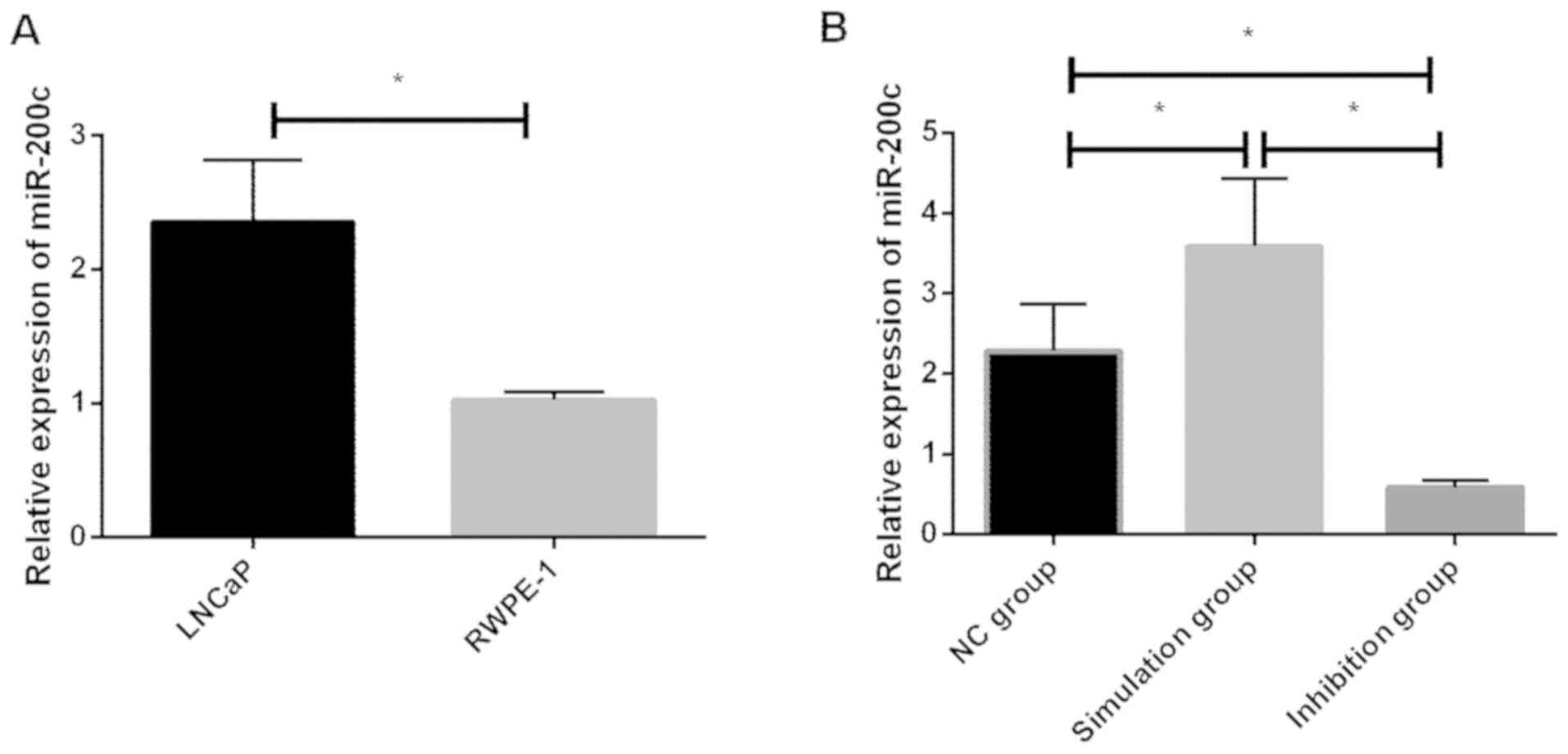
In this study, the relative expression of miR-200c
in LNCaP and RWPE-1 cells before transfection and in the NC,
simulation and inhibition groups of cells after transfection were
detected, and it was found that the relative expression of miR-200c
in LNCaP cells before transfection was significantly increased
compared with RWPE-1 cells, and the difference was statistically
significant (P<0.05). The difference was statistically
significant in the relative expression of miR-200c cells among the
NC, simulation and inhibition groups after transfection
(P<0.05). In addition, there was a significant decrease in the
inhibition group, as well as a difference between the inhibition,
NC, and simulation groups (P<0.05). A significant decrease in
the NC group of cells compared with the simulation group was also
identified, which was statistically different (P<0.05) (Tables II and III and Fig.
1).
| Table II.Relative expression of miR-200c in
LNCaP and RWPE-1 cells before transfection. |
Table II.
Relative expression of miR-200c in
LNCaP and RWPE-1 cells before transfection.
| Groups | LNCaP cells | RWPE-1 cells | F-value | P-value |
|---|
| Relative expression
of miR-200c | 2.353±0.468 | 1.025±0.058 | 4.878 | 0.008 |
| Table III.Relative expression of miR-200c in
each group of cells after transfection. |
Table III.
Relative expression of miR-200c in
each group of cells after transfection.
| Groups | NC group | Simulation group | Inhibition group | F-value | P-value |
|---|
| Relative expression
of miR-200c | 2.284±0.584 | 3.584±0.845 | 0.584±0.095 | 19.143 | 0.003 |
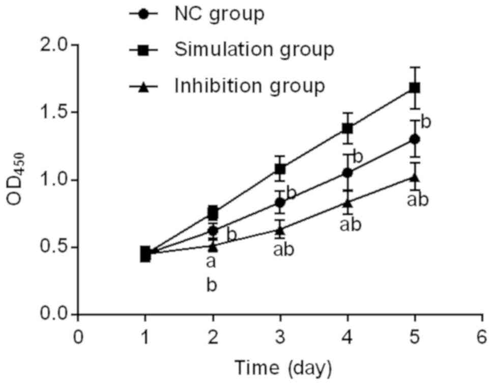
Proliferation in each group of cells
following transfection
Proliferation in each group of cells after
transfection was detected using CCK-8. It was found that there were
differences at the 2nd, 3rd, 4th and 5th days of cell growth among
the NC, simulation and inhibition groups (P<0.05), among which
the growth ability in the inhibition group of cells was
significantly suppressed. Additionally, there was a difference
between the inhibition, NC, and simulation groups (P<0.05), and
the proliferation ability in NC group significantly decreased
compared with simulation group, and there was a statistical
difference (P<0.05) (Fig. 2).
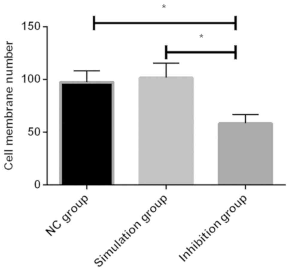
Effect on cell invasion ability after
transfection
The invasion ability of each group of cells after
transfection was detected using Transwell chamber, and it was found
that there was a difference in the three groups of cells
(P<0.05), among which the number of passed membrane cells in the
inhibition group was significantly smaller than that in the
remaining two groups, with a statistical difference (P<0.05).
Additionally, there was no significant decrease in NC group
compared with simulation group (P>0.05) (Table IV and Fig. 3).
| Table IV.Number of passed membrane cells. |
Table IV.
Number of passed membrane cells.
| Groups | No. of passed
membrane cells | F-value | P-value |
|---|
| NC group |
97.51±10.58ab |
|
|
| Simulation group |
119.58±13.9a | 23.058 | 0.002 |
| Inhibition group | 58.36±8.37 |
|
|
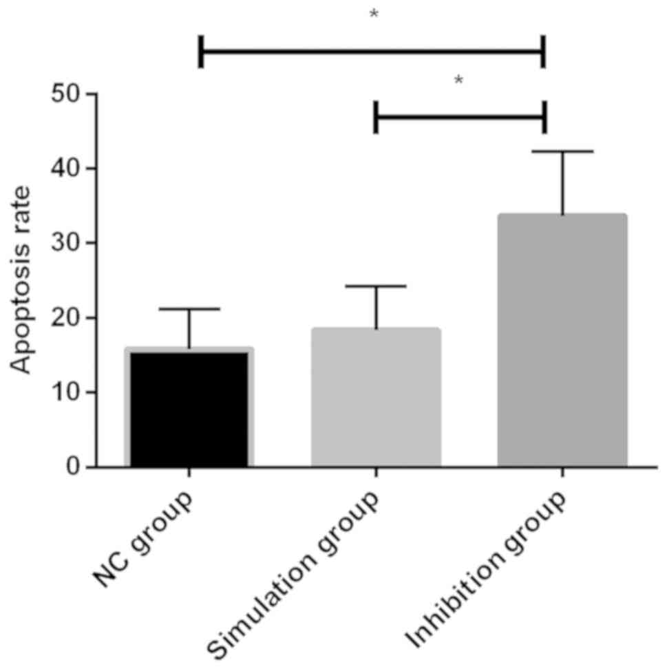
Cell apoptosis after transfection
By flow cytometry detection of the apoptosis ability
of each group of cells after transfection, it was found that there
was a difference in the apoptosis rate among the NC, simulation and
inhibition groups (P<0.05), of which a significant increase was
evident in the inhibition group. Additionally, there was a
difference between the inhibition, NC, and simulation groups
(P<0.05), but there was no statistical difference between the NC
and simulation groups (P>0.05) (Table
V and Fig. 4).
| Table V.Cell apoptosis (%). |
Table V.
Cell apoptosis (%).
| Groups | Apoptosis rate | F-value | P-value |
|---|
| NC group |
12.84±5.32ab |
|
|
| Simulation
group |
18.35±5.94a | 6.088 | 0.036 |
| Inhibition
group | 33.67±8.61 |
|
|
Discussion
PCa is a global malignancy of male with a high
incidence. Statistics have shown that there were approximately
180,890 new PCa patients in the United States in 2016, and
approximately 26,120 patients succumbed to the disease (5). The early clinical symptoms of PCa are
not obvious, and when the tumor is too large to block the urinary
tract or invade to the bladder neck, there is a severe lower
urinary tract infection. In the late stage of tumor, many patients
experience tumor metastasis, the most common of which is bone
metastasis, which is often accompanied by bone pain and fracture
(14). As with other solid tumors,
the main cause of death of PCa is due to cancer metastasis that is
a complex physiological process, including factors such as changes
in cell micro-environment, cell movement and cell growth (15).
At present, the treatment method of PCa at the
middle and late stage is mainly chemotherapy, but it is found
during the treatment of PCa that PCa tumor has a poor sensitivity
to chemotherapy, which leads to the emergence of drug resistance
during long-term chemotherapy, resulting in less than expected
effect (6). The main diagnostic
method of PCa is rectal examination (16) of serum PCa-specific antigen (PSA)
(17). Early screening is based on
rectal examination combined with PSA examination. However, the
subjective nature of rectal examination requires the experience of
clinicians for many years, and the early diagnosis of it before the
formation of nodules is prone to missed diagnosis (16). PSA, as an important serum marker of
PCa, has a high sensitivity that results in a low specificity, and
it may increase in patients with prostatitis or other prostate
diseases (18). Studies have shown
that (19) the diagnosis rate of PCa
is <30% with PSA concentration between 4–10 ng/ml, and PSA can
easily lead to over-diagnosis and over-treatment of PCa. Therefore,
there is a large study space for the diagnosis and treatment of
PCa, and the occurrence and development of it and its molecular
mechanism are still unclear, so it is of great significance for
clinical treatment and diagnosis to probe into the main mechanism
of PCa.
As a class of endogenous non-coding single-stranded
small RNA, miRs mainly present in eukaryotes and can regulate
target genes through transcription (20). Based on the binding of specific
target mRNA 3′-UTR and its target gene specificity, the target gene
mRNA will be degraded or the translation of it will be inhibited,
so as to regulate its post-transcriptional expression (21). Increasing number of studies have
proven that miRs play an important role in the occurrence and
development of a variety of cancers. Studies have shown that
miR-409-3p/-5p significantly increases in PCa tissues but miR-375
expression is low, suggesting that there is a difference in
expression of different miRs in PCa (22,23).
miR-200c, one of the five members of the miR-200 family, is an
important regulator of epithelial-mesenchymal transformation. In
addition to its role in normal cell phenotypic transformation,
miR-200c expression is differential in many cancer cells. For
example, high expression of serum miR-200c is associated with poor
prognosis in lung cancer patients (24). However, there are few studies on
miR-200c and PCa, and it is unclear whether miR-200c regulates PCa.
Therefore, we aimed to discover the biological function of miR-200c
in PCa through this investigation.
The miR-200c expression in PCa cells and normal
prostate cells were detected, and it was found that they
significantly increased in PCa cells compared with normal prostate
cells, and the difference was significant. In study of Vrba et
al (25), it was shown that
miR-200c in normal prostate tissues was lower than that in cancer
tissues. In study of Tao et al (26), it was shown that miR-200c was also
highly expressed in PCa patient tissues, consistent with our
findings. We transfected prostate cancer LNCaP cells and observed
changes in cell biological function after miR-200c. Promoting cell
apoptosis and inhibiting cell proliferation are currently important
ideas for clinical treatment of tumors. Through detection of each
group of cells after transfection, it was found that by inhibition
of the miR-200c expression can effectively reduce cell
proliferation and promote cell apoptosis rate. Ali et al
(27), showed that curcumin
inhibited the proliferation and apoptosis of PCa cells, which was
significantly reduced by detection of the miR-200c expression in
cells, suggesting that low expression of miR-200c can inhibit PCa
proliferation and promote apoptosis. Cancer metastasis is a major
cause of cancer exacerbations in the late stage. Through detection
of cell invasion ability, it was found that inhibition of the
miR-200c expression had a significant inhibitory effect on the
invasion ability of cells. Burk et al (28), showed that inhibition of the miR-200c
expression by ZEB1 inhibited the invasion ability of PCa cells,
which is consistent with our findings, suggesting that
down-regulating miR-200c can reduce the invasion ability of PCa
cells.
However, there are still some defects in this study.
First of all, the number of cells in vitro experiments was
small. Secondly, more in-depth studies were not conducted, and the
target genes and related pathways were not tested and validated.
Therefore, in future studies, we hope to collect clinical specimens
to validate our results, and test their target genes and pathways
to further explore their mechanisms, laying the foundation for
future clinical treatment and diagnosis.
In conclusion, the low expression of miR-200c is
beneficial in inhibiting proliferation and invasion of LNCaP cells
in vitro and promoting apoptosis, which may be a potential
target for PCa biotherapy.
Acknowledgements
Not applicable.
Funding
No funding was received.
Availability of data and materials
The datasets used and/or analyzed during the present
study are available from the corresponding author on reasonable
request.
Authors' contributions
JL drafted the manuscript. JL and YL were
responsible for cell culture and transfection. XZ assisted with
CCK-8 detection. QM contributed to MTT assay. LY performed the PCR.
All authors read and approved the final manuscript.
Ethics approval and consent to
participate
The study was approved by the Ethics Committee of
Shengzhou People's Hospital (Shengzhou, China).
Patient consent for publication
Not applicable.
Competing interests
The authors declare that they have no competing
interests.
References
|
1
|
Schröder FH, Hugosson J, Roobol MJ,
Tammela TL, Zappa M, Nelen V, Kwiatkowski M, Lujan M, Määttänen L,
Lilja H, et al ERSPC Investigators, : Screening and prostate cancer
mortality: Results of the European Randomised Study of Screening
for Prostate Cancer (ERSPC) at 13 years of follow-up. Lancet.
384:2027–2035. 2014. View Article : Google Scholar : PubMed/NCBI
|
|
2
|
Jemal A, Siegel R, Xu J and Ward E: Cancer
statistics, 2010. CA Cancer J Clin. 60:277–300. 2010. View Article : Google Scholar : PubMed/NCBI
|
|
3
|
Wong MC, Goggins WB, Wang HH, Fung FD,
Leung C, Wong SY, Ng CF and Sung JJ: Global incidence and mortality
for prostate cancer: Analysis of temporal patterns and trends in 36
countries. Eur Urol. 70:862–874. 2016. View Article : Google Scholar : PubMed/NCBI
|
|
4
|
Stewart BW and Wild CP: World Cancer
Report 2014. IARC Publications. (Lyon). 2014.
|
|
5
|
Siegel RL, Miller KD and Jemal A: Cancer
statistics, 2015. CA Cancer J Clin. 65:5–29. 2015. View Article : Google Scholar : PubMed/NCBI
|
|
6
|
Watson PA, Arora VK and Sawyers CL:
Emerging mechanisms of resistance to androgen receptor inhibitors
in prostate cancer. Nat Rev Cancer. 15:701–711. 2015. View Article : Google Scholar : PubMed/NCBI
|
|
7
|
Ceder Y, Bjartell A, Culig Z, Rubin MA,
Tomlins S and Visakorpi T: The molecular evolution of
castration-resistant prostate cancer. Eur Urol Focus. 2:506–513.
2016. View Article : Google Scholar : PubMed/NCBI
|
|
8
|
Nelson WG, De Marzo AM and Isaacs WB:
Prostate cancer. N Engl J Med. 349:366–381. 2003. View Article : Google Scholar : PubMed/NCBI
|
|
9
|
Bandara KV, Michael MZ and Gleadle JM:
MicroRNA biogenesis in hypoxia. MicroRNA. 6:80–96. 2017. View Article : Google Scholar : PubMed/NCBI
|
|
10
|
Wu HC, Lai MT, Wu CI, Chen HY, Wan L, Tsai
FJ and Chen WC: E-cadherin gene 3-UTR C/T polymorphism is
associated with prostate cancer. Urol Int. 75:350–353. 2005.
View Article : Google Scholar : PubMed/NCBI
|
|
11
|
Colden M, Dar AA, Saini S, Dahiya PV,
Shahryari V, Yamamura S, Tanaka Y, Stein G, Dahiya R and Majid S:
MicroRNA-466 inhibits tumor growth and bone metastasis in prostate
cancer by direct regulation of osteogenic transcription factor
RUNX2. Cell Death Dis. 8:e25722017. View Article : Google Scholar : PubMed/NCBI
|
|
12
|
Meng X, Müller V, Milde-Langosch K,
Trillsch F, Pantel K and Schwarzenbach H: Diagnostic and prognostic
relevance of circulating exosomal miR-373, miR-200a, miR-200b and
miR-200c in patients with epithelial ovarian cancer. Oncotarget.
7:16923–16935. 2016. View Article : Google Scholar : PubMed/NCBI
|
|
13
|
Livak KJ and Schmittgen TD: Analysis of
relative gene expression data using real-time quantitative PCR and
the 2(-Delta Delta C(T)) method. Methods. 25:402–408. 2001.
View Article : Google Scholar : PubMed/NCBI
|
|
14
|
Freitag MT, Radtke JP, Hadaschik BA,
Kopp-Schneider A, Eder M, Kopka K, Haberkorn U, Roethke M,
Schlemmer HP and Afshar-Oromieh A: Comparison of hybrid (68)Ga-PSMA
PET/MRI and (68)Ga-PSMA PET/CT in the evaluation of lymph node and
bone metastases of prostate cancer. Eur J Nucl Med Mol Imaging.
43:70–83. 2016. View Article : Google Scholar : PubMed/NCBI
|
|
15
|
Gundem G, Van Loo P, Kremeyer B,
Alexandrov LB, Tubio JM, Papaemmanuil E, Brewer DS, Kallio HML,
Högnäs G, Annala M, et al ICGC Prostate Group, : The evolutionary
history of lethal metastatic prostate cancer. Nature. 520:353–357.
2015. View Article : Google Scholar : PubMed/NCBI
|
|
16
|
Catalona WJ, Richie JP, Ahmann FR, Hudson
MA, Scardino PT, Flanigan RC, DeKernion JB, Ratliff TL, Kavoussi
LR, Dalkin BL, et al: Comparison of digital rectal examination and
serum prostate specific antigen in the early detection of prostate
cancer: Results of a multicenter clinical trial of 6,630 men. J
Urol. 197:S200–S207. 2017. View Article : Google Scholar : PubMed/NCBI
|
|
17
|
Jemal A, Fedewa SA, Ma J, Siegel R, Lin
CC, Brawley O and Ward EM: Prostate cancer incidence and PSA
testing patterns in relation to USPSTF screening recommendations.
JAMA. 314:2054–2061. 2015. View Article : Google Scholar : PubMed/NCBI
|
|
18
|
Abd-Alazeez M, Ahmed HU, Arya M, Charman
SC, Anastasiadis E, Freeman A, Emberton M and Kirkham A: The
accuracy of multiparametric MRI in men with negative biopsy and
elevated PSA level - can it rule out clinically significant
prostate cancer? Urol Oncol. 32:45.e17–45.e22. 2014. View Article : Google Scholar
|
|
19
|
Lee SW, Hosokawa K, Kim S, Jeong OC, Lilja
H, Laurell T and Maeda M: A highly sensitive porous silicon
(P-Si)-based human kallikrein 2 (hK2) immunoassay platform toward
accurate diagnosis of prostate cancer. Sensors (Basel).
15:11972–11987. 2015. View Article : Google Scholar : PubMed/NCBI
|
|
20
|
Suzuki HI, Young RA and Sharp PA:
Super-enhancer-mediated RNA processing revealed by integrative
MicroRNA network analysis. Cell. 168:1000–1014.e15. 2017.
View Article : Google Scholar : PubMed/NCBI
|
|
21
|
Li XL, Andersen JB, Ezelle HJ, Wilson GM
and Hassel BA: Post-transcriptional regulation of RNase-L
expression is mediated by the 3-untranslated region of its mRNA. J
Biol Chem. 282:7950–7960. 2007. View Article : Google Scholar : PubMed/NCBI
|
|
22
|
Josson S, Gururajan M, Hu P, Shao C, Chu
GY, Zhau HE, Liu C, Lao K, Lu CL, Lu YT, et al: miR-409-3p/-5p
promotes tumorigenesis, epithelial-to-mesenchymal transition, and
bone metastasis of human prostate cancer. Clin Cancer Res.
20:4636–4646. 2014. View Article : Google Scholar : PubMed/NCBI
|
|
23
|
Huang X, Yuan T, Liang M, Du M, Xia S,
Dittmar R, Wang D, See W, Costello BA, Quevedo F, et al: Exosomal
miR-1290 and miR-375 as prognostic markers in castration-resistant
prostate cancer. Eur Urol. 67:33–41. 2015. View Article : Google Scholar : PubMed/NCBI
|
|
24
|
Liu XG, Zhu WY, Huang YY, Ma LN, Zhou SQ,
Wang YK, Zeng F, Zhou JH and Zhang YK: High expression of serum
miR-21 and tumor miR-200c associated with poor prognosis in
patients with lung cancer. Med Oncol. 29:618–626. 2012. View Article : Google Scholar : PubMed/NCBI
|
|
25
|
Vrba L, Jensen TJ, Garbe JC, Heimark RL,
Cress AE, Dickinson S, Stampfer MR and Futscher BW: Role for DNA
methylation in the regulation of miR-200c and miR-141 expression in
normal and cancer cells. PLoS One. 5:e86972010. View Article : Google Scholar : PubMed/NCBI
|
|
26
|
Tao T, Liu D, Liu C, Xu B, Chen S, Yin Y,
Ang L, Huang Y, Zhang X and Chen M: Autoregulatory feedback loop of
EZH2/miR-200c/E2F3 as a driving force for prostate cancer
development. Biochim Biophys Acta. 1839:858–865. 2014. View Article : Google Scholar : PubMed/NCBI
|
|
27
|
Ali S, Ahmad A, Banerjee S, Padhye S,
Dominiak K, Schaffert JM, Wang Z, Philip PA and Sarkar FH:
Gemcitabine sensitivity can be induced in pancreatic cancer cells
through modulation of miR-200 and miR-21 expression by curcumin or
its analogue CDF. Cancer Res. 70:3606–3617. 2010. View Article : Google Scholar : PubMed/NCBI
|
|
28
|
Burk U, Schubert J, Wellner U, Schmalhofer
O, Vincan E, Spaderna S and Brabletz T: A reciprocal repression
between ZEB1 and members of the miR-200 family promotes EMT and
invasion in cancer cells. EMBO Rep. 9:582–589. 2008. View Article : Google Scholar : PubMed/NCBI
|