Introduction
Lung cancer is one of the most common cancers in
clinic, it is often found in mucosal epithelium of bronchus
(1). Studies showed that in 2016 new
patients with lung cancer increased ~2.67 million worldwide, and
the morbidity of lung cancer ranked the second among all cancers
(2). Lung adenocarcinoma is a lung
cancer where the age of onset tends to be younger (3). At present, the pathogenesis of lung
adenocarcinoma is still unclear. Tumor metastasis easily occurs in
lung adenocarcinoma and ~30% of patients with advanced lung
adenocarcinoma have metastasis, which makes the fatality rate of
lung adenocarcinoma the highest among all cancers (4,5). It has
been shown that without effective treatment, the median survival
time of this type of patients is only 1 month (6). The most effective means of treating
lung adenocarcinoma in clinic are resection, radiotherapy and
chemotherapy (7). At present, with
the development of the treatment of tumor diseases, lung cancer
resection has been improved and nidus separation can be finished
preferably in patients during resection (8), however, in the process of postoperative
chemotherapy, side effects easily appear when normal lung tissue in
the radiation field is injured (9).
Patients with slight injury have difficulty in breathing and
impairment of lung function, while in severe cases patients present
extensive pulmonary fibrosis, which directly threatens their life
(10). Therefore, the way to improve
the therapeutic effective rate of lung adenocarcinoma is a major
focus in current clinical research. Molecular targeted therapy has
become a hotspot in research. Some data have demonstrated that
AFAP1-AS1, CDK4, CDK6 and NAT8L may become potential targets of
future clinical treatment of lung cancer (11–13).
Epidermal growth factor receptor-tyrosine kinase inhibitor
(EGFR-TKI) has an extremely important influence on lung
adenocarcinoma (14). Cancer factors
enhance the susceptibility of tumor tissue for programmed cell
death protein 1 (PD-1) blockade via EGFR pathway, and the key of
EGFR-TKI is inhibiting the combination of PD-1 and EGFR (15). It has been proven that lung
adenocarcinoma receives the largest beneficial effect when EGFR-TKI
is applied in the treatment of non-small cell carcinoma (16,17), but
it is still controversial how to better predict the clinical
efficacy and prognosis of patients. Carcinoembryonic antigen (CEA)
is currently the most sensitive tumor marker (18), and lactate dehydrogenase (LDH) is a
prognostic indicator that is closely related to tumor cell load and
distant metastasis (19). There are
few studies on the two in the targeted therapy of lung
adenocarcinoma, therefore, this report retrospectively analyzed
patients with lung adenocarcinoma who were admitted and treated in
The First People's Hospital of Chuzhou (Chuzhou, China) since 2014
to investigate the significance of CEA and LDH in the targeted
therapy of lung adenocarcinoma.
Patients and methods
General data
Eighty-nine patients with lung adenocarcinoma, who
were admitted to The First People's Hospital of Chuzhou from
January 2014 to February 2015, were the study subjects and were
analyzed retrospectively. Among the patients, there were 38
patients with EGFR sensitive mutation, accounting for 42.70% of all
subjects. There were 34 males and 55 females, aged from 37 to 69
years, with an average age of 51.63±11.74 years.
Inclusion and exclusion criteria
Inclusion criteria: all patients who were diagnosed
with lung adenocarcinoma via biopsy in the Pathology Department of
The First People's Hospital of Chuzhou; patients who received lung
tissue resection or EGFR-TKI targeted therapy in The First People's
Hospital of Chuzhou; patients with complete clinical data; patients
40–65 years of age. Exclusion criteria: patients with other
abnormalities in cardiopulmonary functions before the preoperative
diagnosis; with other tumor diseases, blood diseases, immune
diseases, or physical disabilities; who were chronically ill;
patients in gestation period; patients who had surgical
contraindications; patients who had received chemotherapy before
operation; patients pathologically diagnosed with local or distant
metastasis after operation. The study was approved by the Ethics
Committee of The First People's Hospital of Chuzhou. Patients had
complete clinical data and signed informed consents were obtained
from the patients and/or the guardians.
Methods
Among the 89 study subjects, 51 patients received
resection in The First People's Hospital of Chuzhou and were set as
the operation group; the other 38 patients received EGFR-TKI
targeted therapy in The First People's Hospital of Chuzhou, and
were considered the targeted group. The operations were performed
by the senior chief clinicians. The therapeutic drug was AZD9291,
also known as Osimertinib, which is the third generation of
EGFR-TKI. This compound is an irreversible mutant selective
EGFR-TKI (exon 19 deletion type EGFRIC50=12.92 nM,
L858R/T790M EGFR IC50=11.44 nM, wild-type EGFR
IC50=493.8 nM), oral administration, 80 mg, PO QD.
Venous blood of patients (5.0 ml) was taken before
surgery, 4 weeks after the targeted therapy, or 1 week after
surgery. CEA electrochemiluminescence kit (cat. no. LT13001;
Quanzhou Lantu Biotechnology Co., Ltd., Quanzhou, China) was used
to detect the expression of CEA in serum with UniCel DxI 800
immunoassay system (Beckman Coulter, Inc., Brea, CA, USA), and an
automatic biochemical analyzer (Beckman Coulter, Inc., Shanghai,
China) was used to detect the expression of LDH in serum.
Observation indicators
The clinical data of patients in the two groups
included sex, age, weight, and course of disease. The therapeutic
effective rate was calculated using the Response Evaluation
Criteria in Solid Tumors (RECIST) of 2012 as a reference (20) and the patients were separated into
four stages, based on complete response (CR), partial response
(PR), stable disease (SD) and progressive disease (PD). All
patients took imageological examination at the 5th week after the
treatment, and the imageological results were used for the
evaluation and reference of the final efficacy. Therapeutic
effective rate = (the number of patients who were graded in
CR+PR)/total number ×100%. Patients in the targeted group were
given a 3-year prognostic follow-up in the form of call, letter,
hospital review and visit. The deadline of the follow-up was April
2018 and the ultimate event was the death of the patient. The
3-year survival rate of the patients was recorded.
Statistical analysis
SPSS 24.0 statistical software (Shanghai Yuchuang
Network Technology Co., Ltd., Shanghai, China) was used to analyze
and process all data; the enumeration data were expressed in the
form of rate, and Chi-square (χ2) test was used for
their comparison between groups; the measurement data were
expressed as the mean ± standard deviation, and paired t-test was
used for their comparison between groups. Kaplan-Meier survival
analysis was used for the survival curves and log-rank test was
used for the comparison of the survival rates. P<0.05 was
considered to indicate a statistically significant difference.
Results
Comparison of the clinical data
There were no significant differences concerning
age, weight, course of disease, sex, smoking, drinking, place of
residence, tumor metastasis and differentiated degree of patients
between the two groups (P>0.050). This proves that there was
comparability between the patients of the two groups. Details are
shown in Table I.
 | Table I.Comparison of the patients' clinical
data between the two groups [n (%)]. |
Table I.
Comparison of the patients' clinical
data between the two groups [n (%)].
| Characteristics | Targeted group
(n=38) | Operation group
(n=51) | χ2 or
t | P-value |
|---|
| Age (years) | 51.67±12.33 | 50.84±11.96 | 0.320 | 0.750 |
| Weight (kg) | 72.96±15.68 | 74.24±16.07 | 0.376 | 0.708 |
| Course of disease
(weeks) |
3.52±1.66 |
3.70±1.54 | 0.528 | 0.599 |
| Sex |
|
| 0.045 | 0.831 |
| Male | 15 (39.47) | 19 (37.25) |
|
|
|
Female | 23 (60.53) | 32 (62.75) |
|
|
| Smoking |
|
| 0.163 | 0.686 |
| Yes | 8
(21.05) | 9
(17.65) |
|
|
| No | 30 (78.95) | 42 (82.35) |
|
|
| Drinking |
|
| 0.112 | 0.738 |
| Yes | 5
(13.16) | 8
(15.69) |
|
|
| No | 33 (86.84) | 43 (84.31) |
|
|
| Place of
residence |
|
| 0.362 | 0.547 |
| City | 31 (81.58) | 44 (86.27) |
|
|
|
Countryside | 7
(18.42) | 7
(13.73) |
|
|
| Tumor metastasis |
|
| 0.056 | 0.813 |
| Yes | 9
(23.68) | 11 (21.57) |
|
|
| No | 29 (76.32) | 40 (78.43) |
|
|
| Differentiated
degree |
|
| 0.159 | 0.924 |
| High | 13 (34.21) | 16 (31.37) |
|
|
|
Middle | 20 (52.63) | 29 (56.86) |
|
|
| Low | 5
(13.16) | 6
(11.76) |
|
|
Comparison of the therapeutic
effective rate
In the targeted group, 52.63% of the cases were CR,
28.95% were PR, 13.16% were SD, and 5.26% were PD; the therapeutic
effective rate was 81.58%. In the operation group, 56.86% of the
cases were CR, 25.49% were PR, 9.80% were SD, and 5.88% were PD;
the therapeutic effective rate was 82.35%. The differences were
found to be statistically not significant when the therapeutic
effective rates of the two groups were compared (P>0.050)
(Table II).
| Table II.Comparison of the therapeutic
effective rate [n (%)]. |
Table II.
Comparison of the therapeutic
effective rate [n (%)].
| Items | Targeted group
(n=38) | Operation group
(n=51) | χ2 | P-value |
|---|
| CR | 20 (52.63) | 29 (56.86) | 0.787 | 0.375 |
| PR | 11 (28.95) | 13 (25.49) | 0.132 | 0.716 |
| SD | 5 (13.16) | 5 (9.80) | 0.246 | 0.620 |
| PD | 2 (5.26) | 4 (7.84) | 0.016 | 0.900 |
| Effective rate
(%) | 81.58 | 82.35 | 0.009 | 0.925 |
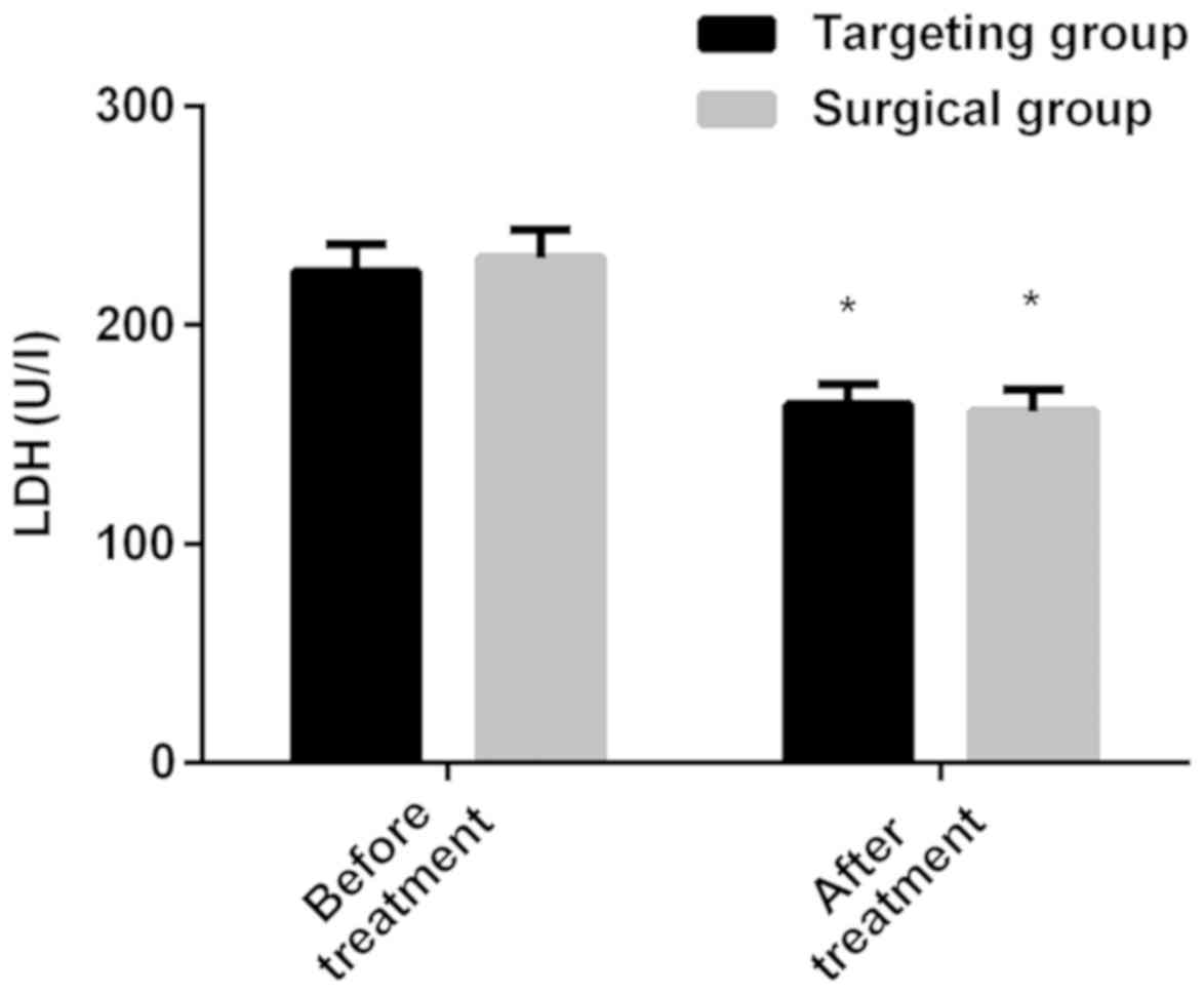
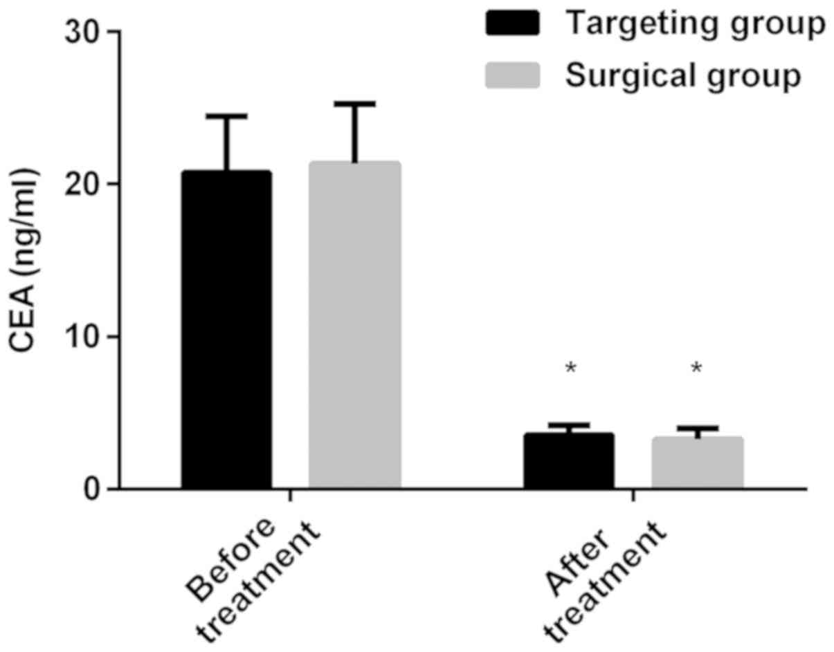
Comparison of the expression levels of
LDH and CEA
The expression levels of LDH in the targeted group
before and after treatment were 224.56±12.67 and 163.75±9.24 U/l,
respectively; the expression levels of LDH in the operation group
before and after treatment were 230.64±13.08 and 160.54±9.87 U/l,
respectively. There was no significant difference in the LDH of the
patients in the two groups before or after treatment (P>0.050).
The expression level of LDH after treatment was lower than that
before treatment (P<0.050) in both groups. The expression levels
of CEA in the targeted group before and after treatment were
20.77±3.68 and 3.54±0.62 ng/ml, respectively; the expression levels
of CEA in the operation group before and after treatment were
21.34±3.92 and 3.27±0.70 ng/ml, respectively. There was no
significant difference in CEA of patients in the two groups before
or after treatment (P>0.050). The expression level of CEA after
treatment was lower than that before treatment (P<0.050) in both
groups (Figs. 1 and 2).
Difference of efficacy of the targeted
groups
According to the medians of LDH and CEA expression
levels in patients of the two groups before treatment, 20 cases
were in LDH high-expression group (LDH≥224.56 U/l); 18 patients
were in LDH low-expression group (LDH<224.56 U/l); 17 patients
were in CEA high-expression group (CEA≥20.77 ng/ml); and 21
patients were in CEA low-expression group (CEA<20.77 ng/ml). The
therapeutic effective rate of LDH high-expression group was 65.00%,
which was significantly lower than that of LDH low-expression group
(100.00%) (P=0.004). The therapeutic effective rate of CEA
high-expression group was 64.71%, which was also significantly
lower than that of CEA low-expression group (95.24%) (P=0.016).
Details are shown in Tables III
and IV.
| Table III.Therapeutic effective rate of LDH
high and low-expression groups [n (%)]. |
Table III.
Therapeutic effective rate of LDH
high and low-expression groups [n (%)].
| Items | LDH high-expression
group (n=20) | LDH low-expression
group (n=18) | χ2 | P-value |
|---|
| CR | 5 (25.00) | 14 (77.78) |
|
|
| PR | 8 (40.00) | 4 (22.22) |
|
|
| SD | 5 (25.00) | 0 (0.00) |
|
|
| PD | 2 (10.00) | 0 (0.00) |
|
|
| Effective rate
(%) | 65.00 | 100.00 | 8.485 | 0.004 |
| Table IV.Therapeutic effective rate of CEA
high and low-expression groups [n (%)]. |
Table IV.
Therapeutic effective rate of CEA
high and low-expression groups [n (%)].
| Items | CEA high-expression
group (n=17) | CEA low-expression
group (n=21) | χ2 | P-value |
|---|
| CR | 5 (29.41) | 15 (71.43) |
|
|
| PR | 6 (35.29) | 5 (23.81) |
|
|
| SD | 4 (23.53) | 1 (4.76) |
|
|
| PD | 2 (11.76) | 0 (0.00) |
|
|
| Effective rate
(%) | 64.71 | 95.24 | 5.828 | 0.016 |
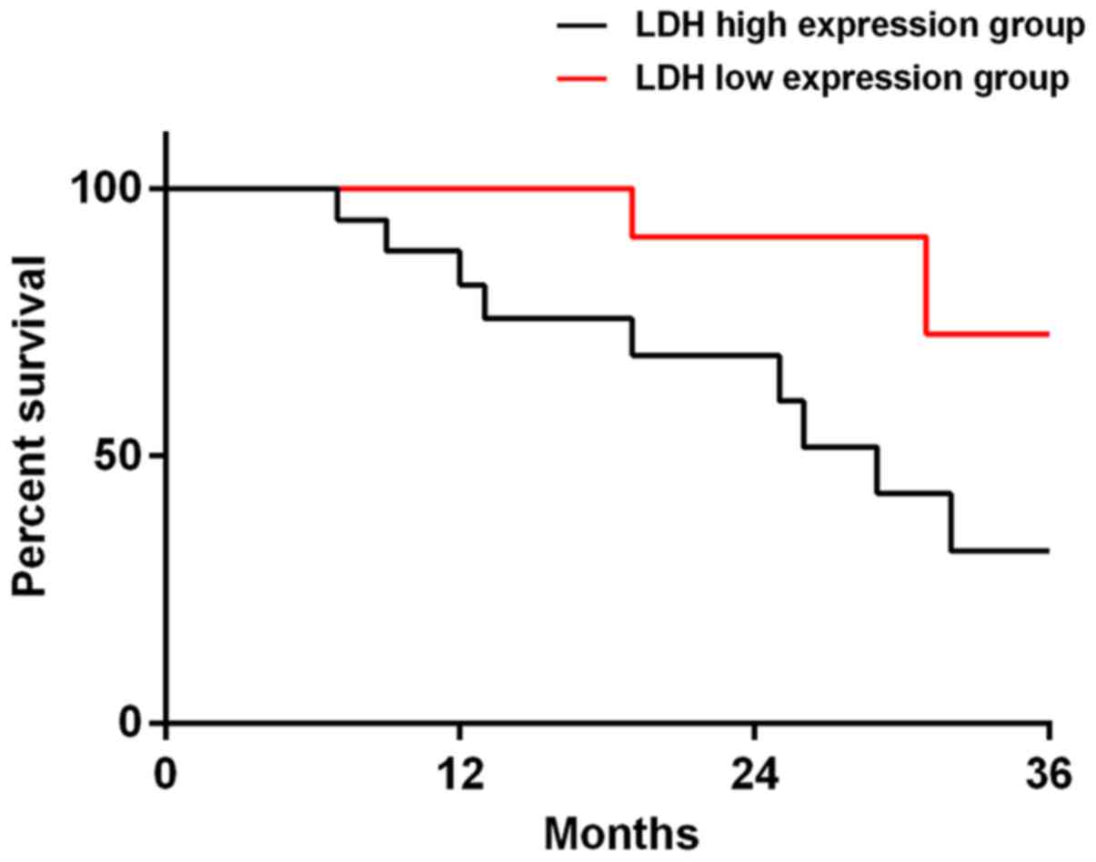
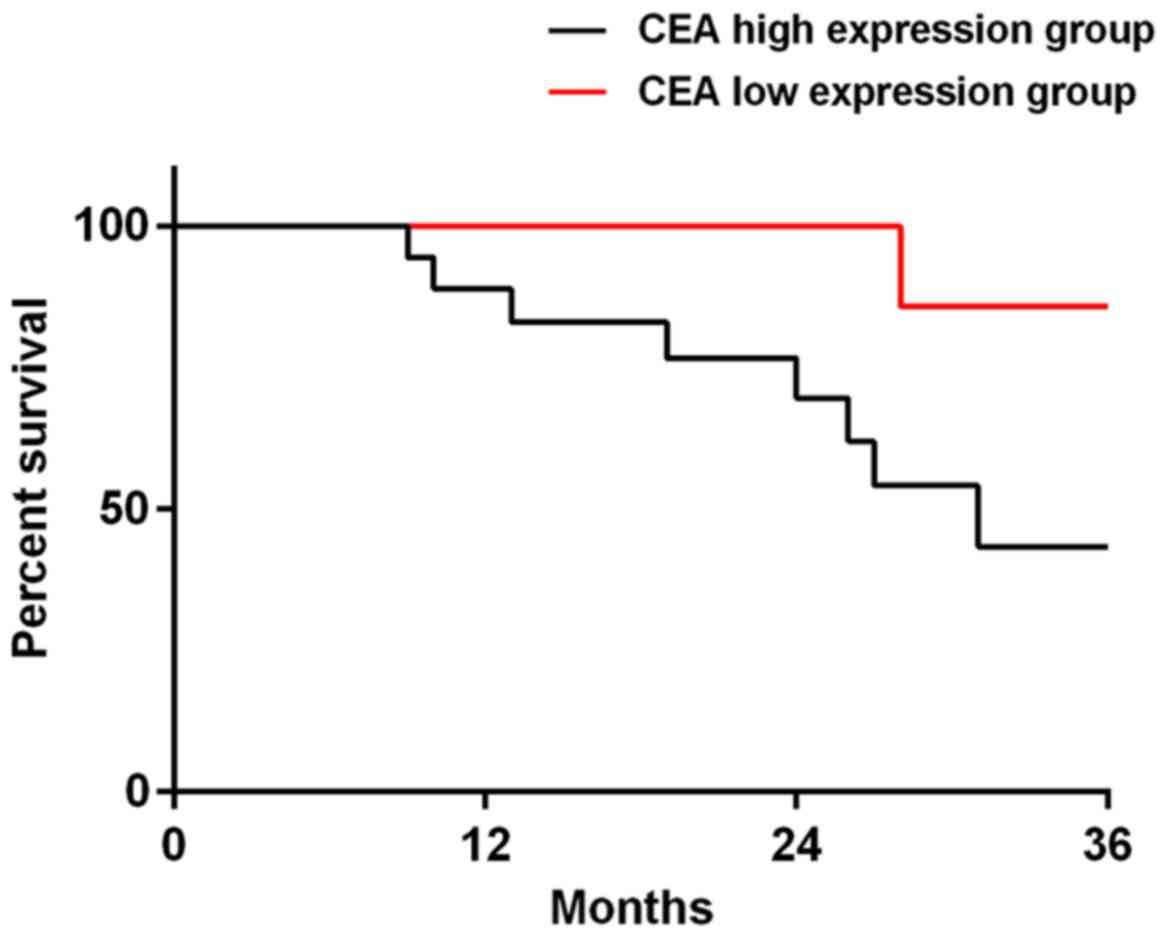
Prognosis
Among the 38 patients in the targeted group, 37
patients were successfully followed up, and the success rate of the
follow-up visit was 97.37%. There were 19 patients in LDH
high-expression group, 18 patients in LDH low-expression group, 16
patients in CEA high-expression group and 21 patients in CEA
low-expression group. The 3-year total mortality of LDH
high-expression group was 47.37%, which was significantly higher
than that of LDH low-expression group (11.11%) (P=0.034). The
3-year total mortality of CEA high-expression group was 56.25%,
which was significantly higher than that of CEA low-expression
group (4.76%) (P=0.020). When the data of all the deceased patients
were compared, it was concluded that 14 patients (77.78%) had
high-expression of LDH and CEA (Figs.
3 and 4).
Discussion
The morbidity and mortality of lung adenocarcinoma
are extremely high in clinic (21).
The scheme of diagnosis and treatment of lung adenocarcinoma has
long been a research hotspot in clinic, and targeted therapy has
gradually become popularized in recent years. Compared with
traditional resection, radiotherapy and chemotherapy, the targeted
therapy is safer in ensuring the effective remission of patients'
tumors, and it achieves the purpose of treating tumors by
regulating one or more factors directly related to tumors (21,22).
While EGFR-TKI is applied in lung adenocarcinoma, the mechanism is
using the combination between gefitinib or erlotinib and EGFR in
lung adenocarcinoma cells, and disrupting the signal transduction
of the downstream cells by inhibiting the activation of tyrosine
kinase, thereby preventing the spread, infiltration and metastasis
of cancer cells (23). However, some
related drug resistance usually appears in most patients in the
process of targeted therapy, which weakens the therapeutic effect
of targeted drugs (24). So it is
particularly important to monitor the therapeutic effect and
estimate the condition of prognosis in the process of targeted
therapy.
In this study, the difference of the efficacy
between EGFR-TKI targeted therapy and traditional resection for the
patients with lung adenocarcinoma, and the expression levels of LDH
and CEA were investigated. The significance of the expression
levels of LDH and CEA and the effect of prognosis of LDH and CEA in
patients with lung adenocarcinoma who were treated with EGFR-TKI
targeted therapy were also investigated. Results showed that there
is no significant difference in the therapeutic effective rates and
the expression of LDH and CEA between the targeted and the
operation group, which suggests that EGFR-TKI targeted therapy has
an extremely high application value for treating lung
adenocarcinoma. LDH mainly belongs to the enzyme in the glycolytic
pathway and plays a great role in facilitating the formation of
lactic acid, which originates from pyruvate (25). In general, LDH exists in almost every
cell of the organisms, and its activity is generally high in human
body. Therefore, LDH does not have high specificity for the early
diagnosis of diseases, and it is often used as a reference to
prognostically monitor and estimate the state of the disease of
patients (26). CEA is a
glycoprotein that only exists in cancer and embryonic tissues, and
directly acts on the adhesion reaction between tumor cells, which
has a strong regulatory ability on the differentiation, apoptosis,
infiltration and metastasis of cancer cells (27). In this study, there was no difference
between the targeted and the operation group, which proves that
EGFR-TKI targeted therapy could effectively improve the condition
of tumor nidus of lung adenocarcinoma in patients, reduce the tumor
volume in patients, and achieve the purpose of treating tumors. In
addition, there was no difference in the expression of CEA and LDH
before or after treatment between the two groups, which
preliminarily suggests that these indicators could be generally
applied for the treatment of lung cancer, and errors in expression
levels would not be caused by traumatic surgery. However, more
in-depth research is required for comparison in order to obtain
most accurate experiment results. The contingency could not be
excluded due to the small sample size in this study. Analysis of
the efficacy and the difference of prognosis of patients in CEA
high-expression group, CEA low-expression group, LDH
high-expression group and LDH low-expression group in the targeted
group showed that CEA and LDH high-expression groups were both
significantly worse than CEA and LDH low-expression groups. This
suggests that CEA and LDH both have a high application value for
monitoring the efficacy and prognosis of patients who receive
EGFR-TKI targeted therapy. Tumor cells can perform unlimited
aerobic glycolysis with the support of enough nutrition, while the
main substance that provides nutrition for them is LDH (28). It is speculated that due to the
incomplete elimination of tumor nidus, cancer cells consume a large
amount of oxygen in patient's body, thus the patient's body is
chronically in a state of hypoxia, which induces the enhancement of
glycolysis and raises the level of LDH, allowing tumor cells to get
enough energy again to further metastasize and invade. So, the
prognosis of LDH high-expression group is significantly worse than
that of LDH low-expression group. CEA, a tumor marker, reflects the
active ability of tumors. The higher its expression is, the
stronger the activation ability of tumors is, thereby, the
prognosis is worse.
In this study, the difference of efficacy between
EGFR-TKI targeted therapy and traditional resection in the
treatment of patients with lung adenocarcinoma and the expression
of LDH and CEA were investigated, and the significance of the
expression and the effect of prognosis of LDH and CEA in patients
with lung adenocarcinoma treated with EGFR-TKI targeted therapy
were also investigated. There were some limitations because of the
lack of experimental conditions. For example, the sample size was
small, and the experimental population was uniform. Also, the time
of the follow-up was short. LDH and CEA are both reactants with low
specificity, and the abnormal expression of them could occur in
patients with severe infections and organ damage.
In conclusion, the levels of CEA and LDH in serum
are abnormally expressed in the process of EGFR-TKI targeted
therapy of lung adenocarcinoma, which is of great significance for
monitoring the efficacy and prognosis of the therapy of lung
adenocarcinoma targeted by EGFR-TKI.
Acknowledgements
Not applicable.
Funding
No funding was received.
Availability of data and materials
The datasets used and/or analyzed during the present
study are available from the corresponding author on reasonable
request.
Authors' contributions
GH, ZJ and SX recorded and analyzed the observation
indicators. XS and WW collected and interpreted the patients' data
using electrochemiluminescence, the automatic biochemical analyzer
and SPSS 24.0 statistical software. GH wrote and revised the
manuscript. All authors read and approved the final manuscript.
Ethics approval and consent to
participate
The study was approved by the Ethics Committee of
The First People's Hospital of Chuzhou (Chuzhou, China). Patients
who participated in this research had complete clinical data and
signed informed consents were obtained from the patients and/or the
guardians.
Patient consent for publication
Not applicable.
Competing interests
The authors declare that they have no competing
interests.
References
|
1
|
Reck M, Rodríguez-Abreu D, Robinson AG,
Hui R, Csőszi T, Fülöp A, Gottfried M, Peled N, Tafreshi A, Cuffe
S, et al KEYNOTE-024 Investigators, : Pembrolizumab versus
chemotherapy for PD-L1 - positive non-small-cell lung cancer. N
Engl J Med. 375:1823–1833. 2016. View Article : Google Scholar : PubMed/NCBI
|
|
2
|
Herbst RS, Baas P, Kim DW, Felip E,
Pérez-Gracia JL, Han JY, Molina J, Kim JH, Arvis CD, Ahn MJ, et al:
Pembrolizumab versus docetaxel for previously treated,
PD-L1-positive, advanced non-small-cell lung cancer (KEYNOTE-010):
A randomised controlled trial. Lancet. 387:1540–1550. 2016.
View Article : Google Scholar : PubMed/NCBI
|
|
3
|
Lou Y, Diao L, Cuentas ERP, Denning WL,
Chen L, Fan YH, Byers LA, Wang J, Papadimitrakopoulou VA, Behrens
C, et al: Epithelial-mesenchymal transition is associated with a
distinct tumor microenvironment including elevation of inflammatory
signals and multiple immune checkpoints in lung adenocarcinoma.
Clin Cancer Res. 22:3630–3642. 2016. View Article : Google Scholar : PubMed/NCBI
|
|
4
|
Takada K, Okamoto T, Shoji F, Shimokawa M,
Akamine T, Takamori S, Katsura M, Suzuki Y, Fujishita T, Toyokawa
G, et al: Clinical significance of PD-L1 protein expression in
surgically resected primary lung adenocarcinoma. J Thorac Oncol.
11:1879–1890. 2016. View Article : Google Scholar : PubMed/NCBI
|
|
5
|
Johung KL, Yeh N, Desai NB, Williams TM,
Lautenschlaeger T, Arvold ND, Ning MS, Attia A, Lovly CM, Goldberg
S, et al: Extended survival and prognostic factors for patients
with ALK-rearranged non-small-cell lung cancer and brain
metastasis. J Clin Oncol. 34:123–129. 2016. View Article : Google Scholar : PubMed/NCBI
|
|
6
|
Schuler M, Wu YL, Hirsh V, O'Byrne K,
Yamamoto N, Mok T, Popat S, Sequist LV, Massey D, Zazulina V, et
al: First-line afatinib versus chemotherapy in patients with
non-small cell lung cancer and common epidermal growth factor
receptor gene mutations and brain metastases. J Thorac Oncol.
11:380–390. 2016. View Article : Google Scholar : PubMed/NCBI
|
|
7
|
Ambrogio C, Gómez-López G, Falcone M,
Vidal A, Nadal E, Crosetto N, Blasco RB, Fernández-Marcos PJ,
Sánchez- Céspedes M, Ren X, et al: Combined inhibition of DDR1 and
Notch signaling is a therapeutic strategy for KRAS-driven lung
adenocarcinoma. Nat Med. 22:270–277. 2016. View Article : Google Scholar : PubMed/NCBI
|
|
8
|
Martin JT, Durbin EB, Chen L, Gal T, Mahan
A, Ferraris V and Zwischenberger J: Nodal upstaging during lung
cancer resection is associated with surgical approach. Ann Thorac
Surg. 101:238–244; discussion 44–45. 2016. View Article : Google Scholar : PubMed/NCBI
|
|
9
|
Kwon Y: Mechanism-based management for
mucositis: Option for treating side effects without compromising
the efficacy of cancer therapy. OncoTargets Ther. 9:2007–2016.
2016. View Article : Google Scholar
|
|
10
|
Chen QY, Jiao DM, Wang J, Hu H, Tang X,
Chen J, Mou H and Lu W: miR-206 regulates cisplatin resistance and
EMT in human lung adenocarcinoma cells partly by targeting MET.
Oncotarget. 7:24510–24526. 2016.PubMed/NCBI
|
|
11
|
Zeng Z, Bo H, Gong Z, Lian Y, Li X, Li X,
Zhang W, Deng H, Zhou M, Peng S, et al: AFAP1-AS1, a long noncoding
RNA upregulated in lung cancer and promotes invasion and
metastasis. Tumour Biol. 37:729–737. 2016. View Article : Google Scholar : PubMed/NCBI
|
|
12
|
Patnaik A, Rosen LS, Tolaney SM, Tolcher
AW, Goldman JW, Gandhi L, Papadopoulos KP, Beeram M, Rasco DW,
Hilton JF, et al: Efficacy and safety of abemaciclib, an inhibitor
of CDK4 and CDK6, for patients with breast cancer, non-small cell
lung cancer, and other solid tumors. Cancer Discov. 6:740–753.
2016. View Article : Google Scholar : PubMed/NCBI
|
|
13
|
Lou TF, Sethuraman D, Dospoy P, Srivastva
P, Kim HS, Kim J, Ma X, Chen PH, Huffman KE, Frink RE, et al:
Cancer-specific production of N-acetylaspartate via NAT8L
overexpression in non-small cell lung cancer and its potential as a
circulating biomarker. Cancer Prev Res (Phila). 9:43–52. 2016.
View Article : Google Scholar : PubMed/NCBI
|
|
14
|
Wu SG, Liu YN, Tsai MF, Chang YL, Yu CJ,
Yang PC, Yang JC, Wen YF and Shih JY: The mechanism of acquired
resistance to irreversible EGFR tyrosine kinase inhibitor-afatinib
in lung adenocarcinoma patients. Oncotarget. 7:12404–12413.
2016.PubMed/NCBI
|
|
15
|
Ahn MJ, Sun JM, Lee SH, Ahn JS and Park K:
EGFR TKI combination with immunotherapy in non-small cell lung
cancer. Expert Opin Drug Saf. 16:465–469. 2017. View Article : Google Scholar : PubMed/NCBI
|
|
16
|
Zheng D, Ye X, Zhang MZ, Sun Y, Wang JY,
Ni J, Zhang HP, Zhang L, Luo J, Zhang J, et al: Plasma EGFR T790M
ctDNA status is associated with clinical outcome in advanced NSCLC
patients with acquired EGFR-TKI resistance. Sci Rep. 6:209132016.
View Article : Google Scholar : PubMed/NCBI
|
|
17
|
Gainor JF, Shaw AT, Sequist LV, Fu X,
Azzoli CG, Piotrowska Z, Huynh TG, Zhao L, Fulton L, Schultz KR, et
al: EGFR mutations and ALK rearrangements are associated with low
response rates to PD-1 pathway blockade in non-small cell lung
cancer (NSCLC): A retrospective analysis. Clin Cancer Res.
22:4585–4593. 2016. View Article : Google Scholar : PubMed/NCBI
|
|
18
|
Bacac M, Fauti T, Sam J, Colombetti S,
Weinzierl T, Ouaret D, Bodmer W, Lehmann S, Hofer T, Hosse RJ, et
al: A novel carcinoembryonic antigen T-cell bispecific antibody
(CEA TCB) for the treatment of solid tumor. Clin Cancer Res.
22:3286–3297. 2016. View Article : Google Scholar : PubMed/NCBI
|
|
19
|
Valvona CJ, Fillmore HL, Nunn PB and
Pilkington GJ: The regulation and function of lactate dehydrogenase
A: Therapeutic potential in brain tumor. Brain Pathol. 26:3–17.
2016. View Article : Google Scholar : PubMed/NCBI
|
|
20
|
Shiono S and Yanagawa N: Spread through
air spaces is a predictive factor of recurrence and a prognostic
factor in stage I lung adenocarcinoma. Interact Cardiovasc Thorac
Surg. 23:567–572. 2016. View Article : Google Scholar : PubMed/NCBI
|
|
21
|
Minguet J, Smith KH and Bramlage P:
Targeted therapies for treatment of non-small cell lung cancer -
recent advances and future perspectives. Int J Cancer.
138:2549–2561. 2016. View Article : Google Scholar : PubMed/NCBI
|
|
22
|
Cha YJ, Kim HR, Lee CY, Cho BC and Shim
HS: Clinicopathological and prognostic significance of programmed
cell death ligand-1 expression in lung adenocarcinoma and its
relationship with p53 status. Lung Cancer. 97:73–80. 2016.
View Article : Google Scholar : PubMed/NCBI
|
|
23
|
Park K, Tan EH, O'Byrne K, Zhang L, Boyer
M, Mok T, Hirsh V, Yang JC8 Lee KH, Lu S, et al: Afatinib versus
gefitinib as first-line treatment of patients with EGFR
mutation-positive non-small-cell lung cancer (LUX-Lung 7): A phase
2B, open-label, randomised controlled trial. Lancet Oncol.
17:577–589. 2016. View Article : Google Scholar : PubMed/NCBI
|
|
24
|
Yang JCH, Sequist LV, Zhou C, Schuler M,
Geater SL, Mok T, Hu CP, Yamamoto N, Feng J, O'Byrne K, et al:
Effect of dose adjustment on the safety and efficacy of afatinib
for EGFR mutation-positive lung adenocarcinoma: Post hoc analyses
of the randomized LUX-Lung 3 and 6 trials. Ann Oncol. 27:2103–2110.
2016. View Article : Google Scholar : PubMed/NCBI
|
|
25
|
Zhou M, Snedecor BR, Ng CKD and Shen A:
Decreasing lactate level and increasing polypeptide production by
downregulating the expression of lactate dehydrogenase and pyruvate
dehydrogenase kinase. US Patent 9,487,809, Filed November 28 2012.
November 8–2016.
|
|
26
|
Mohammad GH, Olde Damink SW, Malago M,
Dhar DK and Pereira SP: Pyruvate kinase M2 and lactate
dehydrogenase A are overexpressed in pancreatic cancer and
correlate with poor outcome. PLoS One. 11:e01516352016. View Article : Google Scholar : PubMed/NCBI
|
|
27
|
Sørensen CG, Karlsson WK, Pommergaard HC,
Burcharth J and Rosenberg J: The diagnostic accuracy of
carcinoembryonic antigen to detect colorectal cancer recurrence - A
systematic review. Int J Surg. 25:134–144. 2016. View Article : Google Scholar : PubMed/NCBI
|
|
28
|
Koukourakis MI, Kakouratos C, Kalamida D,
Bampali Z, Mavropoulou S, Sivridis E and Giatromanolaki A:
Hypoxia-inducible proteins HIF1α and lactate dehydrogenase LDH5,
key markers of anaerobic metabolism, relate with stem cell markers
and poor post-radiotherapy outcome in bladder cancer. Int J Radiat
Biol. 92:353–363. 2016. View Article : Google Scholar : PubMed/NCBI
|