Introduction
Bladder cancer is the most common malignant tumor in
the bladder mucosa in the urinary system (1). The morbidity and mortality of bladder
cancer head the list in urogenital tumors and rank in ninth place
in all malignant tumors worldwide. The number of patients who
survive for 5 years is less than 62%, and with the changes in
lifestyle and environmental effects, the morbidity and detection
rate of bladder cancer globally have shown a rapid upward trend
(2,3). Treatment strategy for bladder cancer is
mainly surgical resection in clinic. Although surgery can excise
bladder cancer partially, there is still no effective control
method for the high recurrence and high metastasis rate of bladder
cancer following surgery (4,5). Bladder cancer has the characteristics
of easy recurrence and easy metastasis, clarifying that the
pathogenic mechanism of bladder cancer has important clinical
significance for treating bladder cancer and improving the survival
time of patients (6,7). MicroRNAs (miRNAs/miRs) are an
endogenous and non-coding RNA, which has regulatory functions and
is always involved in the proliferation, differentiation and
apoptosis of cells and some other processes. An increasing number
of studies have shown that the abnormal expression of miRNA is
associated with the development and metastasis of bladder cancer,
and researchers have found abnormal profiles of miRNA in a variety
of tumors, including bladder cancer. An in-depth study of its
mechanism is expected to provide new ideas and targets for the
diagnosis and treatment of bladder cancer (8,9).
miR-490-5p is abnormally expressed in many types of
cancer such as colorectal and bladder cancer, and there is an
obvious downward trend in its expression level in tumor tissues. We
speculated that it plays a similar role as a tumor suppressor gene
in bladder cancer (10). It is
currently believed that miR-148a-3p and miR-608 mainly play a role
as anti-cancer miRNA in solid tumors and inhibit the growth and
progression of tumors (11). In
addition, the overexpression of miR-608 can significantly inhibit
the proliferation, cell cycle progression and migration ability of
cancer cells in colon cancer (12).
However, the expression features of miR-490-5p, miR-148a-3p and
miR-608 in bladder cancer and their specific effects on the
biological characteristics of bladder cancer cells are still
unclear. Therefore, this experimental study was carried out on the
expression features of miR-490-5p, miR-148a-3p and miR-608 in
bladder cancer and their effects on the biological characteristics
of bladder cancer cells in order to provide a new theoretical basis
for the diagnosis and treatment of bladder cancer in molecular
biology.
Materials and methods
Collection of the data
A total of 30 patients with bladder cancer who had
surgical resection in the Hunan Provincial People's Hospital
(Changsha, China) from April 2015 to August 2016 were selected.
There were 20 males and 10 females, aged 35–83 years, with an
average age of 59.01±8.93 years. During the operation, 30 samples
of bladder cancer tissues and 30 of normal adjacent tissues were
excised and collected after obtaining patient consent. Inclusion
criteria for the study were: the patient tissue sections were
diagnosed as bladder cancer tissues or normal adjacent tissues by
the Pathology Department of the hospital; and all the specimens
were immediately placed and reserved in liquid nitrogen at −180°C..
Exclusion criteria were: patients who received chemotherapy,
immunotherapy, radiotherapy and any other treatments before the
surgery.
Patients and their families were informed before the
study was carried out and they signed the informed consent. The
study was approved by the Ethics Committee of the Hunan Provincial
People's Hospital.
Main reagents and instruments
Human bladder cancer T24 cells (Cell Bank of Chinese
Academy of Science, Shanghai, China), TRIzol reagent (Applied
Biosystems; Thermo Fisher Scientific, Inc., Waltham, MA, USA),
RT-qPCR kit and minScript reverse transcription kit (Takara
Biotechnology Co., Ltd., Dalian, China), HBS-1096A enzyme analyzer
(Nanjing Detie Laboratory Equipment Co., Ltd., Nanjing, China),
qPCR instrument (Bio-Rad Laboratories, Inc., Hercules, CA, USA),
DMEM medium (Gibco; Thermo Fisher Scientific, Inc.), fetal bovine
serum (FBS) and trypsin (HyClone; GE Healthcare Life Sciences,
Logan, UT, USA), CCK8 kit (Beijing Zhijie Fangyuan Technology Co.,
Ltd., Beijing, China), Transwell Chamber (BD Biosciences, Franklin
Lakes, NJ, USA), and CyFlow Cube 8 flow cytometer (Sysmex Partec
GmbH, Görlitz, Germany) were used in the study. Primer sequences of
miR-490-5p, miR-148a-3p, miR-608 and internal reference U6 and
miRNA negative control were produced and designed by Shanghai Jima
Industrial Co., Ltd., Shanghai, China (Table I).
 | Table I.Primer sequences of miR-490-5p,
miR-148a-3p, miR-608 and internal reference U6. |
Table I.
Primer sequences of miR-490-5p,
miR-148a-3p, miR-608 and internal reference U6.
| Group | Sequences of upstream
primer | Sequences of
downstream primer |
|---|
| miR-490-5p |
5′-CATGGATCTCCAGGTGG-3′ |
5′-TGGTGTCGTGGAGTCG-3′ |
| miR-148a-3p |
5′-TCAGTGCACTACAGAACTTTGT-3′ |
5′-GTCACCCCTGTTTCTGGCAC-3′ |
| miR-608 |
5′-ATTTTATTTTTTAAGTTGGGTTAGG |
5′-CTAACCTCAATCTCTACTACTACAACTC-3′ |
| U6 |
5′-CTCGCTTCGGCAGCACA-3′ |
5′-AACGCTTCACGAATTTGCGT-3′ |
Detection of miR-490-5p, miR-148a-3p
and miR-608
RT-qPCR technology was used to detect the expression
of miR-490-5p, miR-148a-3p and miR-608 in bladder cancer tissues
and normal adjacent tissues. Total RNA in the tissues was extracted
and dissolved in 20 µl of DEPC water according to the instructions
of TRIzol reagent. The reverse transcription kit was used to
reverse transcribe total RNA, reaction system: 1 µl of M-MLV, 1 µl
of Olig (dT), 0.5 µl of RNA enzyme inhibitor, 1 µl of dNTPs. RNAse
free water was replenished to 15 µl. The mixture was incubated for
60 min at 38°C. Subsequently, 1 µl of cDNA was taken, at 85°C for 5
sec and the synthesized cDNA was used as a template for the
amplification of RT-qPCR. The prepared PCR reaction system
comprised: 2.5 µl of 10X PCR buffer solution, 1 µl of dNTPs, 1 µl
of the upstream and downstream primers for each, 0.25 µl of Taq DNA
Polymerase, and ddH2O which was replenished in 25 µl.
The reaction procedure was: 94°C for 1 min; 94°C for 15 sec, 60°C
for 20 sec, a total of 39 cycles. Three replicate wells were set in
each sample and the experiment was repeated 3 times. miR-490-5p,
miR-148a-3p and miR-608 all used U6 as an internal reference. After
the reaction was finished, the amplification curve and the
solubility curve of qPCR were confirmed, and the relative amount of
the target gene was calculated according to the result
parameters.
Culture and cell transfection
The human bladder cancer T24 cells were placed in
DMEM medium containing 10% PBS and cultured at 37°C in 5%
CO2. When the adherent growth and fusion of the cells
reached 85%, 25% trypsin was added for digestion. After the
digestion was finished, the cell line was placed in the medium and
cultured further. The cells in log phase were selected and
transfected, and grouped prior to the transfection. Cells that were
not transfected were divided into 5 groups: blank group, negative
RNA control (NC group), mR148a-3p mimics (group B), miR-490-5p
mimics (group A), and miR-608 mimics (group C). Lipofectamine 2000
and DNA were diluted and mixed according to the protocol of the
Lipofectamine 2000 manufacturer kit, and liposome Lipofectamine
2000 was used to, respectively, transfect NC, miR-490-5p mimics,
miR-148a-3p mimics, miR-608 mimics into the bladder cancer cells,
then it was incubated for 5 min at room temperature, and finally
the mixed solution was mixed with the cells, prior to transfection
at 37°C in 5% CO2. After transfection for 48 h, RT-qPCR
technology was used to detect the condition of the expression of
miR-490-5p, miR-148a-3p and miR-608 of T24 cells in which
miR-490-5p, miR-148a-3p, miR-608 and miR-NC were transfected.
Cell proliferation detected by
CCK8
Bladder cancer T24 cells transfected for 48 h in
each group were inoculated into a 96-well plate, and there was 100
µl of bladder cancer T24 cells in each well, which was diluted at
4×10³ cells/ml after digestion by trypsin, and then the
culture plate was placed in a cell culture incubator for 24 h.
Next, the culture plate was taken out and the original culture
solution was discarded, prior to the addition of NC, miR-490-5p
mimics, miR-148a-3p mimics, and miR-608 mimics onto the culture
plate. After culturing for 48 and 72 h, the culture plate was taken
out and the original culture solution was discarded, followed by
the addition of 100 µl of CCK8 solution into each well.
Subsequently, the culture plate was incubated for 4 h at 37°C, and
the microplate reader (Thermo Fisher Scientific, Inc.) was used to
measure the absorbance at 450 mm to detect the condition of cell
proliferation. The experiment was repeated 3 times.
Apoptosis detected by flow cytometry
in each group
After digestion by trypsin, the cells treated with
miR-490-5p, miR-148a-3p, miR-608 and NC for 48 h were collected,
and a concentration of 75% ethanol was used to fix them for 24 h at
20°C. The cells were centrifuged at 3,000 × g for 5 min at a
constant temperature of 4°C, the ethanol was discarded and PBS was
used to rinse them once. They were again centrifuged at 3,000 × g
for 5 min at a constant temperature of 4°C and the supernatant was
discarded. DNA Staining Solution (500 µl) was added into the
samples and the samples were mixed adequately. Finally, the
prepared solution was transferred to the flow tube. After
incubation for 30 min in the dark, CyFlow Cube 8 flow cytometer was
used for detection.
Extracorporeal invasive ability of the
cells detected by Transwell chambers
Firstly, trypsin was used to digest the cells, and
the culture solution was discarded after centrifugation for 10,000
× g at 4°C for 5 min. PBS was used to rinse the solution twice, and
then in serum-free medium containing BsA resuspend, and the cell
density was adjusted to 5×10⁴/ml, after which 1 ml of
medium containing FBS was added to the lower chamber plate of 6
wells and 2 ml of the cell suspension was added into the Transwell
chamber. After 24 h of routine culture, the cells in the Transwell
chamber were wiped off with a cotton swab. After the Transwell
chamber was dried, the film was made and the slice was sealed, and
microscope (Olympus; Tokyo, Japan) was used for observation.
Statistical analysis
Statistical analysis was carried out using the SPSS
17.0 (Beijing Sichuang Weida Information Technology Co., Ltd.,
Beijing, China) software system; [n(%)] was used to express the
enumeration data, and χ2 test was applied in the
comparison between two groups. Mean ± standard deviation was used
to express the measurement data, t-test or F-test was used for
comparisons between the groups. P<0.05 was considered to
indicate a statistically significant difference.
Results
Expression of miR-490-5p, miR-148a-3p
and miR-608 in bladder cancer tissues and normal adjacent
tissues
The expression level of miR-490-5p in bladder cancer
tissues and normal adjacent tissues was 1.19±0.71 and 8.42±2.01,
respectively. When the two groups were compared with each other,
the expression level of miR-490-5p in bladder cancer tissues was
significantly lower than that in normal adjacent tissues, and the
difference was statistically significant (P<0.001). The
expression level of miR-148a-3p in bladder cancer tissues and
normal adjacent tissues was 0.12±0.06 and 1.01±0.12, respectively;
when the two groups were compared with each other, the expression
level of miR-148a-3p in bladder cancer tissues was significantly
lower than that in normal adjacent tissues, and the difference was
statistically significant (P<0.001). The expression level of
miR-608 in bladder cancer tissues and normal adjacent tissues was
0.07±0.03 and 0.31±0.02, respectively; when the two groups were
compared with each other, the expression level of miR-608 in
bladder cancer tissues was significantly lower than that in normal
adjacent tissues, and the difference was statistically significant
(P<0.001; Table II).
| Table II.The expression of miR-490-5p,
miR-148a-3p and miR-608 in bladder cancer and normal adjacent
tissues. |
Table II.
The expression of miR-490-5p,
miR-148a-3p and miR-608 in bladder cancer and normal adjacent
tissues.
| Group | Bladder cancer
tissues (n=30) | Normal adjacent
tissues (n=30) | t | P-value |
|---|
| miR-490-5p | 1.19±0.71 | 8.42±2.01 | 18.580 | <0.001 |
| miR-148a-3p | 0.12±0.06 | 1.01±0.12 | 36.330 | <0.001 |
| miR-608 | 0.07±0.03 | 0.31±0.02 | 36.460 | <0.001 |
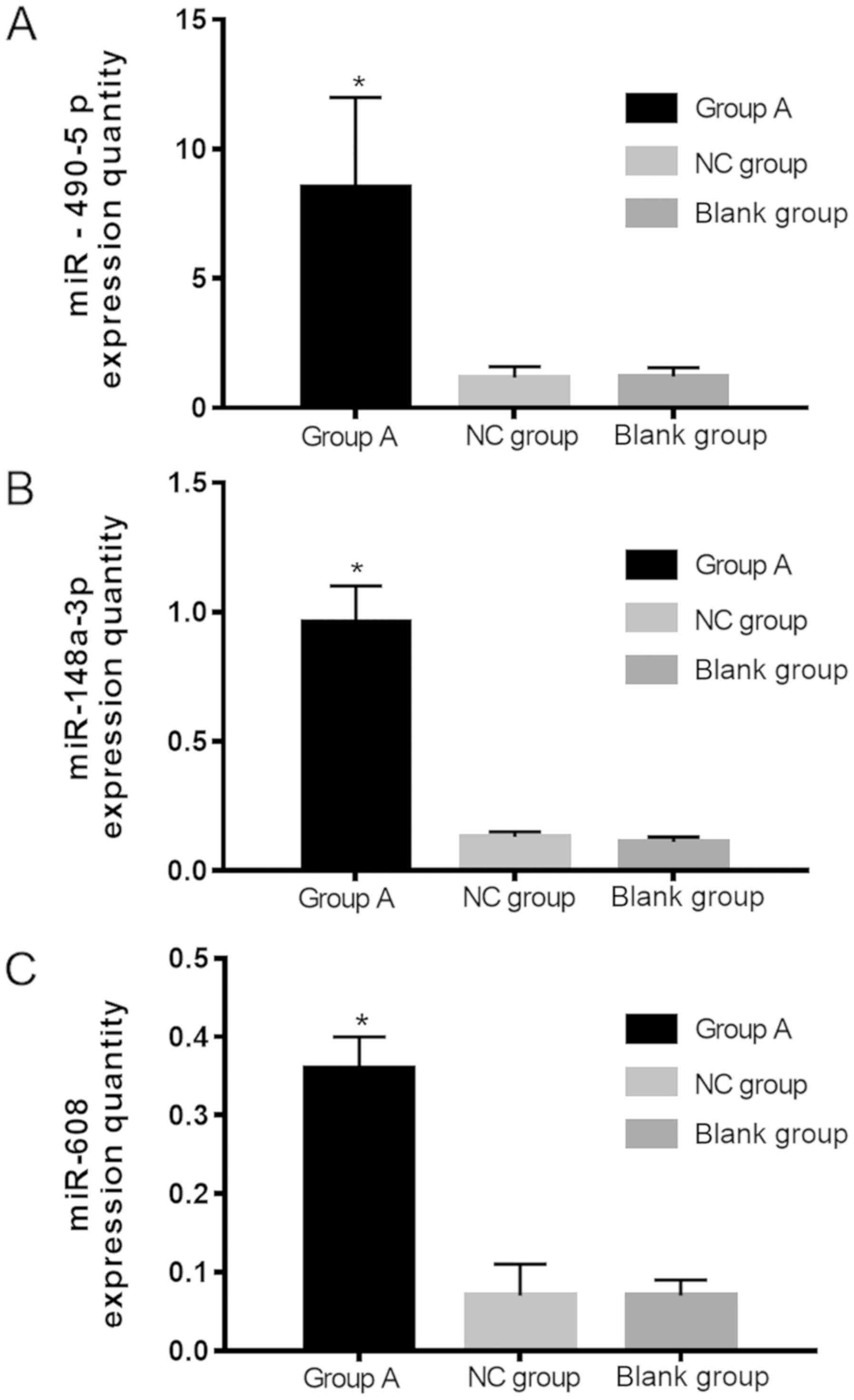
Relative expression level of
miR-490-5p, miR-148a-3p and miR-608 in the cells in each group
after transfection
The expression level of miR-490-5p in group A, group
NC and blank group was 8.50±3.49, 1.17±0.43 and 1.21±0.35,
respectively; the expression level of miR-490-5p in group A was
significantly higher than that in group NC and blank group; the
difference of the expression level was statistically significant
(P<0.001), and there was no significant difference in the
expression level of miR-490-5p in group NC and blank group
(P>0.05). The expression level of miR-148a-3p in group B, group
NC and blank group was 0.96±0.14, 0.13±0.02 and 0.11±0.02,
respectively; the expression level of miR-148a-3p in group B was
significantly higher than that in group NC and blank group; the
difference of the expression level was statistically significant
(P<0.001). No significant difference in the expression level of
miR-148a-3p in NC and blank groups was found (P>0.05). The
expression level of miR-608 in group C, group NC and blank group
was 0.36±0.04, 0.07±0.04 and 0.07±0.02, respectively. The
expression level of miR-608 in group C was significantly higher
than that in the NC and blank groups, and the difference was
statistically significant (P<0.001), but there was no
significant difference in the expression of miR-608 in the NC and
blank groups (P>0.05; Fig.
1A-C).
Comparison of the proliferation
ability of T24 cells in each group after transfection
The results of the comparison between the groups
showed that the differences were not statistically significant when
the survival rate of the cells at 24 h among groups A-C, group NC
and blank group was compared (P>0.05); there was no significant
difference in the survival rate of the cells among groups A-C
(P>0.05); at 48 and 72 h, there was no significant difference in
the survival rate of the cells among groups A-C (P>0.05), but
the survival rate of the cells in these groups was significantly
lower than that in group NC and blank group (P<0.001), and there
was no significant difference in the survival rate of the cells
between group NC and blank group at 48 and 72 h (P>0.05). The
results of the comparison within the groups showed that the
survival rate of the cells within groups A-C showed a gradual
downward trend from 24 to 72 h, and the differences were
statistically significant when compared at different time points
within the three groups (P<0.001); the survival rate of the
cells within group NC and blank group also showed a gradual
downward trend from 24 to 72 h, but the differences were not
statistically significant (P>0.05; Table III).
| Table III.Comparison of the survival rate of the
cells (%) in each group. |
Table III.
Comparison of the survival rate of the
cells (%) in each group.
| Group | Group A | Group B | Group C | Group NC | Blank group | F | P-value |
|---|
| 24 h | 97.42±3.01 | 97.87±3.68 | 96.99±3.54 | 97.12±1.04 | 97.78±3.02 | 0.501 | 0.735 |
| 48 h | 85.24±2.03 | 84.92±3.58 | 85.16±2.57 | 97.01±1.56 | 96.89±2.91 | 183.300 | <0.001 |
| 72 h | 79.82±2.48 | 80.08±2.73 | 80.27±2.58 | 96.25±2.22 | 96.33±3.58 | 312.100 | <0.001 |
| F | 378.300 | 225.200 | 257.900 | 2.392 | 1.583 |
|
|
| P-value | <0.001 | <0.001 | <0.001 | 0.052 | 0.211 |
|
|
Comparison of the condition of the
invasion of T24 cells in each group
The number of the invasive cells in groups A-C was
200.56±21.78, 202.32±20.41, and 199.56±23.53, respectively, and
these numbers were significantly lower than that in blank group
(498.38±50.25) and that in group NC (500.47±46.91). The differences
were statistically significant (P<0.05). However, there was no
significant difference among groups A-C, and there was no
significant difference among blank group and group NC also
(P>0.05; Table IV).
| Table IV.Comparison of the condition of the
invasion of T24 cells in each group. |
Table IV.
Comparison of the condition of the
invasion of T24 cells in each group.
| Group | Group A | Group B | Group C | Group NC | Blank group | F | P-value |
|---|
| Number of the
invasive cells | 200.56±21.78 | 201.32±20.41 | 199.56±23.53 | 498.38±50.25 | 500.47±46.91 | 651.800 | <0.001 |
Comparison of apoptosis ability of T24
cells in each group after the transfection
Apoptosis rate in groups A-C was 20.14±3.32%,
19.92±4.47% and 19.73±4.01%, respectively, and there was no
significant difference in the apoptosis rate of the cells among
groups A-C (P>0.05); but the apoptosis rates were significantly
higher than that in group NC (3.75±0.65%) and blank group
(3.67±0.91%) (P<0.05), and there was no significant difference
in the apoptosis rate between group NC and blank group (P>0.05;
Table V).
| Table V.Comparison of the apoptosis rate (%)
in each group. |
Table V.
Comparison of the apoptosis rate (%)
in each group.
| Group | Group A | Group B | Group C | Group NC | Blank group | F | P-value |
|---|
| Apoptosis rate
(%) | 20.14±3.32 | 19.92±4.47 | 19.73±4.01 | 3.75±0.65 | 3.67±0.91 | 651.800 | <0.001 |
Discussion
The development of bladder cancer is affected by the
biological characteristics of bladder cancer cells, such as
proliferation, invasion and apoptosis, and is closely related to
the regulation of miRNA (13). A
large number of studies have shown that both proliferation and
apoptosis of tumor cells determine the growth and decline of tumor
to control the growth rate of tumors (14). Cell proliferation is the basis of the
growth, development and reproduction of organism and is an
important life feature of organisms (15). The proliferation of normal cells will
stop naturally when it reaches a certain extent, but the
proliferation of cancer cells is generally not affected by
neurohumoral and environmental factors, which can divide and
proliferate unlimitedly and has the characteristics of being out of
control (16). The unceasing
proliferation of cancer cells will also carry out the invasion and
metastasis of cancer cells through blood or lymph, and the invasion
and metastasis of cancer cell are often the main reasons of death
of patients who have malignant tumors (17). miRNA plays an important role in the
process of carcinogenesis and tumor progression, and the expression
of miRNA affects the invasiveness of tumor cells (18). Studies have demonstrated that as
members of miRNA, the expression changes of miR-490-5p, miR-148a-3p
and miR-608 have been confirmed to be associated with the
occurrence and development of malignant tumors (11,19,20); but
the specific effect and mechanism of miR-490-5p, miR-148a-3p and
miR-608 in bladder cancer cells are unclear; therefore, this
experimental study on the expression features of miR-490-5p,
miR-148a-3p and miR-608 in bladder cancer and their effects on the
biological characteristics of bladder cancer cells was carried out
in order to provide a new theoretical basis for the diagnosis and
treatment of bladder cancer in molecular biology.
In this study, we first used RT-qPCR technology to
detect the expression of miR-490-5p, miR-148a-3p and miR-608 in
bladder cancer tissues and normal adjacent tissues. The results
showed that the expression levels of miR-490-5p, miR-148a-3p and
miR-608 in bladder cancer tissues were significantly lower than
those in normal adjacent tissues, and the differences were
statistically significant (P<0.001); there are now a large
number of similar studies that have confirm the expression levels
of miR-490-5p, miR-148a-3p and miR-608 in bladder cancer tissues
were significantly lower than those in normal adjacent tissues and
they have a low expression in bladder cancer tissues (10,12,21).
Next, we investigated the condition of the proliferation, invasion
and apoptosis of bladder cancer cells affected by miR-490-5p,
miR-148a-3p and miR-608, and the results showed that after the
transfection, the expression level of miR-490-5p in group A was
significantly higher than that in group NC and blank group, the
expression level of miR-148a-3p in group B was significantly higher
than that in group NC and blank group, and the difference of the
expression level was statistically significant; the expression
level of miR-608 in group C was significantly higher than that in
group NC and blank group, and the difference was statistically
significant (P<0.001). After the transfection, miR-490-5p,
miR-148a-3p and miR-608 were overexpressed in T24 cells. Then we
continued to analyze the effects of miR-490-5p, miR-148a-3p and
miR-608 on the ability of proliferation, invasion and apoptosis of
bladder cancer T24 cells, and the results showed that at 48 and 72
h, the survival rate of the cells in groups A-C was significantly
lower than that in group NC and blank group (P<0.001); the
results of the comparison within the groups showed that the
survival rate of the cells within groups A-C showed a gradual
downward trend from 24 to 72 h, and the differences were
statistically significant when compared at different time points
within the three groups (P<0.001). After the transfection, the
number of invasive cells in groups A-C was significantly lower than
that in blank group and group NC, and the difference was
statistically significant (P<0.05). However, there was no
significant difference among groups A-C, and there was no
significant difference among blank group and group NC (P>0.05).
After the transfection, the apoptosis rate in groups A-C was
significantly higher than that in group NC and blank group
(P<0.05). Based on the above results, we considered that the
overexpression of miR-490-5p, miR-148a-3p and miR-608 could reduce
the survival rate of bladder cancer T24 cells and inhibit the
ability of proliferation and invasion of bladder cancer cells to
some extent as well as promoting apoptosis of bladder cancer cells.
He et al (22) used CCK8
method for detection after they transfected miR-608 Inhibitor,
found that the proliferation ability of tumor cells was enhanced to
a certain extent after miR608 was inhibited, which reversely proved
that the expression of miR608 could inhibit the proliferation
ability of tumor cells. Regarding the effects of miR-490-5p and
miR-148a-3p on bladder cancer cells, there have been related
studies which carried out experimental observation on the ability
of proliferation, invasion and apoptosis of miR-490-5p or
miR-148a-3p which have been transfected, and the results of the
related studies show that the transfected miR-490-5p and
miR-148a-3p are overexpressed in bladder cancer T24 cells, and the
ability of the proliferation and invasion of T24 cells was
inhibited at this moment and the apoptosis rate increased (23,24);
this is similar to the results of our study.
This study has some shotcomings, for example, the
number of patients included were small, which was not satisfactory
for statistical data. Increasing the number of subjects at a later
date is anticipated.
In summary, miR-490-5p, miR-148a-3p and miR-608 are
lowly expressed in bladder cancer T24 cells, and the overexpression
of miR-490-5p, miR-148a-3p and miR-608 has inhibitory effect on the
proliferation and invasion of cancer T24 cells and promotes
apoptosis of bladder cancer T24 cells. This experiment showed that
miR-490-5p, miR-148a-3p and miR-608 are involved in the biological
process of bladder cancer cells and they show potential to be used
as a diagnostic marker and therapeutic target of bladder
cancer.
Acknowledgements
Not applicable.
Funding
No funding was received.
Availability of data and materials
The datasets used and/or analyzed during the present
study are available from the corresponding author on reasonable
request.
Authors' contributions
MX and WY conceived and designed the study. WY
collected the patients' data. WZ analyzed and interpreted the data.
JH and MX performed the experiment. MX was a major contributor in
writing the manuscript. JH reviewed the manuscript and helped with
cell culture and transfection. All authors read and approved the
final manuscript.
Ethics approval and consent to
participate
The study was approved by the Ethics Committee of
Hunan Provincial People's Hospital (Changsha, China). Each patient
who participated in this research had complete clinical data.
Signed informed consents were obtained from the patients or
guardians.
Patient consent for publication
Not applicable.
Competing interests
The authors declare that they have no competing
interests.
References
|
1
|
Grayson M: Bladder cancer. Nature.
551:S332017. View
Article : Google Scholar : PubMed/NCBI
|
|
2
|
Antoni S, Ferlay J, Soerjomataram I, Znaor
A, Jemal A and Bray F: Bladder cancer incidence and mortality: A
global overview and recent trends. Eur Urol. 71:96–108. 2017.
View Article : Google Scholar : PubMed/NCBI
|
|
3
|
Tuccori M, Filion KB, Yin H, Yu OH, Platt
RW and Azoulay L: Pioglitazone use and risk of bladder cancer:
Population based cohort study. BMJ. 352:i15412016. View Article : Google Scholar : PubMed/NCBI
|
|
4
|
Sato Y, Kondo T, Takagi T, Junpei I and
Tanabe K: Treatment strategy for bladder cancer in patients on
hemodialysis: A clinical review of 28 cases. Int Urol Nephrol.
48:503–509. 2016. View Article : Google Scholar : PubMed/NCBI
|
|
5
|
Yang GL, Zhang LH, Liu Q, Wang ZL, Duan
XH, Huang YR and Bo JJ: Commentary on ‘A novel treatment strategy
for newly diagnosed high-grade T1 bladder cancer: gemcitabine and
cisplatin adjuvant chemotherapy-A single-institution experience’.
Urol Oncol. 36:346–347. 2018. View Article : Google Scholar : PubMed/NCBI
|
|
6
|
Heidari F, Abbas Zade S, Mir Hosseini SH
and Ghadian A: Metformin for the prevention of bladder cancer
recurrence: Is it effective? Nephrourol Mon. 8:e302612016.
View Article : Google Scholar : PubMed/NCBI
|
|
7
|
Parodi A, Traverso P, Kalli F, Conteduca
G, Tardito S, Curto M, Grillo F, Mastracci L, Bernardi C, Nasi G,
et al: Residual tumor micro-foci and overwhelming regulatory T
lymphocyte infiltration are the causes of bladder cancer
recurrence. Oncotarget. 7:6424–6435. 2016. View Article : Google Scholar : PubMed/NCBI
|
|
8
|
Mearini E, Poli G, Cochetti G, Boni A,
Egidi MG and Brancorsini S: Expression of urinary miRNAs targeting
NLRs inflammasomes in bladder cancer. OncoTargets Ther.
10:2665–2673. 2017. View Article : Google Scholar
|
|
9
|
Wang C, Chen Z, Ge Q, Hu J, Li F, Hu J, Xu
H, Ye Z and Li LC: Up-regulation of p21(WAF1/CIP1) by miRNAs and
its implications in bladder cancer cells. FEBS Lett. 588:4654–4664.
2014. View Article : Google Scholar : PubMed/NCBI
|
|
10
|
Lan G, Yang L, Xie X, Peng L and Wang Y:
MicroRNA-490-5p is a novel tumor suppressor targeting c-FOS in
human bladder cancer. Arch Med Sci. 11:561–569. 2015. View Article : Google Scholar : PubMed/NCBI
|
|
11
|
Wang X, Liang Z, Xu X, Li J, Zhu Y, Meng
S, Li S, Wang S, Xie B, Ji A, et al: miR-148a-3p represses
proliferation and EMT by establishing regulatory circuits between
ERBB3/AKT2/c-myc and DNMT1 in bladder cancer. Cell Death Dis.
7:e25032016. View Article : Google Scholar : PubMed/NCBI
|
|
12
|
Liang Z, Wang X, Xu X, Xie B, Ji A, Meng
S, Li S, Zhu Y, Wu J, Hu Z, et al: MicroRNA-608 inhibits
proliferation of bladder cancer via AKT/FOXO3a signaling pathway.
Mol Cancer. 16:962017. View Article : Google Scholar : PubMed/NCBI
|
|
13
|
Zhao F, Ge YZ, Zhou LH, Xu LW, Xu Z, Ping
WW, Wang M, Zhou CC, Wu R and Jia RP: Identification of hub miRNA
biomarkers for bladder cancer by weighted gene coexpression network
analysis. OncoTargets Ther. 10:5551–5559. 2017. View Article : Google Scholar
|
|
14
|
Wang Y, Du C, Zhang N, Li M, Liu Y, Zhao
M, Wang F and Luo F: TGF-β1 mediates the effects of aspirin on
colonic tumor cell proliferation and apoptosis. Oncol Lett.
15:5903–5909. 2018.PubMed/NCBI
|
|
15
|
Chen W, Wang J, Liu S, Wang S, Cheng Y,
Zhou W, Duan C and Zhang C: MicroRNA-361-3p suppresses tumor cell
proliferation and metastasis by directly targeting SH2B1 in NSCLC.
J Exp Clin Cancer Res. 35:762016. View Article : Google Scholar : PubMed/NCBI
|
|
16
|
Wen Z, Zhang Y, Wang X, Zeng X, Hu Z, Liu
Y, Xie Y, Liang G, Zhu J, Luo H, et al: Novel 3′,5′-diprenylated
chalcones inhibited the proliferation of cancer cells in vitro by
inducing cell apoptosis and arresting cell cycle phase. Eur J Med
Chem. 133:227–239. 2017. View Article : Google Scholar : PubMed/NCBI
|
|
17
|
Yano S, Mii S, Tome Y, Hiroshima Y, Uehara
F, Miwa S and Hoffman RM: Abstract A41: Cancer cells invade or
divide, not both. Cancer Res. 73 Suppl 3:A412013. View Article : Google Scholar
|
|
18
|
Baumgart S, Hölters S, Ohlmann CH, Bohle
R, Stöckle M, Ostenfeld MS, Dyrskjøt L, Junker K and Heinzelmann J:
Exosomes of invasive urothelial carcinoma cells are characterized
by a specific miRNA expression signature. Oncotarget.
8:58278–58291. 2017. View Article : Google Scholar : PubMed/NCBI
|
|
19
|
Chen W, Ye L, Wen D and Chen F: MiR-490-5p
inhibits hepatocellular carcinoma cell proliferation, migration and
invasion by directly regulating ROBO1. Pathol Oncol Res. 2:1–9.
2017.
|
|
20
|
Othman N, In LL, Harikrishna JA and Hasima
N: Bcl-xL silencing induces alterations in hsa-miR-608 expression
and subsequent cell death in A549 and SK-LU1 human lung
adenocarcinoma cells. PLoS One. 8:e817352013. View Article : Google Scholar : PubMed/NCBI
|
|
21
|
Lombard A, Mooso B, Libertini S, Lim R,
Costanzo N, Ghosh P and Mudryj M: Abstract 5239: MiR-148a promotes
apoptosis in urothelial cell carcinoma of the bladder cells in part
by targeting DNMT1. Cancer Res. 74 Suppl 19:52392014. View Article : Google Scholar
|
|
22
|
He L, Meng D, Zhang SH, Zhang Y, Deng Z
and Kong LB: microRNA-608 inhibits human hepatocellular carcinoma
cell proliferation via targeting the BET family protein BRD4.
Biochem Biophys Res Commun. 501:1060–1067. 2018. View Article : Google Scholar : PubMed/NCBI
|
|
23
|
Li S, Xu X, Xu X, Hu Z, Wu J, Zhu Y, Chen
H, Mao Y, Lin Y, Luo J, et al: MicroRNA-490-5p inhibits
proliferation of bladder cancer by targeting c-Fos. Biochem Biophys
Res Commun. 441:976–981. 2013. View Article : Google Scholar : PubMed/NCBI
|
|
24
|
Mao LM, Chen YH and Liao ZP: The abnormal
expression of miR-148a in bladder cancer and its clinical
significance. Chin J Health Lab Tech. 15:2143–2145. 2017.(In
Chinese).
|