Introduction
Soft tissue sarcoma (STS) is a malignant tumor
derived from mesenchymal tissues. The worldwide prevalence rate of
STS is as high as 1.8–5.0/100,000 every year, and it accounts for
only 1% in malignant tumors of adults, but as high as 10% in
children's tumors. Currently, more than 50 kinds of STS subtypes
have been definitely diagnosed clinically (1,2). There
are approximately 11,000 new cases of STS each year in the United
States (3), but as many as
2.0/100,000 new cases each year in China (4). In all STS, pleomorphic sarcoma, also
known as malignant fibrous histiotoma in clinic, has the highest
incidence rate, accounting for 25–35%, followed by liposarcoma
(25%), and the incidence rates of malignant schwannoma and
leiomyosarcoma are relatively lower. The incidence rate of STS in
males is higher than that in females, and it mainly occurs in the
age group of 30–50 years and in any part of the body, mainly the
trunk, head and neck, and limbs (5).
STS has not only complex typing, but also high heterogeneous
degree. Moreover, the growth of STS is characterized by destruction
and infiltration, and its prognosis is poor and postoperative
recurrence occurs easily because of its strong invasion (6).
p16 protein, a kind of tumor suppressor protein, can
bind to cyclin-dependent kinase 4 (CDK4), and its function of
preventing the normal cell cycle is exerted by inhibiting pRB
protein phosphorylation and blocking CDK4 complex formation
(7). nm23 gene is a key gene
involved in the invasion of malignant tumors, and was isolated by
Steeg et al (8) in the
complementary deoxyribonucleic acid (cDNA) library with mouse
melanoma K-1735 cell lines as the research objects. Studies have
shown that the nm23 gene plays inhibitory and regulatory roles in
the outcome of cancer cells, significantly inhibiting the
proliferation and invasion of tumor cells (9). In addition, abnormal changes in the
nm23 gene widely exist in malignant tumors, which are closely
related to cancer cell invasion (10).
The occurrence and development of osteosarcoma is a
process of gradual evolution, which eventually develops into a
highly invasive tumor. The existence of periodic dysfunction and
changes in the expression of invasion-associated genes are worthy
of full study. Therefore, we selected to study the expression of
p16 and nm23-H1 and its relationship with the pathogenesis and
prognosis of soft tissue sarcoma. In order to investigate the
expression levels of p16 and nm23-H1 in STS and the correlations of
their expression levels with the incidence and prognosis of STS,
messenger ribonucleic acid (mRNA) and protein expression levels of
p16 and nm23-H1 in STS tissues were detected via reverse
transcription-quantitative polymerase chain reaction (RT-qPCR) and
immunohistochemical method, respectively, in this study, and the
relationship between the expression levels of p16 and nm23-H1 was
analyzed.
Patients and methods
Materials
Frozen and paraffin specimens of tumor tissues and
corresponding para-carcinoma normal tissues were collected from 64
STS patients treated in the General Hospital of Chinese People's
Liberation Army (Beijing, China), from January 2007 to January
2012. Patients were aged 11–72 years, including 35 males and 29
females, all of them were pathologically diagnosed with STS, and
they had complete clinical data and received surgical treatment in
the hospital.
This study was reviewed and approved by the Clinical
Ethics Committee of General Hospital of Chinese People's Liberation
Army, and all patients or their families signed an informed
consent.
TRIzol RNA extraction kit (Takara, Dalian, China),
primer synthesis, RT kits and quantitative SYBR-Green PCR kits
(Ambion; Thermo Fisher Scientific, Inc., Waltham, MA, USA), primary
rabbit anti-human p16 and nm23-H1 antibodies (1:100; cat. nos.
10883-1-AP and 11086-2-AP, respectively; Proteintech Group, Inc.,
Wuhan, China), immunohistochemical staining kit SP-9001 (Beijing
Zhongshan Goldenbridge Biotechnology Co., Ltd., Beijing,
China).
RT-qPCR
Approximately 50 mg tumor tissues and para-carcinoma
normal tissues cryopreserved in liquid nitrogen were used, and the
total RNA was extracted from specimens using the TRIzol kit.
Qualified RNA specimens [absorbance
(A)260/A280 ratio of 1.8–2.0] were selected
for reverse transcription to obtain cDNA. PCR was carried out with
cDNA as a template according to the manufacturer's instructions.
The specific reaction conditions were as follows: pre-denaturation
at 94°C for 4 min, denaturation at 94°C for 1 min, annealing at
50°C for 50 sec, extension at 72°C for 1 min, amplification for 35
cycles, and extension for 10 min. Glyceraldehyde-3-phosphate
dehydrogenase (GAPDH) mRNA was used as a control, the primer
sequences are shown in Table I, and
the experimental results were analyzed using the 2−∆Cq
method (11).
 | Table I.RT-qPCR primer sequences. |
Table I.
RT-qPCR primer sequences.
| Genes | Primer sequences |
|---|
| p16 | F:
5′-GAAGAAAGAGGAGGGGCTG-3′ |
|
| R:
5′-GCGCTACCTGATTCCAATTC-3′ |
| nm23-H1 | F:
5′-GTGAAAAGCAATGTGGT-3′ |
|
| R:
5′-TTGCCATGGTCTGGGAG-3′ |
| GAPDH | F:
5′-ATGGCACCGTCAAGGCTGAG-3′ |
|
| R:
5′-GCAGTGATGGCATGGACTGT-3′ |
Immunohistochemistry
Streptavidin peroxidase (SP) method was used in
immunohistochemical assay in accordance with instructions of the
SP-9001 kit. The tissue was fixed with 40% formaldehyde at 20°C for
12 h. The paraffin sections were dewaxed and inactivated with 3%
H2O2, followed by antigen retrieval via
microwave twice (750 W, 95°C, once every 5 min). A total of 5 µl
sections was prepared and blocked with 5% milk at 20°C for 1 h.
Then it was incubated with the primary antibody (dilution, 1:100)
at 4°C overnight. After sections were rinsed with tris-buffered
saline with Tween-20 (TBST) 3 times, biotin-labeled secondary
polyclonal antibody (cat. no. SA00004-11; Proteintech Group, Inc.)
was added dropwise for incubation for 30 min, and sections were
rinsed again with TBST. Then the color was developed via
diaminobenzidine (DAB) in the dark, and the development time was
controlled, followed by re-staining using hematoxylin and sealing
with gum.
Five fields of vision were randomly selected, and
cells were graded and scored according to the percentage of
positive cells and the staining intensity. No staining, light
yellow, brown yellow and dark brown were denoted as 0, 1, 2 and 3
points, respectively. The number of positive cells <5%, 5–25%,
26–50% and >50% were denoted as 0, 1, 2 and 3 points,
respectively. The two scores were added up, and the final score ≥3
points indicated positive expression, while <3 points indicated
negative expression.
Correlation analysis
According to the expression levels of p16 and
nm23-H1 in STS tissues, 64 STS patients were divided into the
p16-positive group, the p16-negative group, the nm23-H1-positive
group and the nm23-H1-negative group. Spearman's correlation
analysis was used to study the correlation between p16 and nm23-H1
mRNA expression and protein expression and the relationship between
expression levels of p16 and nm23-H1 in STS tissues. Patients were
followed up once a month from the first day after operation for a
total of 5 years. Moreover, correlations of p16 and nm23-H1
expression levels with pathological parameters and prognosis of
patients were analyzed combined with clinical and follow-up
data.
Statistical analysis
Statistical Product and Service Solutions (SPSS)
17.0 (SPSS, Inc., Chicago, IL, USA) software was used for data
processing. Measurement data were presented as mean ± standard
deviation, and t-test was used for the intergroup comparison.
Chi-square test was used for the intergroup comparison of
enumeration data, and Spearman's test was used for correlation
analysis. Kaplan-Meier survival curves was used for survival curve
analysis of STS patients with log rank test. P≤0.05 was considered
to indicate a statistically significant difference.
Results
p16 and nm23-H1 mRNA expression levels
in tissue specimens
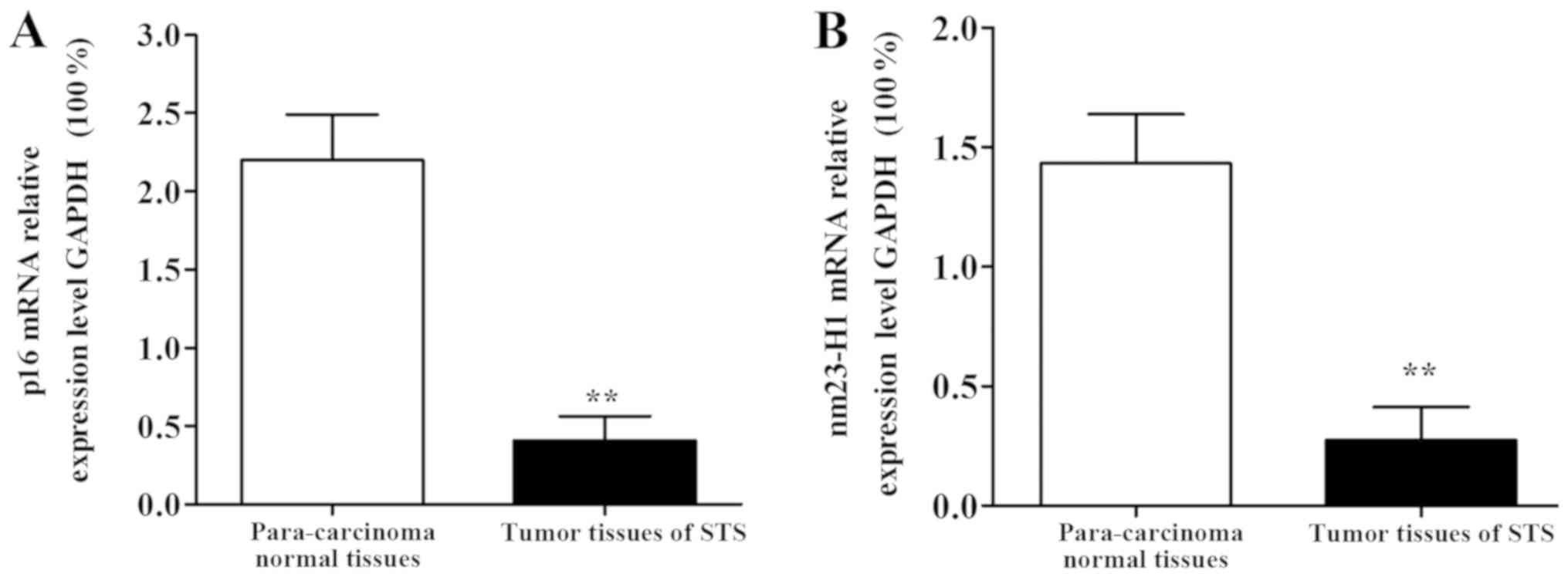
The results of RT-qPCR showed that p16 and nm23-H1
mRNA expression levels in tumor tissues of STS patients were
significantly decreased compared with those in para-carcinoma
normal tissues, and differences were statistically significant
(P<0.01) (Fig. 1).
p16 and nm23-H1 protein expression
levels in STS tissues
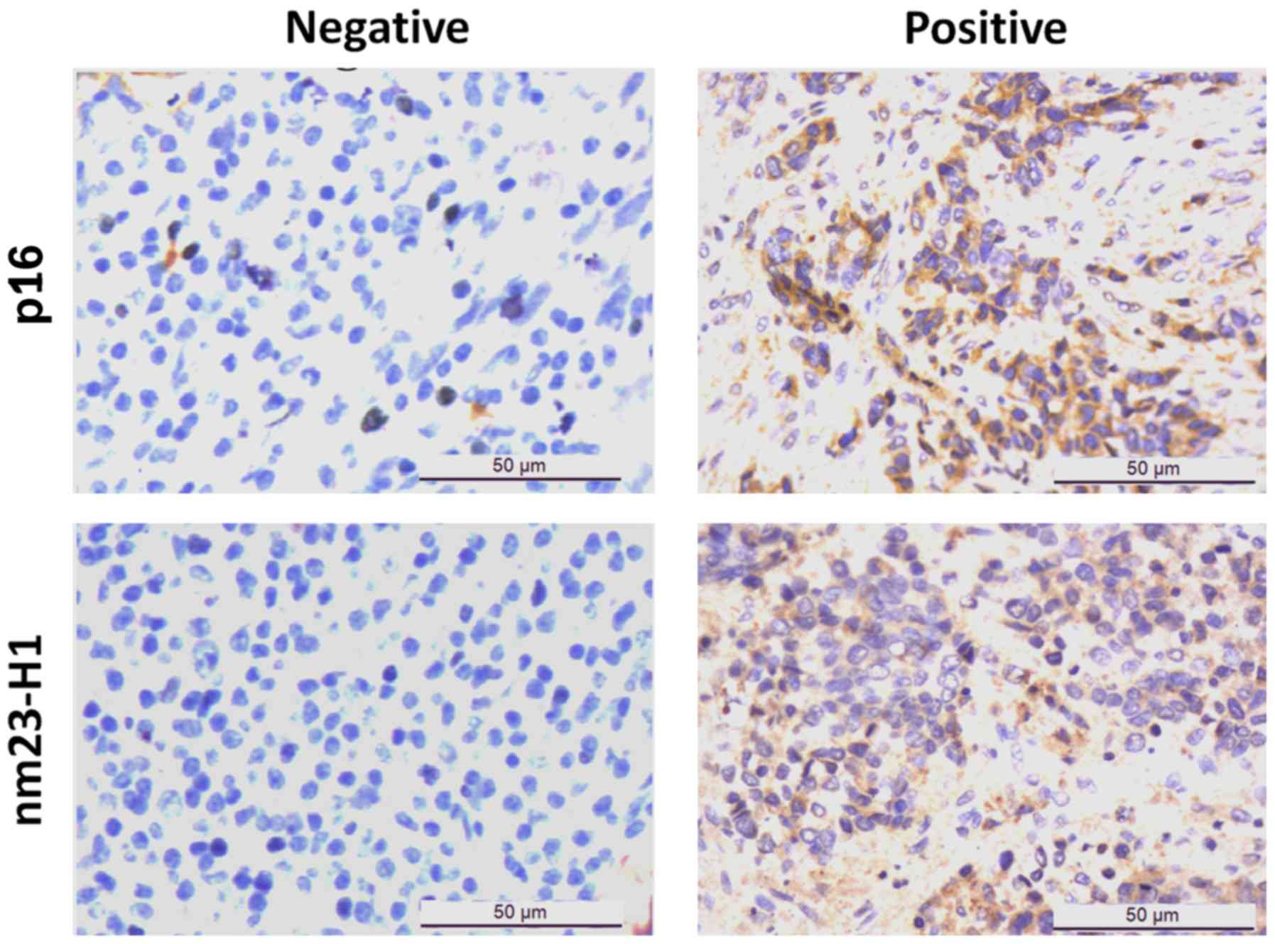
Results of immunohistochemical detection showed that
the positive immunohistochemical staining of p16 and nm23-H1 was
dark brown, and p16 staining was mainly located in the nucleus and
cytoplasm. nm23-H1 staining was mainly located in the cytoplasm
(Fig. 2). Immunohistochemical scores
showed that the positive expression rate of p16 in STS tissues was
43.75% (28/64), which was significantly lower than that in
para-carcinoma normal tissues (85.93%, 55/64). The positive
expression rate of nm23-H1 in STS tissues was 39.06% (25/64), which
was also significantly lower than that in para-carcinoma normal
tissues (89.06%, 57/64). Differences were statistically significant
(P<0.01).
Correlation between p16 and nm23-H1
mRNA expression and protein expression
Correlation between the expression of p16 and
nm23-H1 mRNA and the expression of protein was analyzed by
Spearman's test. The results showed that the expression of p16 and
nm23-H1 mRNA was positively correlated with the expression of
protein (p16: r=0.92, P<0.01; nm23-H1: r=0.95, P<0.01).
Correlation between p16 and nm23-H1
protein expression levels in STS tissues
Among the 64 cases of STS tissues, p16 and nm23-H1
proteins were positive in 20 cases, p16 and nm23-H1 proteins were
negative in 31 cases, p16 protein was positive and nm23-H1 protein
was negative in 8 cases, p16 protein was negative and nm23-H1
protein was positive in 5 cases. Results of Spearman's correlation
analyses showed that there was a positive correlation between the
expression levels of p16 and nm23-H1 proteins (correlation
coefficient r=0.722), and the correlation had statistical
significance (P<0.01) (Table
II).
| Table II.Correlation between p16 and nm23-H1
protein expression levels in STS tissues. |
Table II.
Correlation between p16 and nm23-H1
protein expression levels in STS tissues.
|
| nm23-H1 |
|
|
|---|
|
|
|
|
|
|---|
| p16 | Positive | Negative | r value | P-value |
|---|
| Positive | 20 | 8 | 0.722 | <0.01 |
| Negative | 5 | 31 |
|
|
Correlation of p16 and nm23-H1
expression levels with pathological parameters of STS patients
Analysis results of the relationship of pathological
parameters of STS patients with p16 and nm23-H1 expression levels
are shown in Table III. Chi-square
test showed that the negative expression of p16 was correlated with
tumor size, metastasis and staging in patients (P<0.01), but had
no correlation with sex and age of patients (P>0.05). Besides,
the negative expression of nm23-H1 was correlated with tumor
metastasis and staging in patients (P<0.01), but had no
correlations with sex, age and tumor size (P>0.05).
| Table III.Correlation of abnormal expression
levels of p16 and nm23-H1 with pathological parameters of STS
patients. |
Table III.
Correlation of abnormal expression
levels of p16 and nm23-H1 with pathological parameters of STS
patients.
|
|
| p16 (n=36) | nm23-H1 (n=39) |
|---|
|
|
|
|
|
|---|
| Pathological
parameters | n | Negative [n (%)] | χ2
value | P-value | Negative [n (%)] | χ2
value | P-value |
|---|
| Sex |
|
| 0.12 | >0.05 |
| 0.74 | >0.05 |
| Male | 35 | 19 (54.29) |
|
| 23 (65.71) |
|
|
|
Female | 29 | 17 (58.62) |
|
| 16 (55.17) |
|
|
| Age (years) |
|
| 0.70 | >0.05 |
| 0.19 | >0.05 |
| ≥60 | 26 | 13 (50.00) |
|
| 15 (57.69) |
|
|
|
<60 | 38 | 23 (60.52) |
|
| 24 (63.16) |
|
|
| Tumor size (cm) |
|
| 9.80 | <0.01 |
| 0.42 | >0.05 |
| ≥5 | 39 | 28 (71.79) |
|
| 25 (64.10) |
|
|
|
<5 | 25 | 8
(32.00) |
|
| 14 (56.00) |
|
|
| Tumor metastasis |
|
| 7.52 | <0.01 |
| 9.12 | <0.01 |
| Yes | 33 | 25 (75.76) |
|
| 26 (78.79) |
|
|
| No | 31 | 12 (38.71) |
|
| 13 (41.94) |
|
|
| Clinical
staging |
|
| 8.13 | <0.01 |
| 8.50 | <0.01 |
| I | 22 | 7
(31.82) |
|
| 8
(36.36) |
|
|
|
II–III | 42 | 29 (69.05) |
|
| 31 (73.81) |
|
|
Correlation of p16 and nm23-H1 protein
expression levels with survival of STS patients
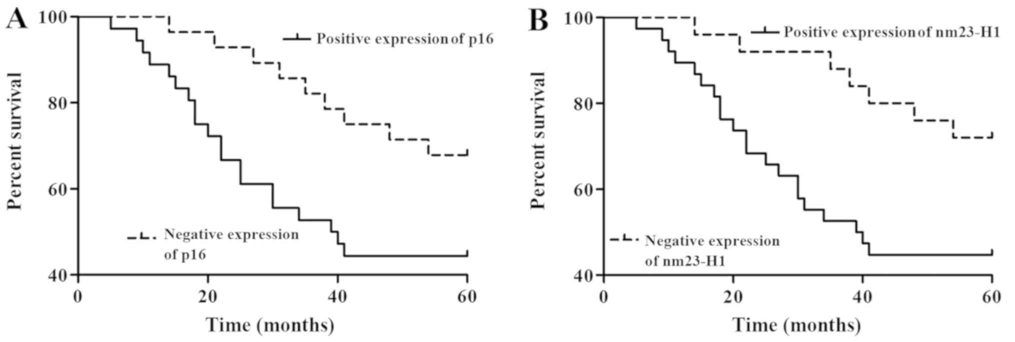
Results of the 5-year follow-up revealed that of the
64 patients, 35 cases survived and 29 cases had died, and the
overall 5-year survival rate was 54.68%. Kaplan-Meier survival
curves was used for survival curve analysis of STS patients with
log rank test. Patients with negative protein expression levels of
p16 and nm23-H1 had poorer survival. Univariate survival analyses
showed that both p16 and nm23-H1 could significantly affect the
overall survival rate of STS patients (P<0.05; Fig. 3).
Discussion
Soft tissue sarcoma (STS), as a kind of malignant
tumor derived from mesoderm, has higher heterogeneity and many
different subtypes (12). Although
its incidence rate is not high, STS has higher recurrence and
metastasis rates. Clinical statistics have shown that the overall
3-year and 5-year survival rates of STS patients were 77 and 75%,
the recurrence rates were 28 and 37%, and the metastasis rates were
35 and 43%, respectively (13).
Studies have shown that the positive margin is the most important
factor affecting the recurrence and metastasis of STS (14,15).
P16 gene, a CDK inhibitor, encodes the p16 protein
that could specifically bind to CDK4 or CDK6 and competitively
affect the binding of cyclin D, thereby inhibiting the activity of
CDK4 and CDK6, leading to cell cycle arrest, and inhibiting tumor
cell growth (16,17). Studies have proven that there are
gene deletion, mutation, methylation and other abnormalities in p16
in a variety of tumor cell lines and solid tumors, so it is
speculated that p16 gene plays a key role in tumor formation and
development processes (18).
Nm23 gene is located on human chromosome 17q22,
which encodes a protein containing 152 amino acids and having a
high homology to the amino acid sequence of nucleoside diphosphate
kinase (NDPK) (19,20). Therefore, it is speculated that nm23
gene may exert a similar effect to NDPK, and that it can play an
important role in regulating cell signal transduction and cell
differentiation. There are 2 subtypes of human nm23 gene, namely
nm23-H1 and nm23-H12, the former of which plays an important role
in inhibiting tumor metastasis (21). It was found that nm23-H1 can inhibit
the metastasis of a variety of malignant tumors, and its expression
level is negatively correlated with tumor invasion and metastasis.
Moreover, tumor patients with high nm23-H1 expression have better
prognosis (8).
In order to further investigate the expression
levels of p16 and nm23-H1 in tumor tissues of STS patients and
their influence on the pathological parameters and prognosis of STS
patients, RT-qPCR and immunohistochemical assay were performed in
this study. Detection results showed that the expression levels of
p16 and nm23-H1 in tumor tissues of STS patients were significantly
lower than those in para-carcinoma normal tissues (P<0.01).
Results of immunohistochemical assay showed that the positive
expression rates of p16 and nm23-H1 in tumor tissues of STS
patients (43.75 and 39.06%, respectively) were significantly lower
than those in para-carcinoma normal tissues (85.93 and 89.06%,
respectively). The results of Spearman's test showed that the
expression of p16 and nm23-H1 mRNA was positively correlated with
the expression of protein. p16 is a CDK4 inhibitor protein, and
some scholars have proposed that nm23 may regulate the function of
p53 gene at CDK level. Correlation analyses in this study showed
that there was a significantly positive correlation between the
expression levels of p16 and nm23-H1, indicating that p16 and nm23
genes may have a close correlation in STS, and its specific
molecular process and mechanism need to be studied more profoundly.
Along with clinicopathological parameters and the survival
statistical analysis of patients, it was found that the negative
expression of p16 was correlated with tumor size, metastasis and
staging in patients, but had no correlation with the sex and age of
patients. Besides, the negative expression of nm23-H1 was
correlated with tumor metastasis and staging in patients, but had
no correlation with sex, age and tumor size in patients. The 64 STS
patients were followed up for 5 years, and results showed that the
overall 5-year survival rate of patients was 54.68%, and patients
with positive expression levels of p16 and nm23-H1 had a better
prognosis. Moreover, univariate survival analyses showed that both
p16 and nm23-H1 could affect the overall survival rate of STS
patients.
In conclusion, the expression of p16 and nm23-H1 is
low in STS, and their expression levels are closely related to the
pathological parameters and prognosis of STS patients, so they can
serve as reference indexes for prognosis estimation of STS.
Acknowledgements
Not applicable.
Funding
No funding was received.
Availability of data and materials
All data generated or analyzed during this study are
included in this published article.
Authors' contributions
JJ performed the data analyses and wrote the
manuscript. PY and XZha contributed significantly to data analysis
and manuscript preparation. FY performed RT-qPCR. GS and WB helped
with data analysis. GH and MX collected the data and were
responsible for statistical analysis. WW was a major contributor in
performing immunohistochemical assay. XZhe performed the data
analysis with constructive discussions. All authors read and
approved the final manuscript.
Ethics approval and consent to
participate
This study was reviewed and approved by the Clinical
Ethics Committee of General Hospital of Chinese People's Liberation
Army (Beijing, China) and all patients or their families signed an
informed consent.
Patient consent for publication
Not applicable.
Competing interests
The authors declare that they have no competing
interests.
References
|
1
|
Doyle LA: Sarcoma classification: An
update based on the 2013 World Health Organization Classification
of Tumors of Soft Tissue and Bone. Cancer. 120:1763–1774. 2014.
View Article : Google Scholar : PubMed/NCBI
|
|
2
|
Fletcher CDM, Bridge JA, Hogendoorn PCW
and Mertens F: WHO Classification of Tumours of Soft Tissue and
Bone. (Fourth). 5:IARC Press. (Lyon). 95–104. 2013.
|
|
3
|
Jemal A, Bray F, Center MM, Ferlay J, Ward
E and Forman D: Global cancer statistics. CA Cancer J Clin.
61:69–90. 2011. View Article : Google Scholar : PubMed/NCBI
|
|
4
|
Shi YQ, Zong XY, Wang J and Li S: Clinical
analysis of 251 cases of soft tissue sarcomas. Zhonghua Wai Ke Za
Zhi. 41:116–118. 2003.(In Chinese). PubMed/NCBI
|
|
5
|
Bannasch H, Eisenhardt SU, Grosu AL, Heinz
J, Momeni A and Stark GB: The diagnosis and treatment of soft
tissue sarcomas of the limbs. Dtsch Arztebl Int. 108:32–38.
2011.PubMed/NCBI
|
|
6
|
Ruymann FB and Grovas AC: Progress in the
diagnosis and treatment of rhabdomyosarcoma and related soft tissue
sarcomas. Cancer Invest. 18:223–241. 2000. View Article : Google Scholar : PubMed/NCBI
|
|
7
|
Kashiwabara K, Oyama T, Sano T, Fukuda T
and Nakajima T: Correlation between methylation status of the
p16/CDKN2 gene and the expression of p16 and Rb proteins in primary
non-small cell lung cancers. Int J Cancer. 79:215–220. 1998.
View Article : Google Scholar : PubMed/NCBI
|
|
8
|
Steeg PS, Bevilacqua G, Kopper L,
Thorgeirsson UP, Talmadge JE, Liotta LA and Sobel ME: Evidence for
a novel gene associated with low tumor metastatic potential. J Natl
Cancer Inst. 80:200–204. 1988. View Article : Google Scholar : PubMed/NCBI
|
|
9
|
Parhar RS, Shi Y, Zou M, Farid NR, Ernst P
and al-Sedairy ST: Effects of cytokine-mediated modulation of nm23
expression on the invasion and metastatic behavior of B16F10
melanoma cells. Int J Cancer. 60:204–210. 1995. View Article : Google Scholar : PubMed/NCBI
|
|
10
|
Okada K, Urano T, Goi T, Baba H, Yamaguchi
A, Furukawa K and Shiku H: Isolation of human nm23 genomes and
analysis of loss of heterozygosity in primary colorectal carcinoma
using a specific genomic probe. Cancer Res. 54:3979–3982.
1994.PubMed/NCBI
|
|
11
|
Livak KJ and Schmittgen TD: Analysis of
relative gene expression data using real time quantitative PCR and
the 2(Delta Delta C(T)) method. Methods. 25:402–408. 2001.
View Article : Google Scholar : PubMed/NCBI
|
|
12
|
Jemal A, Siegel R, Ward E, Hao Y, Xu J and
Thun MJ: Cancer statistics, 2009. CA Cancer J Clin. 59:225–249.
2009. View Article : Google Scholar : PubMed/NCBI
|
|
13
|
Casali PG and Blay JY:
ESMO/CONTICANET/EUROBONET Consensus Panel of experts: Soft tissue
sarcomas: ESMO Clinical Practice Guidelines for diagnosis,
treatment and follow-up. Ann Oncol. 21 Suppl 5:v198–v203. 2010.
View Article : Google Scholar : PubMed/NCBI
|
|
14
|
Campos M, De Campos SG, Ribeiro GG, Eguchi
FC, Silva SR, De Oliveira CZ, Da Costa AM, Curcelli EC, Nunes MC,
Penna V, et al: Ki-67 and CD100 immunohistochemical expression is
associated with local recurrence and poor prognosis in soft tissue
sarcomas, respectively. Oncol Lett. 5:1527–1535. 2013. View Article : Google Scholar : PubMed/NCBI
|
|
15
|
Ch'ng E, Tomita Y, Zhang B, He J, Hoshida
Y, Qiu Y, Morii E, Nakamichi I, Hamada K, Ueda T, et al: Prognostic
significance of CD100 expression in soft tissue sarcoma. Cancer.
110:164–172. 2007. View Article : Google Scholar : PubMed/NCBI
|
|
16
|
Merlo A, Herman JG, Mao L, Lee DJ,
Gabrielson E, Burger PC, Baylin SB and Sidransky D: 5′ CpG island
methylation is associated with transcriptional silencing of the
tumour suppressor p16/CDKN2/MTS1 in human cancers. Nat Med.
1:686–692. 1995. View Article : Google Scholar : PubMed/NCBI
|
|
17
|
Reimers N, Kasper HU, Weissenborn SJ,
Stützer H, Preuss SF, Hoffmann TK, Speel EJ, Dienes HP, Pfister HJ,
Guntinas-Lichius O, et al: Combined analysis of HPV-DNA, p16 and
EGFR expression to predict prognosis in oropharyngeal cancer. Int J
Cancer. 120:1731–1738. 2007. View Article : Google Scholar : PubMed/NCBI
|
|
18
|
Fan X, Yu K, Wu J, Shao J, Zhu L and Zhang
J: Correlation between squamous cell carcinoma of the lung and
human papillomavirus infection and the relationship to expression
of p53 and p16. Tumour Biol. 36:3043–3049. 2015. View Article : Google Scholar : PubMed/NCBI
|
|
19
|
Postel EH: NM23-NDP kinase. Int J Biochem
Cell Biol. 30:1291–1295. 1998. View Article : Google Scholar : PubMed/NCBI
|
|
20
|
Valentijn LJ, Koster J and Versteeg R:
Read-through transcript from NM23-H1 into the neighboring NM23-H2
gene encodes a novel protein, NM23-LV. Genomics. 87:483–489. 2006.
View Article : Google Scholar : PubMed/NCBI
|
|
21
|
Lacombe ML, Milon L, Munier A, Mehus JG
and Lambeth DO: The human nm23/nucleoside diphosphate kinases. J
Bioenerg Biomembr. 32:247–258. 2000. View Article : Google Scholar : PubMed/NCBI
|