Introduction
Liver cancer is a leading cause of death worldwide,
and its incidence is high in Asia (1). Approximately 745,500 individuals
succumb to liver cancer annually (2). Currently, treatment for liver cancer is
not satisfactory and generally ineffective (3). As the symptoms of liver cancer become
evident at an advanced stage rendering it a disease that is
untreatable by surgery, only minority of liver cancer patients are
amenable to curative resection. However, due to the high frequency
of metastasis and recurrence, the prognosis of the treated liver
cancer patients remains poor (4).
These challenges necessitate the identification of potential
biomarkers and finding of new targets to design more powerful
treatments. In the last decade, many studies have been devoted to
exploring the etiopathogenesis of liver cancer.
miRNAs can promote or inhibit gene expression by
recognizing specific binding sites on the target mRNA 3′UTR playing
important roles in various types of cancer (5,6). miRNAs
comprise a type of conserved non-coding RNAs (7)by regulating its direct target mRNA, and
it acts as a crucial post-transcriptional regulator and plays a
critical part in multiple biological processes, including cell
formation, growth, apoptosis and metastasis (8). A growing number of studies have
revealed that miRNAs may be used as tissue-specific biomarkers for
liver cancer. miR-188-5p was confirmed to suppress cancer cell
metastasis and proliferation via targeting FGF5 directly in
hepatocellular carcinoma (9).
miR-345 was reported to inhibit hepatocellular carcinoma metastasis
by inhibiting YAP1 (10), and
miR-487a can promote proliferation and metastasis in liver cancer
(11). Even the significance of
different miRNAs in liver cancer has attracted increasing attention
recently. However, the regulatory mechanism as well as clinical
significances of the majority of miRNAs including miR-99a in liver
cancer, remains unclear.
Homeobox A1 (HOXA1), the earliest expressed
HOX gene in mammals, affects numerous cell processes (12). HOXA1 is an oncogene and is
overexpressed in several tumors, such as gastric cancer (13), small cell lung cancer (14) and oral squamous cell carcinoma
(15), and is associated with poor
prognosis. Recently, increasing studies showed that HOXA1
expression was associated with invasion and migration, acting as a
reliable biomarker of cancer (16).
However, the molecular mechanism of HOXA1 in liver cancer is
still unclear. TargetScan database analysis showed that
HOXA1 was a promising candidate target for miR-99a. The aim
of the study was to investigate the expression and clinical
significance of HOXA1 in liver cancer and to identify the
correlation of miR-99a and the molecular targets regulated by
miR-99a in liver cancer.
Materials and methods
Human tissue specimens
We collected liver cancer tissues and matched normal
tissue samples from 55 liver cancer patients who underwent surgical
treatment in Liaocheng People's Hospital (Liaocheng, China) between
2015 and 2017. All the tissues were snap-frozen in liquid nitrogen
immediately and reserved at −80°C. Moreover, all patients involved
in this research offered written informed consents and the study
was approved by the Ethics Committee of Liaocheng People's
Hospital. The clinicopathological characteristics of liver cancer
patients were listed in Table I.
 | Table I.Association of miR-99a expression with
the clinicopathological characteristics of the liver cancer
patients. |
Table I.
Association of miR-99a expression with
the clinicopathological characteristics of the liver cancer
patients.
|
|
| miR-99a
expression |
|
|---|
|
|
|
|
|
|---|
| Clinicopathological
characteristics | Cases (n=55) | High (n=20) | Low (n=35) | P-value |
|---|
| Age (years) |
|
|
| 0.2048 |
|
>60 | 30 | 11 | 19 |
|
|
≤60 | 25 | 9 | 16 |
|
| Sex |
|
|
| 0.3257 |
|
Male | 26 | 8 | 18 |
|
|
Female | 29 | 12 | 17 |
|
| Tumor size
(cm) |
|
|
| 0.0824 |
|
≥5.0 | 28 | 8 | 20 |
|
|
<5.0 | 27 | 12 | 15 |
|
| TNM stage |
|
|
| 0.0239 |
|
I–II | 25 | 15 | 10 |
|
|
III | 30 | 5 | 25 |
|
| AFP (ng/ml) |
|
|
| 0.0620 |
|
≤20 | 27 | 11 | 16 |
|
|
>20 | 28 | 9 | 19 |
|
| HBV |
|
|
|
|
|
Negative | 22 | 7 | 15 | 0.0658 |
|
Positive | 33 | 13 | 20 |
|
| Cirrhosis |
|
|
| 0.0587 |
|
Yes | 33 | 12 | 21 |
|
| No | 22 | 8 | 14 |
|
| BCLC stage |
|
|
| 0.0116 |
|
0-A | 21 | 13 | 8 |
|
|
B-C | 34 | 7 | 27 |
|
Cell culture
The liver cancer cell lines HepG2 (TCHu72) and Huh7
(TCHu182) (the Cell Bank of the Chinese Academy of Sciences,
Shanghai, China) were maintained in DMEM (Invitrogen; Thermo Fisher
Scientific, Inc., Waltham, MA, USA) in a humidified atmosphere at
37°C containing 5% CO2. In addition, the DMEM medium
contained 10% FBS.
Cell transfection
Lipofectamine 2000 (Invitrogen; Thermo Fisher
Scientific, Inc.) was applied to transfected miR-99a mimics or
miR99a inhibitor and the corresponding control into liver cancer
cell lines following the manufacturer's protocol. The transfection
efficacy was evaluated 48 h after transfection.
Reverse transcription-quantitative PCR
(RT-qPCR)
Total RNAs of liver cancer tissue samples and cells
were extracted using TRIzol reagent (Invitrogen; Thermo Fisher
Scientific, Inc.). A miScript Reverse Transcription kit (Qiagen
Inc., Valencia, CA, USA) was used strictly following the
manufacturer's protocol to reverse transcribe 1 µg RNA of each
sample to cDNA. A miScript SYBR-Green PCR kit (Qiagen Inc.) was
next used to amplify cDNA. The expression of miR-99a and
HOXA1 in tissues was normalized to the expression of U6 and
GAPDH, respectively. U6 and GAPDH were used as controls, and the
relative expression was calculated as per the 2−∆∆Cq
equation (17). The primers used
were shown in Table II.
| Table II.Primer sequences for qRT-PCR. |
Table II.
Primer sequences for qRT-PCR.
| Primer | Sequence |
|---|
| miR-99a | F:
5′-AGTGTGACGTTGACATCCGT-3′ |
|
| R:
5′-GCAGCTCAGTAACAGTCCGC-3′ |
| U6 | F:
5′-CTCGCTTCGGCAGCACA-3′ |
|
| R:
5′-AACGCTTCACGAATTTGCGT-3′ |
| HOXA1 | F:
5′-TCCTGGAATACCCCATACTTAGC-3′ |
|
| R:
5′-GCACGACTGGAAAGTTGTAATCC-3′ |
| GAPDH | F:
5′-GAGTCAACGGATTTGGTCGT-3′ |
|
| R:
5′-GACAAGCTTCCCGTTCTCAG-3′ |
Transwell assays
Transwell assays were conducted to assess the
invasion and migration abilities of liver cancer cells using
24-well Transwell chambers. Liver cancer cells transfected with
miR-99a mimics or inhibitor were collected and resuspended with
DMEM. Then, the cell suspensions were added into the upper chamber
while medium containing 10% FBS was added into the bottom chamber.
After incubation for 12 h, the cells adhering to the upper chamber
were removed using cotton swabs completely. The difference between
the invasion and migration assays was the top chamber coated with
Matrigel for invasion assay. Subsequently, the migrated or invaded
cells adherent to the bottom surface were fixed, stained,
photographed and counted. The number of migration or invasion cells
was counted at a magnification of ×200 from 5 different random
fields.
Western blot analysis
HOXA1 protein expression in liver cancer cell
lines with the transfections was measured via western blots. The
transfected liver cancer cells, HepG2 and Huh7, were lysed using
lysis buffer. After centrifugation at 12,000 × g for 20 min at 4°C,
a BCA protein assay kit (Bio-Rad Laboratories, Inc., Hercules, CA,
USA) was used to assess the protein concentrations. Proteins were
separated with 10% SDS-PAGE and then electrophoretically
transferred onto PVDF membrane. After being blocked in 5% non-fat
milk 1 h at room temperature, the membrane was then incubated with
appropriately diluted primary antibodies [rabbit anti-HOXA1
polyclonal antibody (1:500; cat. no. ab230513; Abcam, Cambridge,
UK) and anti-GAPDH polyclonal antibody (1:2,500; cat. no. ab9485;
Abcam)] at 4°C overnight. Subsequently, the membranes were
incubated with horse-radish peroxidase-linked secondary goat
anti-rabbit polyclonal antibody (1:2,000; cat. no. ab6721; Abcam)
for 1 h. The enhanced chemiluminescence (ECL-plus, Amersham
Pharmacia Biotech Inc., Piscataway, NJ, USA) method was used to
detect the proteins.
Luciferase assay
For the luciferase reporter assays, liver cancer
cells (HepG2 and Huh7) were co-transfected with miR-99a mimics and
HOXA1−3′UTR-WT or corresponding mutant reporter with
Lipofectamine 2000 (Invitrogen; Thermo Fisher Scientific, Inc.).
Forty-eight hours after the transfections, the cells were collected
and the luciferase activities were analyzed by the GloMax
fluorescence reader (Promega Corporation, Madison, WI, USA).
Statistical analysis
Data from three separate experiments were presented
as mean ± standard error (SE). The statistical analysis was
conducted using GraphPad Prism 5 (GraphPad Software, Inc., La
Jolla, CA, USA) and SPSS 18.0 version (SPSS Inc., Chicago, IL,
USA). Correlation between expression levels of miR-99a and HOXA1
was estimated using the Pearson's correlation method. Student's
t-test was applied in the present study. P<0.05 was considered
to indicate a statistically significant difference.
Results
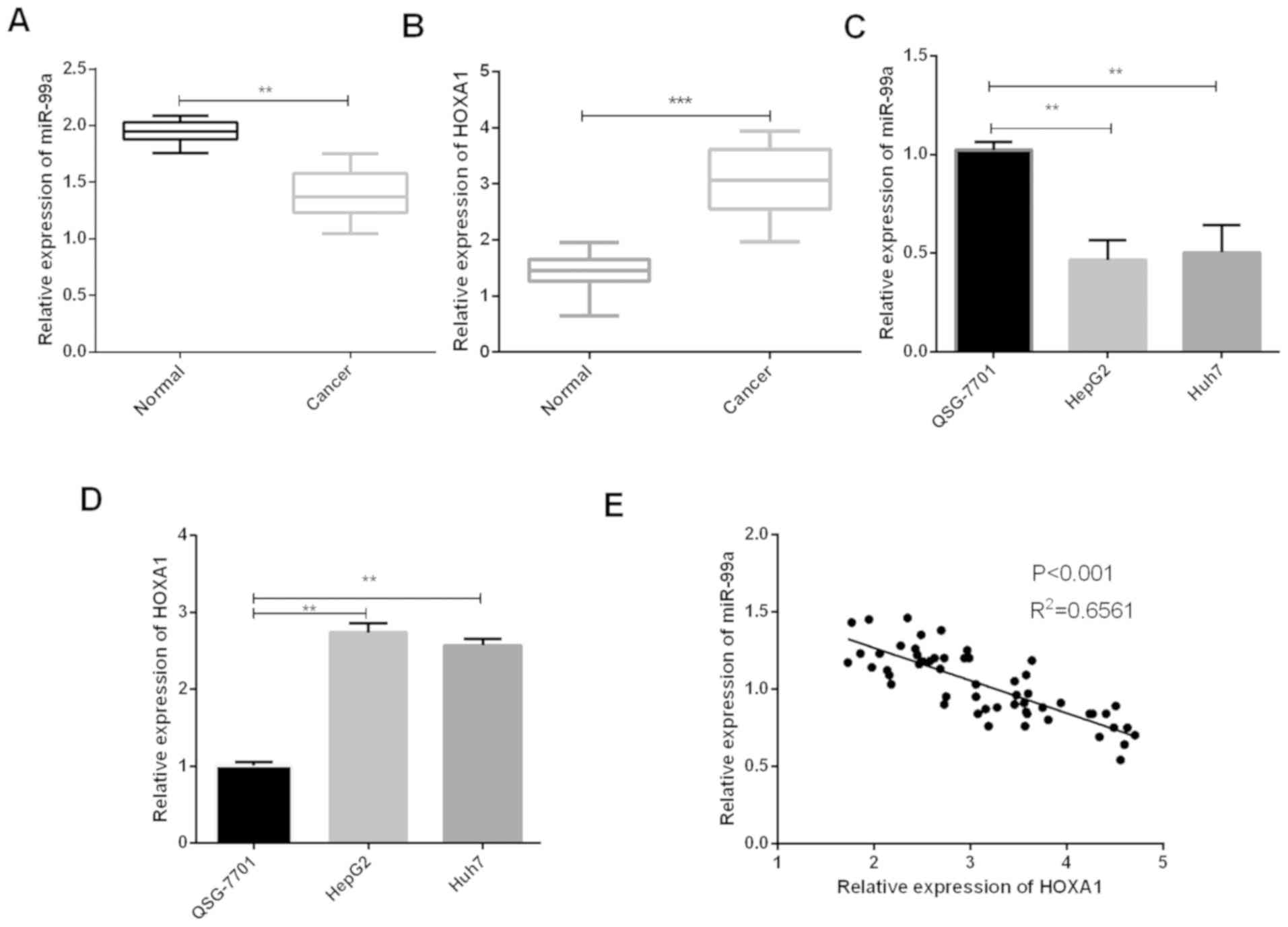
Low expression of miR-99a and
overexpression of HOXA1 in liver cancer
The expression of miR-99a and HOXA1 in liver
cancer tissue samples and cell lines were measured using RT-qPCR to
determine the function of miR-99a in liver cancer tumorigenesis.
Results revealed that miR-99a expression was significantly lower in
liver cancer tissues than those in matched benign tissue samples
(Fig. 1A). Conversely, the
HOXA1 mRNA expression in liver cancer tissue samples was
significantly higher than the normal tissue samples (Fig. 1B). We also verified that miR-99a
expressions in liver cancer cell lines (HepG2 and Huh7) were
decreased in comparison with the normal liver cells (Fig. 1C) while HOXA1 expressions in liver
cancer cell lines (HepG2 and Huh7) were increased compared to the
normal cells (Fig. 1D). In addition,
statistical analysis showed that miR-99a expression was negatively
correlated with HOXA1 in liver cancer tissues (Fig. 1Ε). The association between the
clinicopathological characteristics of 55 liver cancer patients and
miR-99a expression in liver cancer tissue samples is shown in
Table I, showing that miR-99a
expression was significantly associated with TNM stage (P=0.0239)
and BCLC stage (P=0.0116).
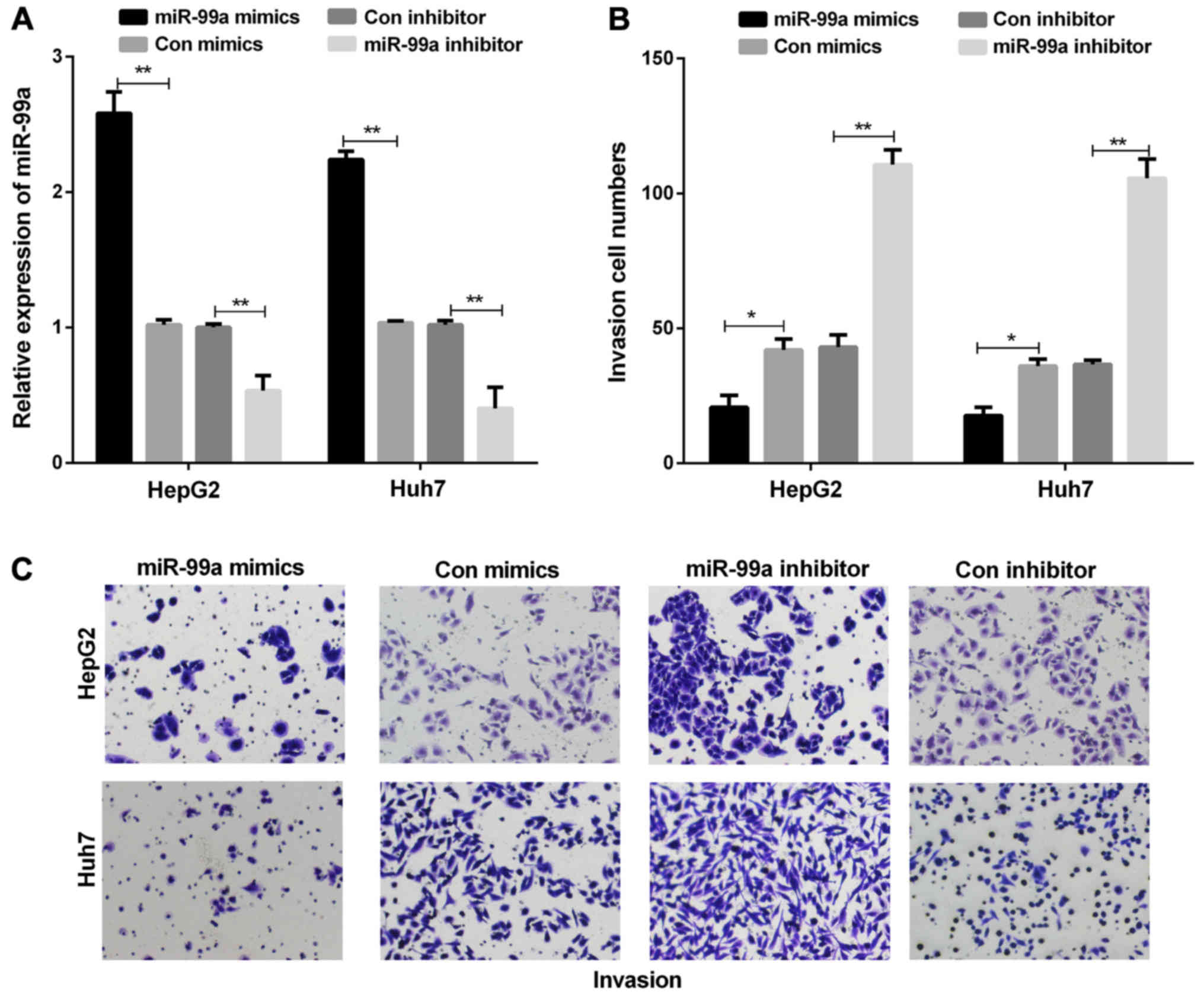
miR-99a suppresses the abilities of
cell invasion in liver cancer cell lines
We established miR-99a overexpression models by
transiently transfecting miR-99a mimics into liver cancer cells to
further investigate the function of miR-99a in liver cancer. The
results showed that miR-99a mimics were significantly upregulated
in both HepG2 and Huh7 cells (Fig.
2A). Next, we investigated the effect of miR-99a on liver
cancer cell invasion. In the cell invasion assays, miR-99a
overexpression suppressed liver cancer cell invasion, whereas
miR-99a inhibition promoted liver cancer cell invasion (Fig. 2B and C).
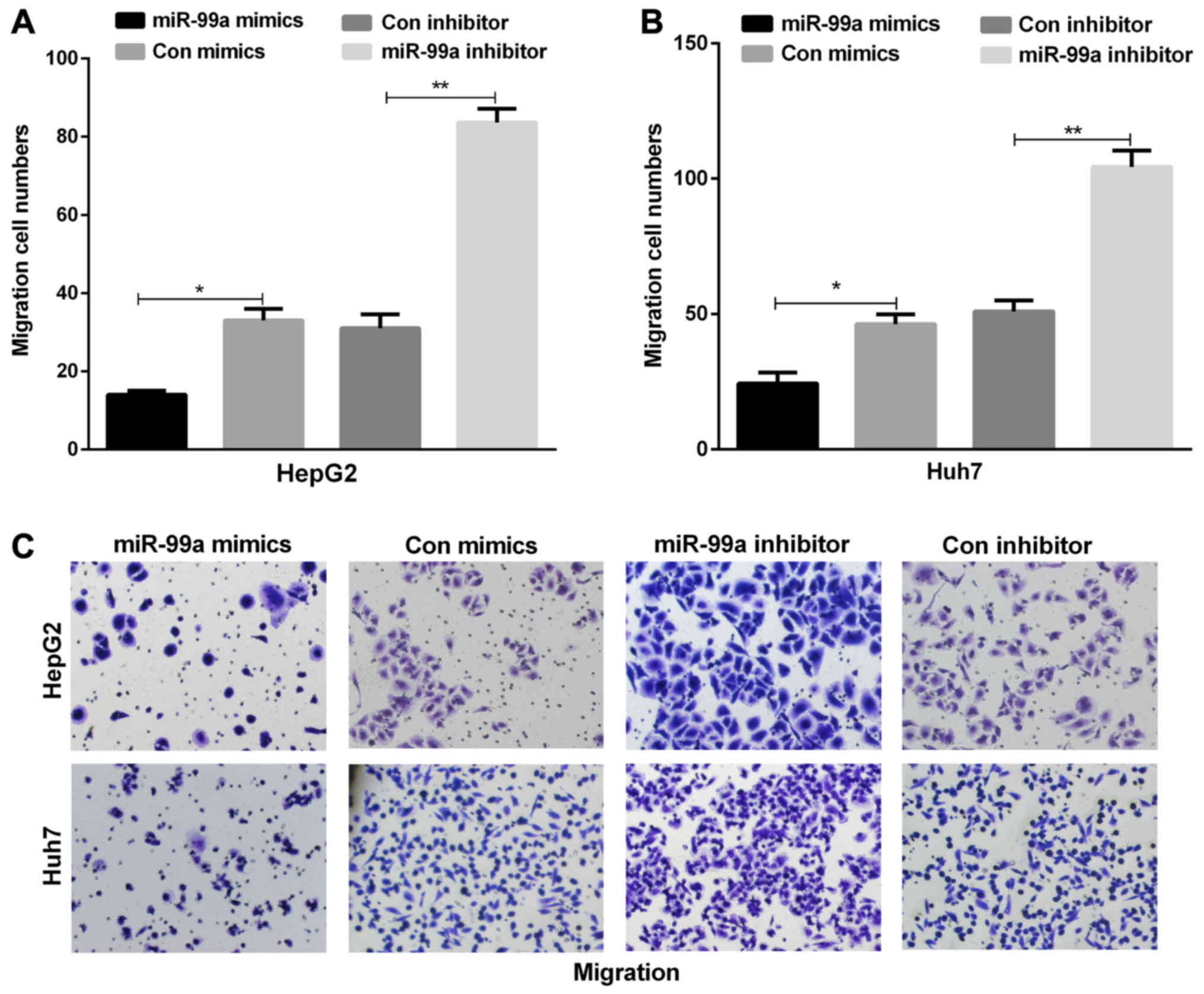
miR-99a inhibits liver cancer cell
migration
To explore the metastastic function of miR-99a in
liver cancer cells, cell migration assay was performed using
Transwell assay. miR-99a overexpression in HepG2 and Huh7 cell
lines contributed to a significant reduction in cell migration in
contrast to the control group. At the same time, we found that the
inhibition of miR-99a increased HepG2 and Huh7 cell migration
(Fig. 3A-C).
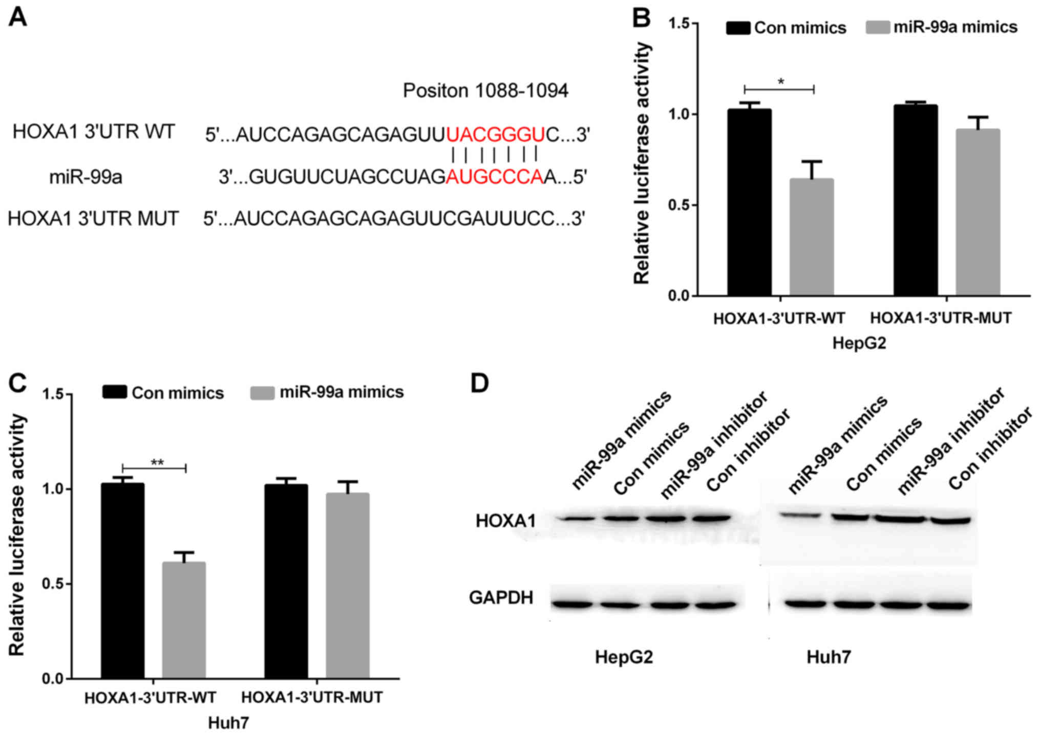
HOXA1 was a direct target for
miR-99a
Targetscan was applied to identify the putative
target of miR-99a. Among all the genes searched, HOXA1 was
selected because of its crucial effect on cell growth. Sequence
analysis showed putative binding sites between miR-99a and
HOXA1 (Fig. 4A). Luciferase
reporter assay was performed on HepG2 and Huh7 cell lines, which
were transfected with miR-99a mimics or miR-control. Then, the
transfected cells were co-transfected with the luciferase reporters
containing WT or MUT of HOXA1 3′UTR, respectively. The
results indicated that miR-99a overexpression inhibited the
luciferase activity of the HOXA1−3′UTR-WT reporter in both
HepG2 and Huh7 cells, not affecting the activity of the
HOXA1−3′UTR-MUT reporter (Fig. 4B
and C). Moreover, the western blots indicated that HOXA1
protein expression was obviously decreased in both HepG2 and Huh7
cells transfected with miR-99a mimics, whereas the protein
expression of HOXA1 was obviously increased in HepG2 and Huh7 cell
lines transfected with miR-99a inhibitor compared with their
respective control group (Fig. 4D).
These results suggested that HOXA1 was a direct target for
miR-99a in liver cancer cells.
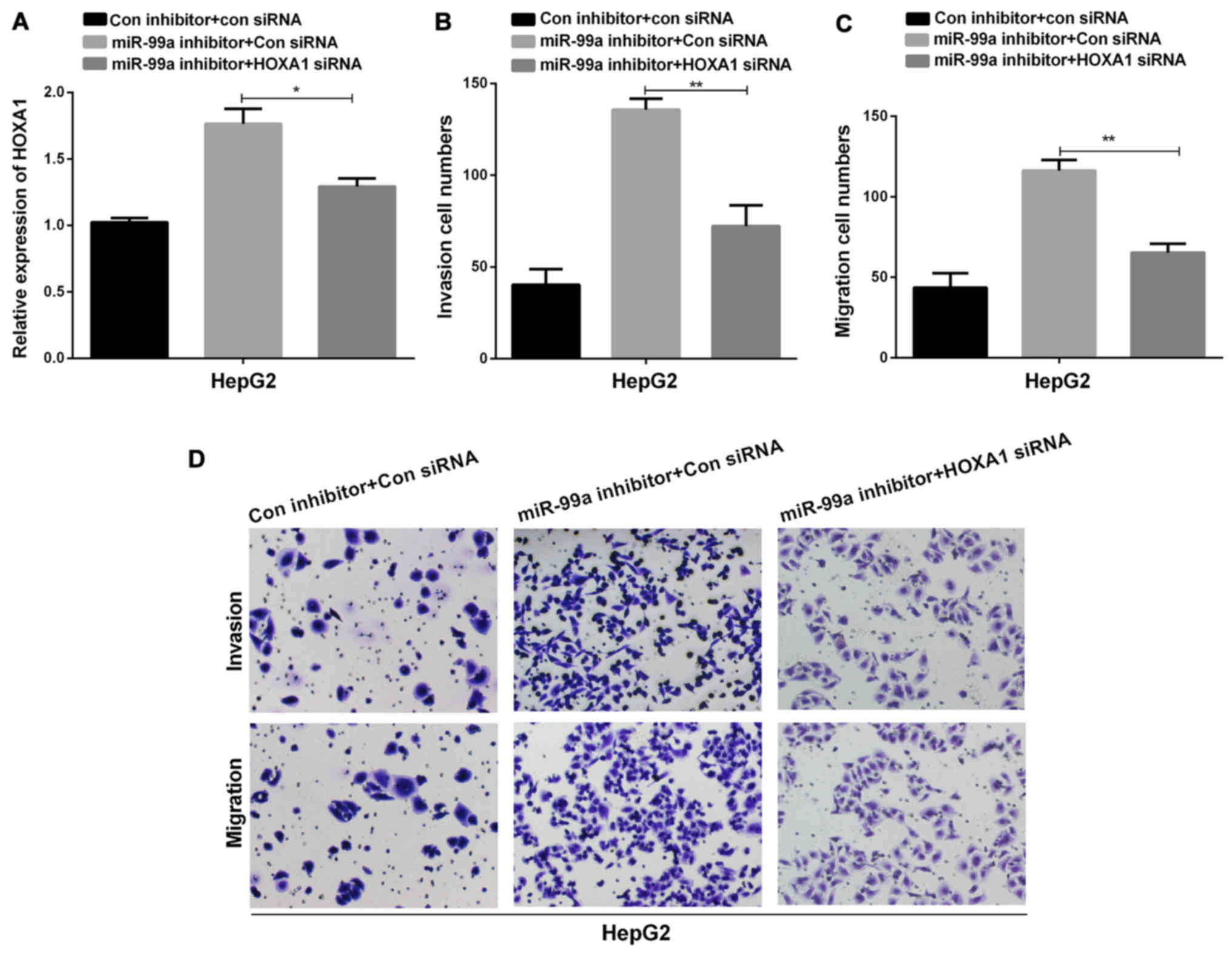
Roles of HOXA1 in regulating the
miR-99a ability in liver cancer cell invasion and migration
We also investigated whether HOXA1 was
essential in regulating the miR-99a ability in liver cancer cell
invasion and migration. HOXA1 siRNAs were transfected into
liver cancer cells to knock down endogenous HOXA1 and
RT-qPCR was used to determine the HOXA1 mRNA expression. The
findings demonstrated that the HOXA1 expression in liver
cancer cells transfected with HOXA1 siRNAs was reduced
significantly compared to the control group (Fig. 5A). Subsequently, Transwell assays
were carried out to observe the migration and invasion capacities
of liver cancer cells co-transfected with HOXA1 siRNAs and
miR-99a inhibitor. From the results, it was evident that deletion
of HOXA1 markedly reversed miR-99a-medicated inhibitory
effect on cell invasion and migration in liver cancer cells
(Fig. 5B-D).
Discussion
Liver cancer is one of the most lethal cancer types,
posing an increasing burden on global health (18). Currently, studies on mechanism have
focused primarily on liver cancer. Although liver cancer has been
widely researched, the possible mechanisms involved remain to be
elucidated because of its complexity. Emerging evidence has shown
that the two main biological characteristics of malignant cancers
are invasion and metastasis, which affect the prognosis of tumor
patients (19). Accommodative
dysfunction of miRNA frequently occurs in multifarious human
tumors, and the aberrant expression of miRNA often contributes to
carcinogenesis by affecting expression levels of multiple genes
(20). Thus, it is valuable to fully
understand the functions of specific miRNAs in the development of
tumor for the diagnosis and treatment of liver cancer.
Recently, as miRNAs have become crucial regulators
in the development of tumor, the target therapy of tumor using
miRNAs emerges as a fresh diagnostic instrument (21). It has been reported that miR-99a is
involved in various tumors, intervening the processes of cell
proliferation, apoptosis as well as inflammation (22,23).
Yang et al verified that miR-99a exerted a crucial effect on
modulating the CSC phenotype of breast carcinoma mediated via
targeting the mTOR/HIF-α signaling pathway (24). In addition, miR-99a was frequently
downregulated in ATC and miR-99a overexpression could reduce
oncogenicity through targeting mTOR (25). Although miR-99a has been identified
as a tumor suppressor in various types of cancer, however, the
clear functions of miR-99a together with HOXA1 in liver
cancer have not been described yet.
HOXA1 is a pivotal transcriptional regulator
of early embryonic development (26). A growing number of studies have
indicated that the aberrant HOXA1 expression in different
cancers is involved in the regulation of biological processes, such
as proliferation and apoptosis (27). For instance, overexpression of
HOXA1 was found in prostate cancer cells and silencing of
HOXA1 suppressed the progression of prostate cancer
(16). HOXA1 was found to be
a target of miR-10a in pancreatic cancer, and it was involved in
tumor cell invasion (28). The
current study aimed to determine the relationship between the
expression of miR-99a and HOXA1 in liver cancer. The
expression of miR-99a and HOXA1 was measured using RT-qPCR,
and the results showed that miR-99a was downregulated, whereas
HOXA1 was overexpressed in liver cancer. Additionally, we
found that miR-99a expression negatively correlated with
HOXA1 expression in liver cancer tissues. Moreover, data
also showed that miR-99a suppressed cell invasion and migration
through HOXA1 in liver cancer.
Collectively, the findings in our study showed that
miR-99a exerted a crucial suppressive effect in liver cancer.
First, our data revealed that the miR-99a expression was
significantly decreased and HOXA1 expression was
significantly increased in liver cancer tissues in contrast to the
control group. The same result was also found in the liver cancer
cell lines. Second, the Transwell assay results revealed that
miR-99a could suppress liver cancer cell invasion and migration. In
addition, we confirmed that HOXA1 was a direct target of
miR-99a. The prevention of tumor progression observed in this study
support the further investigation of miRNA therapy for liver cancer
prevention and treatment.
Acknowledgements
Not applicable.
Funding
No funding was received.
Availability of data and materials
The datasets used and/or analyzed during the present
study are available from the corresponding author on reasonable
request.
Authors' contributions
CT contributed significantly to data analysis and
manuscript preparation. HS helped perform the statistical analysis
with constructive discussions. WS contributed to the conception of
the study. SL collected, interpreted and analyzed the data. All
authors read and approved the final manuscript.
Ethics approval and consent to
participate
All patients involved in this research gave written
informed consents and the study was approved by the Liaocheng
People's Hospital Ethics Committee (Liaocheng, China).
Patient consent for publication
Not applicable.
Competing interests
The authors declare that they have no competing
interests.
References
|
1
|
Torre LA, Bray F, Siegel RL, Ferlay J,
Lortet-Tieulent J and Jemal A: Global cancer statistics, 2012. CA
Cancer J Clin. 65:87–108. 2015. View Article : Google Scholar : PubMed/NCBI
|
|
2
|
Ho C, Wang C, Mattu S, Destefanis G, Ladu
S, Delogu S, Armbruster J, Fan L, Lee SA, Jiang L, et al: AKT
(v-akt murine thymoma viral oncogene homolog 1) and N-Ras
(neuroblastoma ras viral oncogene homolog) coactivation in the
mouse liver promotes rapid carcinogenesis by way of mTOR (mammalian
target of rapamycin complex 1), FOXM1 (forkhead box M1)/SKP2, and
c-Myc pathways. Hepatology. 55:833–845. 2012. View Article : Google Scholar : PubMed/NCBI
|
|
3
|
Wu Y, Cain-Hom C, Choy L, Hagenbeek TJ, de
Leon GP, Chen Y, Finkle D, Venook R, Wu X, Ridgway J, et al:
Therapeutic antibody targeting of individual Notch receptors.
Nature. 464:1052–1057. 2010. View Article : Google Scholar : PubMed/NCBI
|
|
4
|
Han D, Li J, Wang H, Su X, Hou J, Gu Y,
Qian C, Lin Y, Liu X, Huang M, et al: Circular RNA MTO1 acts as the
sponge of miR-9 to suppress hepatocellular carcinoma progression.
Hepatology. 66:1151–1164. 2017. View Article : Google Scholar : PubMed/NCBI
|
|
5
|
Manikandan J, Aarthi JJ, Kumar SD and
Pushparaj PN: Oncomirs: The potential role of non-coding microRNAs
in understanding cancer. Bioinformation. 2:330–334. 2008.
View Article : Google Scholar : PubMed/NCBI
|
|
6
|
Simon DJ, Madison JM, Conery AL,
Thompson-Peer KL, Soskis M, Ruvkun GB, Kaplan JM and Kim JK: The
microRNA miR-1 regulates a MEF-2-dependent retrograde signal at
neuromuscular junctions. Cell. 133:903–915. 2008. View Article : Google Scholar : PubMed/NCBI
|
|
7
|
Yates LA, Norbury CJ and Gilbert RJ: The
long and short of microRNA. Cell. 153:516–519. 2013. View Article : Google Scholar : PubMed/NCBI
|
|
8
|
Hoffend NC, Magner WJ and Tomasi TB: The
epigenetic regulation of Dicer and microRNA biogenesis by
Panobinostat. Epigenetics. 12:105–112. 2017. View Article : Google Scholar : PubMed/NCBI
|
|
9
|
Fang F, Chang RM, Yu L, Lei X, Xiao S,
Yang H and Yang LY: MicroRNA-188-5p suppresses tumor cell
proliferation and metastasis by directly targeting FGF5 in
hepatocellular carcinoma. J Hepatol. 63:874–885. 2015. View Article : Google Scholar : PubMed/NCBI
|
|
10
|
Zhang H, Liu H and Bi H: MicroRNA-345
inhibits hepatocellular carcinoma metastasis by inhibiting YAP1.
Oncol Rep. 38:843–849. 2017. View Article : Google Scholar : PubMed/NCBI
|
|
11
|
Chang RM, Xiao S, Lei X, Yang H, Fang F
and Yang LY: miRNA-487a promotes proliferation and metastasis in
hepatocellular carcinoma. Clin Cancer Res. 23:2593–2604. 2017.
View Article : Google Scholar : PubMed/NCBI
|
|
12
|
Makki N and Capecchi MR: Cardiovascular
defects in a mouse model of HOXA1 syndrome. Hum Mol Genet.
21:26–31. 2012. View Article : Google Scholar : PubMed/NCBI
|
|
13
|
Yuan C, Zhu X, Han Y, Song C, Liu C, Lu S,
Zhang M, Yu F, Peng Z and Zhou C: Elevated HOXA1 expression
correlates with accelerated tumor cell proliferation and poor
prognosis in gastric cancer partly via cyclin D1. J Exp Clin Cancer
Res. 35:152016. View Article : Google Scholar : PubMed/NCBI
|
|
14
|
Xiao F, Bai Y, Chen Z, Li Y, Luo L, Huang
J, Yang J, Liao H and Guo L: Downregulation of HOXA1 gene affects
small cell lung cancer cell survival and chemoresistance under the
regulation of miR-100. Eur J Cancer. 50:1541–1554. 2014. View Article : Google Scholar : PubMed/NCBI
|
|
15
|
Bitu CC, Destro MF, Carrera M, da Silva
SD, Graner E, Kowalski LP, Soares FA and Coletta RD: HOXA1 is
overexpressed in oral squamous cell carcinomas and its expression
is correlated with poor prognosis. BMC Cancer. 12:1462012.
View Article : Google Scholar : PubMed/NCBI
|
|
16
|
Wang H, Liu G, Shen D, Ye H, Huang J, Jiao
L and Sun Y: HOXA1 enhances the cell proliferation, invasion and
metastasis of prostate cancer cells. Oncol Rep. 34:1203–1210. 2015.
View Article : Google Scholar : PubMed/NCBI
|
|
17
|
Livak KJ and Schmittgen TD: Analysis of
relative gene expression data using real-time quantitative PCR and
the 2(-Delta Delta C(T)) Method. Methods. 25:402–408. 2001.
View Article : Google Scholar : PubMed/NCBI
|
|
18
|
Llovet JM, Villanueva A, Lachenmayer A and
Finn RS: Advances in targeted therapies for hepatocellular
carcinoma in the genomic era. Nat Rev Clin Oncol. 12:408–424. 2015.
View Article : Google Scholar : PubMed/NCBI
|
|
19
|
Gumireddy K, Li A, Cao L, Yan J, Liu L, Xu
X, Pazoles C and Huang Q: NOV-002, a glutathione disulfide mimetic,
suppresses tumor cell invasion and metastasis. J Carcinog Mutagen.
2013:S7–002. 2013.PubMed/NCBI
|
|
20
|
Kloosterman WP and Plasterk RH: The
diverse functions of microRNAs in animal development and disease.
Dev Cell. 11:441–450. 2006. View Article : Google Scholar : PubMed/NCBI
|
|
21
|
Hummel R, Hussey DJ and Haier J:
MicroRNAs: Predictors and modifiers of chemo- and radiotherapy in
different tumour types. Eur J Cancer. 46:298–311. 2010. View Article : Google Scholar : PubMed/NCBI
|
|
22
|
Tao Z, Zhao H, Wang R, Liu P, Yan F, Zhang
C, Ji X and Luo Y: Neuroprotective effect of microRNA-99a against
focal cerebral ischemia-reperfusion injury in mice. J Neurol Sci.
355:113–119. 2015. View Article : Google Scholar : PubMed/NCBI
|
|
23
|
Lin KY, Ye H, Han BW, Wang WT, Wei PP, He
B, Li XJ and Chen YQ: Genome-wide screen identified
let-7c/miR-99a/miR-125b regulating tumor progression and stem-like
properties in cholangiocarcinoma. Oncogene. 35:3376–3386. 2016.
View Article : Google Scholar : PubMed/NCBI
|
|
24
|
Yang Z, Han Y, Cheng K, Zhang G and Wang
X: miR-99a directly targets the mTOR signalling pathway in breast
cancer side population cells. Cell Prolif. 47:587–595. 2014.
View Article : Google Scholar : PubMed/NCBI
|
|
25
|
Huang HG, Luo X, Wu S and Jian B: MiR-99a
inhibits cell proliferation and tumorigenesis through targeting
mTOR in human anaplastic thyroid cancer. Asian Pac J Cancer Prev.
16:4937–4944. 2015. View Article : Google Scholar : PubMed/NCBI
|
|
26
|
Li Q, Zhang X, Li N, Liu Q and Chen D:
miR-30b inhibits cancer cell growth, migration, and invasion by
targeting homeobox A1 in esophageal cancer. Biochem Biophys Res
Commun. 485:506–512. 2017. View Article : Google Scholar : PubMed/NCBI
|
|
27
|
López-Romero R, Marrero-Rodríguez D,
Romero-Morelos P, Villegas V, Valdivia A, Arreola H, Huerta-Padilla
V and Salcedo M: The role of developmental HOX genes in cervical
cancer. Rev Med Inst Mex Seguro Soc. 53 (Suppl 2):S188–S193.
2015.(In Spanish). PubMed/NCBI
|
|
28
|
Ohuchida K, Mizumoto K, Lin C, Yamaguchi
H, Ohtsuka T, Sato N, Toma H, Nakamura M, Nagai E, Hashizume M, et
al: MicroRNA-10a is overexpressed in human pancreatic cancer and
involved in its invasiveness partially via suppression of the HOXA1
gene. Ann Surg Oncol. 19:2394–2402. 2012. View Article : Google Scholar : PubMed/NCBI
|