Introduction
Osteosarcoma (OS) is the most common primary
malignant type of bone malignancy in adolescence and is
characterized by the formation of immature bone or osteoid tissues
from spindle matrix cells (1). Due
to the high degree of malignancy of OS and early metastasis to the
lungs, the majority of the patients present pulmonary
micrometastasis at the primary diagnosis (2). At present, the combination of surgical
resection and chemotherapy is the most effective treatment
(3,4). Despite a significant improvement in the
5-year overall survival for patients, the cure rate for patients
with OS has not improved and patients with metastatic or relapsed
disease have a poor prognosis (5,6).
Therefore, identifying more precise prognostic biomarkers and novel
approaches to the treatment of OS is necessary to improve the
outcome of patients with OS.
MicroRNAs (miRNAs) are a group of short non-coding
RNAs (18–25 nucleotides in length) that regulate
post-transcriptional gene expression by targeting the 3′
untranslated region (3′-UTR) of mRNA (7). An increasing number of studies have
demonstrated that miRNAs serve crucial roles in various biological
processes, including inflammation, cell proliferation, migration,
invasion, apoptosis and differentiation (8–10).
Depending on their dysregulation, miRNAs may serve as tumor
suppressors or oncogenes in tumorigenesis processes, which
contribute to cancer metastasis by influencing cell proliferation
and invasion (11,12). Overexpression of miRNA-34a inhibited
the migratory and invasive ability of OS cells by repressing the
expression of CD44 antigen, serving as a tumor suppressor in the
metastasis of OS cells (13). A
previous study by Mosakhani et al (14) identified miRNA-136 (miR-136) and its
target gene, nuclear factor 1 B-type, as novel biomarkers that may
aid in distinguishing primary giant cell tumors of bone with an
increased risk for metastasis. Previous studies have additionally
demonstrated that miR-136 is involved in the progression of a
number of different types of cancer, including cervical carcinoma,
hepatocellular carcinoma, and colon cancer (15–17).
However, the role of miR-136 in OS remains unclear.
In the present study, the expression patterns and
prognostic significance of miR-136 in patients with OS was
investigated. Additionally, the effects of miR-136 on the
biological behaviors of cancer cells were assessed.
Materials and methods
Patients and tissue samples
OS tissue and matched adjacent normal tissue
specimens (located >3 cm away from the tumors) were obtained
between January 2007 and February 2012 from 116 patients with OS
who underwent surgery at The Affiliated Hospital of Qingdao
University (Qingdao, China). All the tissues were verified and
experienced pathologists confirmed the tissue stages, according to
the National Comprehensive Cancer Network guidelines (18,19). The
OS tissue and normal tissue specimens were snap-frozen in liquid
nitrogen following surgery and subsequently stored at −80°C until
use. The inclusion criteria were: All patients were pathologically
diagnosed with OS, none of the enrolled patients received any
therapy prior to surgery. and all patients had complete clinical,
pathological, and follow-up information. All patients agreed to
participate in the present study and provided written informed
consent prior to surgery, and the Ethics Committee of The
Affiliated Hospital of Qingdao University approved the protocol.
The characteristics of the patients are summarized in Table I. The 5-year follow-up information
was updated for the subsequent analysis.
 | Table I.Association between miR-136
expression and clinical characteristics of patients with
osteosarcoma. |
Table I.
Association between miR-136
expression and clinical characteristics of patients with
osteosarcoma.
|
|
| miR-136
expression |
|
|---|
|
|
|
|
|
|---|
| Clinical
characteristics | Cases (n=116) | High (n=50) | Low (n=66) | P-value |
|---|
| Sex |
|
|
| 0.283 |
|
Male | 56 | 27 | 29 |
|
|
Female | 60 | 23 | 37 |
|
| Age |
|
|
| 0.791 |
|
<18 | 61 | 27 | 34 |
|
|
≥18 | 55 | 23 | 32 |
|
| Tumor site |
|
|
| 0.373 |
| Distal
femur | 54 | 27 | 27 |
|
|
Proximal tibia | 41 | 15 | 26 |
|
|
Others | 21 | 8 | 13 |
|
| Enneking staging
(19) |
|
|
| 0.030 |
|
I–II | 68 | 35 | 33 |
|
|
III | 48 | 15 | 33 |
|
| Distant
metastasis |
|
|
| 0.016 |
|
Absent | 57 | 31 | 26 |
|
|
Present | 59 | 19 | 40 |
|
Cell lines and transfection
Human OS cell lines HOS and U2OS and the normal
osteoblast cell line NHOst were obtained from The American Type
Culture Collection (Manassas, VA, USA) and cultured in RPMI 1640
medium (Gibco; Thermo Fisher Scientific, Inc., Waltham, MA, USA)
supplemented with heat-inactivated 10% fetal bovine serum (FBS;
Gibco; Thermo Fisher Scientific, Inc.) at 37°C in a humidified
incubator with 5% CO2. The miR-136 mimic
(5′-ACUCCAUUUGUUUUGAUGAUGG-3′), miR-136 inhibitor
(5′-CCAUCAUCAAAACAAAUGGAGU-3′), mimic negative control (NC)
(5′-TTCTCCGAACGTGTCACGT-3′) or inhibitor NC
(5′-UUCUCCGAACGUGUCACGUTT-3′) were synthesized by Shanghai
GenePharma Co., Ltd. (Shanghai, China). A total of 5×10⁴
cells were seeded into the wells of a 6-well plate. On the
subsequent day, the cells were transfected with miR-136 mimic,
miR-136 inhibitor or the respective miRNA-NCs (mimics NC and
inhibitor NC) using Lipofectamine® 2000 (Invitrogen;
Thermo Fisher Scientific, Inc.) with a final oligonucleotide
concentration of 20 nmol/l, according to the manufacturer's
protocol. As a negative control, transfection reagent alone was
added to the cells as the mock. The transfection efficiency was
detected by reverse transcription-quantitative polymerase chain
reaction (RT-qPCR) analysis after 48 h.
RNA extraction and RT-qPCR
Total RNA was extracted from the tissue samples or
the cells using TRIzol® reagent (Invitrogen; Thermo
Fisher Scientific, Inc.) according to the manufacturer's protocol.
The purity of RNA was obtained by calculating the ratio of optical
density of absorbance at 260/280 nm using NanoDrop ND-1000 (Thermo
Fisher Scientific, Inc.). The purified total RNA was reverse
transcribed to cDNA using M-MLV reverse transcriptase (Promega
Corporation, Madison, WI, USA) according to the manufacturer's
protocol. The thermocycling condition for reverse transcription
were as follows: 25°C for 5 min, 42°C for 60 min, 72°C for 10 min.
The relative miR-136 expression was analyzed by RT-qPCR, which was
performed using SYBR Green Premix Ex Taq (Takara Bio, Inc., Otsu,
Japan) with an ABI 7500 instrument (Applied Biosystems; Thermo
Fisher Scientific, Inc.). Relative expression was calculated using
the 2−ΔΔCq method with U6 as the internal control
(20). The primer sequences and
thermocycling conditions of miR-136 and U6 used were as previously
described (21,22).
Cell proliferation assay
The cell proliferation of HOS or U2OS cells
transfected with NC, miR-136 mimics or miR-136 inhibitor was
measured using the colorimetric MTT method. HOS and U2OS cells were
seeded into 96-well plates at a density of 5×10³
cells/well and transfected. After 48 h, 10 µl MTT (5 mg/ml;
Sigma-Aldrich; Merck KGaA, Darmstadt, Germany) was added to each
well and the cells were incubated at 37°C for 4 h. Subsequent to
incubation, the medium was removed and 100 µl dimethyl sulfoxide
(Sigma-Aldrich; Merck KGaA) was added to the wells to dissolve the
formazan crystals. A wavelength of 490 nm was used for the
colorimetric analysis with a Multiskan MK3 (Thermo Fisher
Scientific, Inc.). Experiments were repeated in triplicate.
Colony-forming assay
Following transfection, cells were plated at a
density of 300 cells/well in a 6-well plate and incubated at 37°C.
The culture medium was replaced according to the change of pH of
the medium, as determined by the phenol red indicator in the
culture medium. After 14 days, cells in each well were washed
carefully with PBS, fixed with methanol for 15 min and stained for
10 min with Giemsa at room temperature. Subsequently, cell colonies
(groups >50 cells) were counted under a light microscope
(magnification, ×40). The test was independently conducted three
times.
Cell migration and invasion
analysis
To examine the effects of miR-136 on cell migration
and invasion, cell assays were performed using a 24-well transwell
chamber (8 µm pore size). For the transwell migration assays, the
cells transfected with mimics, inhibitor or respective miR-negative
controls were added to the top chamber at a density of
1×10⁵ cells/well. For the invasion assays,
2×10⁵ cells were plated in the upper compartment with
Matrigel-coated membranes (Corning Life Sciences, Bedford, MA,
USA). The cells were subsequently incubated in serum-free RPMI-1640
medium at 37°C for 24 h. The lower compartment contained 300 µl
RPMI-1640 medium supplemented with 20% FBS, which was used as the
chemotactic factor. Following incubation for 24 h, the cells that
had migrated into the lower compartment were fixed in 3.7%
formaldehyde for 5 min and stained with 0.1% crystal violet for 15
min at room temperature. The number of cells was counted using a
light microscope (magnification, ×200). Each experiment was
repeated in triplicate.
Dual-luciferase reporter assay
The putative targeting gene, migration and invasion
enhancer 1 (MIEN1), of miR-136 was predicted using web-based miRNA
databases, Targetscan (http://targetscan.org), miRanda (http://www.microrna.org) and miRNA-PicTar (https://dorina.mdc-berlin.de/), all of which used the
3′-UTR as the target region to determine miRNA recognition
elements, and subsequently verified by a dual-luciferase reporter
assay. The 3′-UTR sequence of MIEN1 was amplified and subcloned
into the pGL3 luciferase reporter vector (Promega Corporation).
Cells were cotransfected with wild-type (WT) or mutant (MUT) 3′-UTR
vectors and miR-136 mimics, inhibitors or controls using
Lipofectamine® 2000. After 36 h, the luciferase
activities of the cells were determined with the Dual-Luciferase
Assay System (Promega Corporation) according to the manufacturer's
protocol. The firefly luciferase activities were normalized to
Renilla luciferase activity. All the experiments were
performed in triplicate.
Statistical analysis
Statistical analysis was conducted using SPSS 21.0
(IBM Corp., Armonk, NY, USA) and GraphPad Prism 5 (GraphPad
Software, Inc., La Jolla, CA, USA), and the data are presented as
the mean ± standard deviation. One-way analysis of variance with
Tukey's post hoc test was used for multiple comparisons. The
association between the clinicopathological characteristics of the
patients and miR-136 was analyzed by the χ2 test. The
association between overall survival and miR-136 expression was
estimated using the Kaplan-Meier method with a log-rank test. The
prognostic effects of each clinical characteristic were determined
using a Cox regression analysis. P<0.05 was considered to
indicate a statistically significant difference.
Results
Expression of miR-136 in tissue
specimens and cells
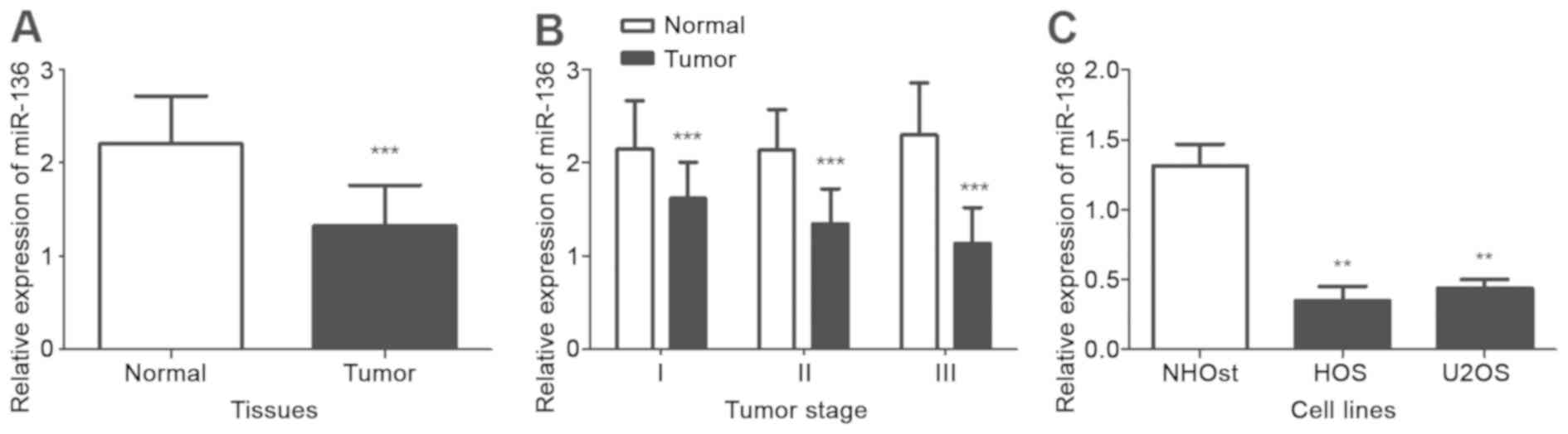
The expression of miR-136 in 116 paired OS tissue
specimens and cell lines were detected by RT-qPCR. As presented in
Fig. 1A, the expression of miR-136
was significantly decreased in OS tissues compared with matched
adjacent non-tumorous tissues (P<0.001). Furthermore, the
expression of miR-136 in tumor tissues at different Enneking stages
(19), including stage I (n=30),
stage II (n=38), stage III (n=48), was analyze and compared with
the expression in matched normal tissues. The expression of miR-136
in tumor tissues at each stage was significantly decreased compared
with matched normal tissues (all P<0.05; Fig. 1B). The expression of miR-136 was
consistently decreased in the OS cell lines, HOS and U2OS, compared
with the normal human OS cell line NHOst (both P<0.05; Fig. 1C). These results suggested that
miR-136 may be a tumor suppressor in OS.
Association of miR-136 expression with
clinical characteristics of patients with OS
Relative miR-136 expression in patients with OS was
associated with specific clinicopathological characteristics.
According to the mean expression level of miR-136 (1.329), the
patients were divided into a low miR-136 expression group (n=66;
0.46–1.31) and a high expression group (n=50; 1.34–2.48). The
analysis results are presented in Table
I. The expression of miR-136 was associated with Enneking stage
(P=0.030) and distant metastasis (P=0.016). There was no
association between miR-136 expression levels and other
clinicopathological characteristics, including sex, age and tumor
site (P>0.05; Table I).
Association between miR-136 expression
and overall survival time of patients with OS
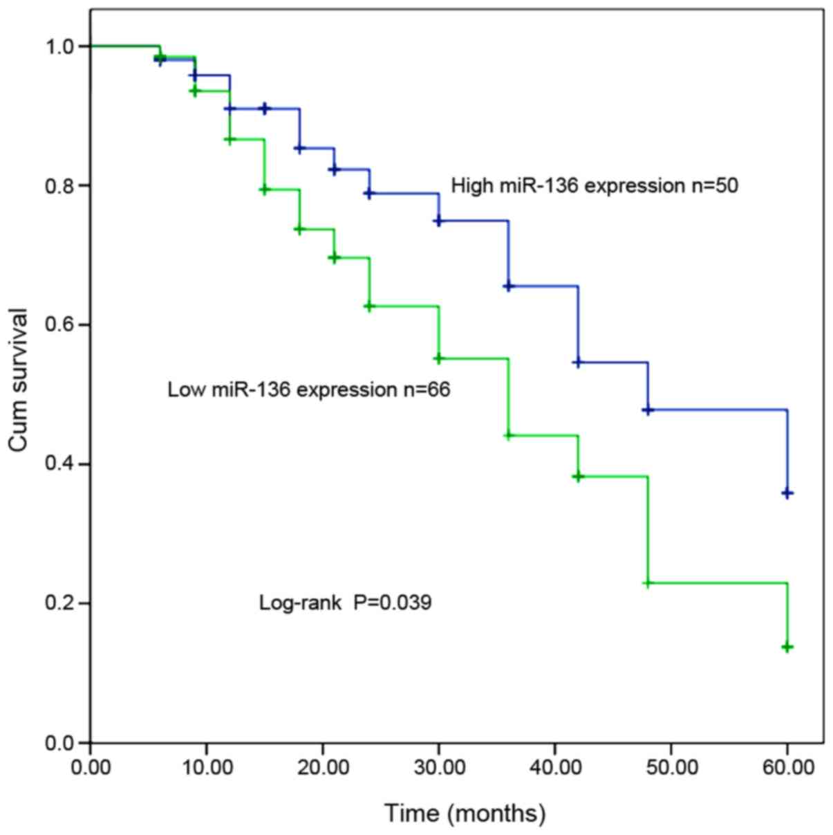
To investigate the prognostic factor of miR-136
expression in OS, Kaplan-Meier and Cox proportional hazard
regression model analyses were performed. As presented in Fig. 2, the survival time of patients with
low miR-136 expression was shorter compared with patients with high
miR-136 expression (log-rank test; P=0.039). Univariate and
multivariate Cox regression analysis results demonstrated that
miR-136 expression (hazard ratio=0.496; 95% confidence
interval=0.250–0.987; P=0.046; Table
II) may be an important prognostic factor and may thus be an
independent biomarker in patients with OS.
| Table II.Univariate and multivariate Cox
analysis of miR-136 expression in patients with osteosarcoma. |
Table II.
Univariate and multivariate Cox
analysis of miR-136 expression in patients with osteosarcoma.
|
| Univariate
analysis | Multivariate
analysis |
|---|
|
|
|
|
|---|
|
Characteristics | HR | 95% CI | P-value | HR | 95% CI | P-value |
|---|
| miR-136 | 0.496 | 0.250–0.987 | 0.046 | 0.496 | 0.250–0.987 | 0.046 |
| Sex | 1.230 | 0.670–2.260 | 0.504 | – | – | – |
| Age | 1.221 | 0.669–2.230 | 0.516 | – | – | – |
| Tumor site | – | – | 0.390 | – | – | – |
| Tumor site
(1) | 1.715 | 0.742–3.964 | 0.207 | – | – | – |
| Tumor site
(2) | 1.702 | 0.738–3.926 | 0.212 | – | – | – |
| Enneking staging
(19) | 1.177 | 0.636–2.179 | 0.604 | – | – | – |
| Distant
metastasis | 0.766 | 0.425–1.380 | 0.375 | – | – | – |
Overexpression of miR-136 inhibits
proliferation of OS cells
As miR-136 expression was negatively associated with
the survival time of the patients, the functional role of miR-136
and its effects on tumor cell proliferation, migration and invasion
were investigated. The normal osteoblast cell line NHOst and two OS
cell lines HOS and U2OS were transfected with miR-136 mimics,
inhibitor or their respective miR-negative controls to regulate the
expression of miR-136 in the tumor cells. The RT-qPCR results
demonstrated that the expression of miR-136 in OS cells transfected
with the miR-136 mimics was significantly increased compared with
the cells transfected with the negative control and mock
(P<0.05; Fig. 3A). The results
additionally demonstrated that the expression of miR-136 in OS
cells transfected with miR-136 inhibitor was decreased compared
with miR-136 expression in the cells with the negative control and
mock (P<0.05; Fig. 3A). The
expression of miR-136 in the normal osteoblast cell line NHOst was
increased in cells transfected with miR-136 mimics and decreased in
cells transfected with miR-136 inhibitor, although these
differences were not considered statistically significant
(P>0.05; Fig. 3A).
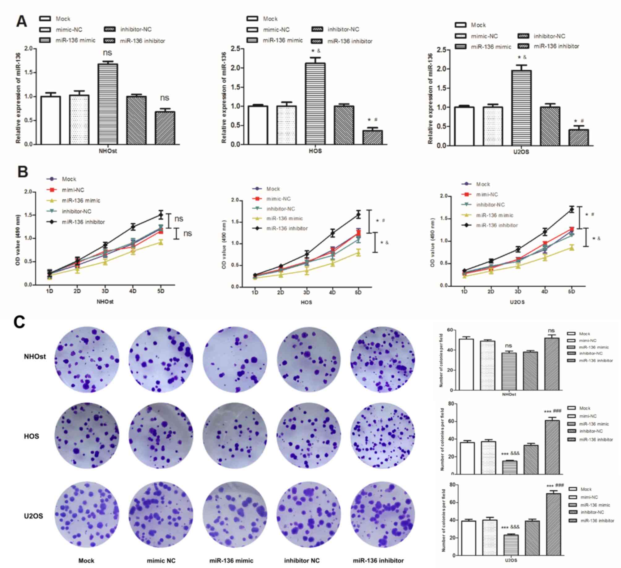 | Figure 3.Effects of miR-136 expression on
proliferation of the OS cells HOS and U2OS compared with the normal
osteoblast cell line NHOst. Each treatment group was measured at
least three times. (A) Expression of miR-136 in NHOst, HOS and U2OS
cells transfected with miR-136 mimics, miR-136 inhibitor or their
respective negative controls. (B) Cell viability was measured in OS
cells that were transfected with miR-136 mimics, inhibitor or
negative controls by an MTT assay. The cell viability in NHOst was
not significantly different. (C) Proliferation of OS cells and the
normal osteoblast cell line NHOst was detected by a colony
formation assay (magnification, ×40). Quantification of the colony
count. &P<0.05,
&&&P<0.001 vs. mimic-NC;
#P<0.05, ###P<0.001 vs. inhibitor-NC;
*P<0.05, ***P<0.001 vs. mock. miR-136, microRNA-136; NC,
negative control; OD, optical density; ns, not significant. |
An MTT assay was used to measure cell proliferation.
The results demonstrated that upregulation of miR-136 inhibited OS
cell proliferation (P<0.05; Fig.
3B). In contrast, an inhibitor of miR-136 had the opposite
effect, increasing OS cell proliferation compared with cells
transfected with respective negative controls or mock (P<0.05;
Fig. 3B). Although cell
proliferation was inhibited in NHOst cells transfected with miR-136
mimics and promoted in cells transfected with miR-136 inhibitor,
the differences were not statistically significant compared with
the controls (P>0.05; Fig. 3B).
Colony formation assays demonstrated that overexpression of miR-136
by miR-136 mimic inhibited colony formation, while inhibition of
miR-136 promoted colony formation (P<0.001; Fig. 3C); however, the differences in colony
count, relative to NHOst cells, was not considered statistically
significant (P>0.05; Fig.
3C).
Overexpression of miR-136 decreases
the migratory and invasive capacities of OS cells
In addition to proliferation, cell migration and
invasion were measured using transwell and Matrigel assays,
respectively. The assay results demonstrated that OS cells
transfected with miR-136 mimics exhibited significantly decreased
migratory and invasive capacity compared with the negative control
and mock groups (P<0.05; Fig. 4).
The OS cells transfected with miR-136 inhibitor promoted the
capacity of migration and invasion, compared with cells transfected
with negative control and mock (P<0.05, Fig. 4). Similar to cell proliferation, cell
migration and invasion in NHOst cells transfected with the miR-136
mimics or inhibitor demonstrated similar trends to the OS cells,
although the differences were not considered statistically
significant (P>0.05; Fig. 4).
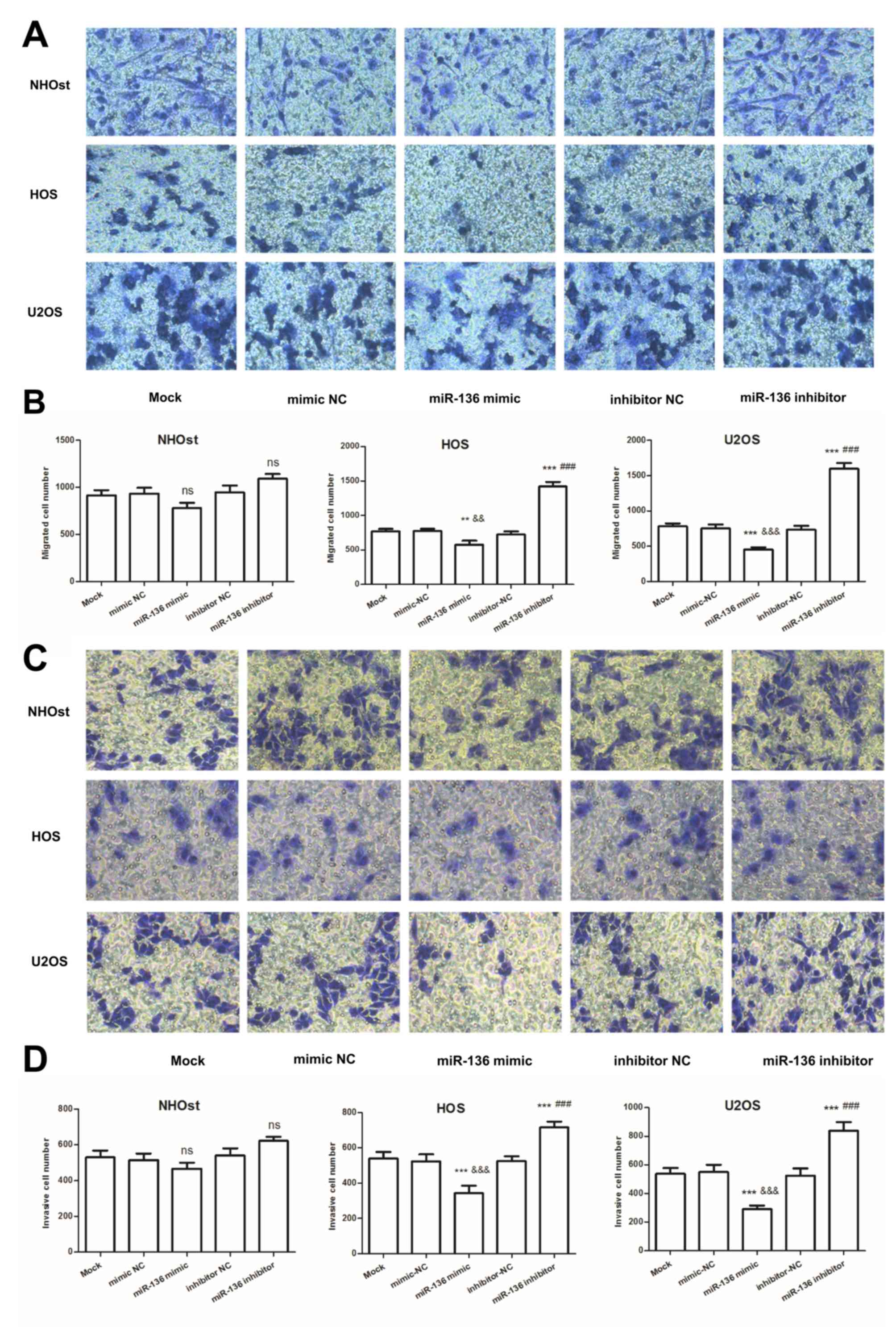 | Figure 4.Effects of miR-136 expression on the
migration and invasion of the OS cell lines, HOS and U2OS, and
normal osteoblast cell line NHOst. (A) Migration analysis of NHOst,
HOS and U2OS cells by transwell assays (magnification, ×200). (B)
Migration of OS cell lines, HOS and U2OS, is suppressed by
increasing miR-136 expression and increased by the downregulation
of miR-136 expression. (C) Invasion analysis of NHOst, HOS and U2OS
cells by Matrigel-precoated transwell assays (magnification, ×200).
(D) Overexpression of miR-136 by miR-136 mimics decreased cell
invasion and downregulation of miR-136 expression increased cell
invasion. &&P<0.01,
&&&P<0.001 vs. mimic-NC;
###P<0.001 vs. inhibitor-NC; **P<0.01,
***P<0.001 vs. mock. miR-136, microRNA-136; NC, negative
control; ns, not significant. |
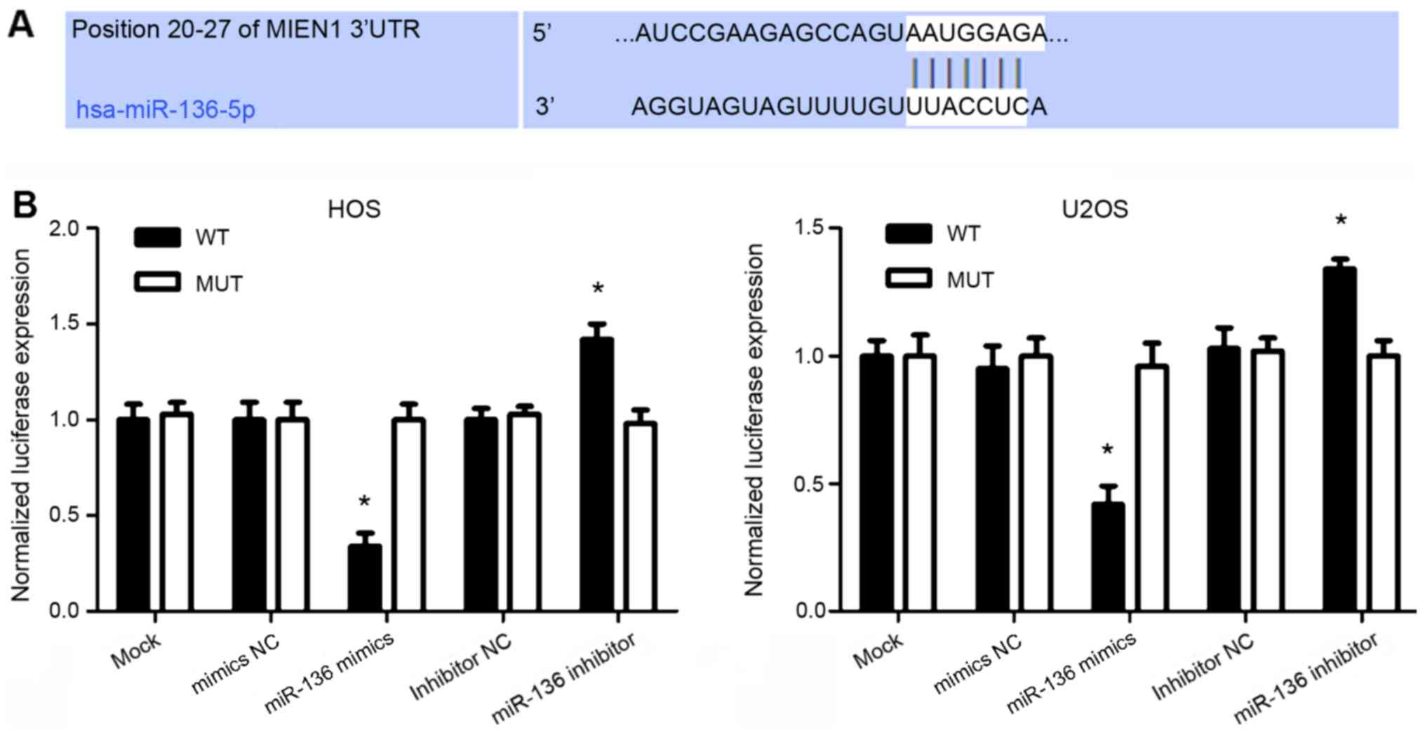
MIEN1 is a direct target of miR-136 in
OS cells
MIEN1 was predicted to be a target of miR-136
(Fig. 5A). A luciferase reporter
assay was used to investigate this hypothesis. As presented in
Fig. 5B, the luciferase activity
assay results demonstrated that cotransfection of the miR-136
mimics inhibited the luciferase activity of the reporter containing
the WT MIEN1 3′-UTR sequence; however, failed to suppress that
containing the MUT MIEN1 3′UTR. In contrast, the luciferase
activity of the reporter with the WT 3′-UTR of MIEN1 was increased
in the cells transfected with the miR-136 inhibitor; however, the
luciferase activity did not alter with the MUT 3′-UTR (Fig. 5B). These results suggested that MIEN1
may be a direct functional target of miR-136 in OS.
Discussion
The low prevalence and large heterogeneity of OS
make it difficult to improve patient survival (23). The specific tumor markers and
prognostic factors of OS have important clinical significance. A
number of previous studies demonstrated a critical role for
molecular biomarkers in tumor pathogenesis (24–26). In
OS, specific prognostic biomarkers have additionally been
identified. Hou et al (27)
demonstrated that cyclin-dependent kinase-1 gene expression was
increased in patients with OS and may thus serve as a biomarker to
predict the occurrence, development and prognosis of OS. Fernanda
Amary et al (28)
demonstrated that fibroblastic growth factor receptor 1 gene
amplification in OS was associated with a poor response to
neoadjuvant chemotherapy. Liang et al (29) demonstrated that Phospholipase A2
Group XVI expression was increased in OS and may thus serve as a
prognostic factor in patients with primary OS for predicting the
development of metastases and poor survival. Together, these
previous studies suggested that identification of cancer-associated
biomarkers for tumor progression and outcome may help to predict
patient prognosis and treatment strategies.
A number of miRNAs have been demonstrated to
contribute to the development of cancer and to serve as biomarkers
for the diagnosis, prognosis or treatment of different types of
cancer (30–32). Taheriazam et al (33) demonstrated that miRNA-130b expression
was increased in OS tissues, whereas, the level of miRNA-218
expression was downregulated; these miRNAs may serve as potential
biomarkers in the early detection of OS. In this study, it was
indicated that the expression of miR-136 was downregulated in the
patients with OS, which is consistent with the results in previous
studies (34,35). For instance, miR-136 was
downregulated and acted as a tumor suppressor in colon cancer
(34). In renal cell carcinoma,
miR-136 was reported to be downregulated and associated with renal
cell carcinoma cellular functions (35). To improve the understanding of the
role of miR-136 in OS, its expression patterns and association with
the clinicopathological features of the patients with OS were
investigated. In the present study, the expression of miR-136 was
decreased in OS tissues and cells compared with the corresponding
normal controls. Furthermore, the expression level of miR-136 was
associated with Enneking staging and distant metastasis in patients
with OS. These results indicated that miR-136 expression is
negatively associated with tumorigenesis and progression of OS,
which suggested that miR-136 may serve a tumor suppressor role in
OS tumorigenesis.
To evaluate the association of miR-136 with the
overall survival of patients with OS, Kaplan-Meier and Cox
regression analyses were used. Kaplan-Meier analysis demonstrated
that patients in the low miR-136 expression group had shorter
survival times compared with patients with high miR-136 expression.
According to Cox regression analysis, miR-136 expression was
associated with the prognosis of OS. Additionally, it may be an
independent prognostic marker in OS.
Previous studies demonstrated the effects of miR-136
on biological behaviors during cancer progression (15,21).
miR-136 inhibited colon cancer cell proliferation and invasion,
which may serve as a potential therapeutic target for colon cancer
(15). However, the functions of
miR-136 in OS cells have not yet been studied, to the best of the
authors' knowledge. In the present study, the effects of miR-136 on
the biological behavior of OS cells were assessed to demonstrate a
functional involvement of miR-136 during OS progression. The
results demonstrated that overexpression of miR-136 may inhibit
tumor cell proliferation, migration and invasion, which suggested a
potential tumor suppressor role for miR-136.
MIEN1 is located in the 17q12 region of the human
chromosome and has been demonstrated to be dysregulated in various
cancer tissues (36,37). A number of miRNAs were demonstrated
to perform biological functions by targeting MIEN1, including
miRNA-26b (38), miRNA-940 (39) and miRNA-136 (34). Ren et al (34) demonstrated that miR-136 directly
targeted the 3′-UTR of MIEN1 in colon cancer. In the present study,
a luciferase reporter assay demonstrated that MIEN1 is a direct
functional target of miR-136 in OS. In a recent study, miR-136
expression was decreased in triple-negative breast cancer, and
suppressed mesenchymal invasion and metastasis by targeting RAS
protein activator-like 2 (40). A
previous study by Yang et al (41) demonstrated that miR-136 directly
targeted mothers against decapentaplegic homolog (Smad)2 and Smad3
in transforming growth factor-β/Smad signaling, leading to
decreased migration and invasiveness of lung adenocarcinoma cell
lines. However, the detailed molecular mechanisms of miR-136
downregulation and the effects of targeting MIEN1 in OS require
further investigation. Additionally, due to the limitation of
sample size, further studies with larger research cohorts are
necessary.
In conclusion, the present findings demonstrated
that miR-136 was downregulated in OS cells and tissues of patients
with OS. Furthermore, overexpression of miR-136 resulted in the
inhibition of cell proliferation, migration and invasion in OS
cells. Further investigation of miR-136 identified that MIEN1 was a
potential target of miR-136. Together, these data suggested that
miR-136 may be a prognostic biomarker and potential therapeutic
target for patients with OS.
Acknowledgements
Not applicable.
Funding
The present study was funded by The Science and
Technology Development Plan Project of Qingdao Economic and
Technological Development Zone (grant no. 2014-1-78; China).
Availability of data and materials
The analyzed datasets generated during the present
study are available from the corresponding author on reasonable
request.
Authors' contributions
YC and YW initiated and designed the work, analyzed
the data, and wrote the manuscript. XH and GW collected clinical
tissues, performed RNA extraction and RT-qPCR assays, and
corresponding data analysis. ZW performed cell experiments. All
authors have read and approved the final version of this
manuscript.
Ethics approval and consent to
participate
All patients agreed to participate in the present
study and provided written informed consent prior to surgery, and
the Ethics Committee of The Affiliated Hospital of Qingdao
University (Qingdao, China) approved the protocol.
Patient consent for publication
Not applicable.
Competing interests
The authors declare that they have no competing
interests.
References
|
1
|
Ottaviani G and Jaffe N: The epidemiology
of osteosarcoma. Cancer Treat Res. 152:3–13. 2009. View Article : Google Scholar : PubMed/NCBI
|
|
2
|
Ritter J and Bielack SS: Osteosarcoma. Ann
Oncol. 21 (Suppl):vii320–325. 2010. View Article : Google Scholar : PubMed/NCBI
|
|
3
|
Jones KB, Ferguson PC, Lam B, Biau DJ,
Hopyan S, Deheshi B, Griffin AM, White LM and Wunder JS: Effects of
neoadjuvant chemotherapy on image-directed planning of surgical
resection for distal femoral osteosarcoma. J Bone Joint Surg Am.
94:1399–1405. 2012. View Article : Google Scholar : PubMed/NCBI
|
|
4
|
Wong KC, Lee V, Shing MM and Kumta S:
Surgical resection of relapse may improve postrelapse survival of
patients with localized osteosarcoma. Clin Orthop Relat Res.
471:814–819. 2013. View Article : Google Scholar : PubMed/NCBI
|
|
5
|
Gill J, Ahluwalia MK, Geller D and Gorlick
R: New targets and approaches in osteosarcoma. Pharmacol Ther.
137:89–99. 2013. View Article : Google Scholar : PubMed/NCBI
|
|
6
|
Meazza C and Scanagatta P: Metastatic
osteosarcoma: A challenging multidisciplinary treatment. Exp Rev
Anticancer Ther. 16:543–556. 2016. View Article : Google Scholar
|
|
7
|
Farazi TA, Hoell JI, Morozov P and Tuschl
T: MicroRNAs in human cancer. Adv Exp Med Biol. 774:1–20. 2013.
View Article : Google Scholar : PubMed/NCBI
|
|
8
|
Wang G, Zhu S, Gu Y, Chen Q, Liu X and Fu
H: MicroRNA-145 and microRNA-133a inhibited proliferation,
migration, and invasion, while promoted apoptosis in hepatocellular
carcinoma cells via targeting FSCN1. Dig Dis Sci. 60:3044–3052.
2015. View Article : Google Scholar : PubMed/NCBI
|
|
9
|
Li C, Zhao L, Chen Y, He T, Chen X, Mao J,
Li C, Lyu J and Meng QH: MicroRNA-21 promotes proliferation,
migration, and invasion of colorectal cancer, and tumor growth
associated with down-regulation of sec23a expression. BMC Cancer.
16:6052016. View Article : Google Scholar : PubMed/NCBI
|
|
10
|
Nishikawa R, Goto Y, Kurozumi A,
Matsushita R, Enokida H, Kojima S, Naya Y, Nakagawa M, Ichikawa T
and Seki N: MicroRNA-205 inhibits cancer cell migration and
invasion via modulation of centromere protein F regulating pathways
in prostate cancer. Int J Urol. 22:867–877. 2015. View Article : Google Scholar : PubMed/NCBI
|
|
11
|
Zhu J, Liu F, Wu Q and Liu X: MiR-221
increases osteosarcoma cell proliferation, invasion and migration
partly through the downregulation of PTEN. Int J Mol Med.
36:1377–1383. 2015. View Article : Google Scholar : PubMed/NCBI
|
|
12
|
Xu M, Jin H, Xu CX, Bi WZ and Wang Y:
MiR-34c inhibits osteosarcoma metastasis and chemoresistance. Med
Oncol. 31:9722014. View Article : Google Scholar : PubMed/NCBI
|
|
13
|
Zhao H, Ma B, Wang Y, Han T, Zheng L, Sun
C, Liu T, Zhang Y, Qiu X and Fan Q: miR-34a inhibits the metastasis
of osteosarcoma cells by repressing the expression of CD44. Oncol
Rep. 29:1027–1036. 2013. View Article : Google Scholar : PubMed/NCBI
|
|
14
|
Mosakhani N, Pazzaglia L, Benassi MS,
Borze I, Quattrini I, Picci P and Knuutila S: MicroRNA expression
profiles in metastatic and non-metastatic giant cell tumor of bone.
Histol Histopathol. 28:671–678. 2013.PubMed/NCBI
|
|
15
|
Yuan Q, Cao G, Li J, Zhang Y and Yang W:
MicroRNA-136 inhibits colon cancer cell proliferation and invasion
through targeting liver receptor homolog-1/Wnt signaling. Gene.
628:48–55. 2017. View Article : Google Scholar : PubMed/NCBI
|
|
16
|
Lu HJ, Jin PY, Tang Y, Fan SH, Zhang ZF,
Wang F, Wu DM, Lu J and Zheng YL: microRNA-136 inhibits
proliferation and promotes apoptosis and radiosensitivity of
cervical carcinoma through the NF-kappaB pathway by targeting E2F1.
Life Sci. 199:167–178. 2018. View Article : Google Scholar : PubMed/NCBI
|
|
17
|
Jia H, Wang H, Yao Y, Wang C and Li P:
miR-136 inhibits malignant progression of hepatocellular carcinoma
cells by targeting cyclooxygenase 2. Oncol Res. 26:967–976. 2018.
View Article : Google Scholar : PubMed/NCBI
|
|
18
|
Biermann JS, Chow W, Reed DR, Lucas D,
Adkins DR, Agulnik M, Benjamin RS, Brigman B, Budd GT, Curry WT, et
al: NCCN guidelines insights: Bone cancer, version 2.2017. J Natl
Compr Canc Netw. 15:155–167. 2017. View Article : Google Scholar : PubMed/NCBI
|
|
19
|
Cates JM: Comparison of the AJCC, MSTS,
and modified spanier systems for clinical and pathologic staging of
osteosarcoma. Am J Surg Pathol. 41:405–413. 2017. View Article : Google Scholar : PubMed/NCBI
|
|
20
|
Livak KJ and Schmittgen TD: Analysis of
relative gene expression data using real-time quantitative PCR and
the 2(-Delta Delta C(T)) method. Methods. 25:402–408. 2001.
View Article : Google Scholar : PubMed/NCBI
|
|
21
|
Shen S, Yue H, Li Y, Qin J, Li K, Liu Y
and Wang J: Upregulation of miR-136 in human non-small cell lung
cancer cells promotes Erk1/2 activation by targeting PPP2R2A.
Tumour Biol. 35:631–640. 2014. View Article : Google Scholar : PubMed/NCBI
|
|
22
|
Zhang H, Cai X, Wang Y, Tang H, Tong D and
Ji F: microRNA-143, down-regulated in osteosarcoma, promotes
apoptosis and suppresses tumorigenicity by targeting Bcl-2. Oncol
Rep. 24:1363–1369. 2010.PubMed/NCBI
|
|
23
|
Poos K, Smida J, Maugg D, Eckstein G,
Baumhoer D, Nathrath M and Korsching E: Genomic heterogeneity of
osteosarcoma-shift from single candidates to functional modules.
PLoS One. 10:e01230822015. View Article : Google Scholar : PubMed/NCBI
|
|
24
|
Hass HG, Jobst J, Vogel U, Scheurlen M and
Nehls O: Overexpression of tumor-associated trypsin inhibitor
(SPINK1/TATI) in hepatitis C-associated hepatocellular carcinoma:
Potential implications for viral hepatocarcinogenesis. Oncol Res
Treat. 37:732–738. 2014. View Article : Google Scholar : PubMed/NCBI
|
|
25
|
Karakus N, Kara N, Ulusoy AN, Ozaslan C,
Tural S and Okan I: Evaluation of CYP17A1 and LEP gene
polymorphisms in breast cancer. Oncol Res Treat. 38:418–422. 2015.
View Article : Google Scholar : PubMed/NCBI
|
|
26
|
Dimberg J, Olsen RS, Skarstedt M, Lofgren
S, Zar N and Matussek A: Polymorphism of the p38β gene in patients
with colorectal cancer. Oncol Lett. 8:1093–1095. 2014. View Article : Google Scholar : PubMed/NCBI
|
|
27
|
Hou G, Chen B, Xu W, Zhao H, Liu K and Yao
H: Expression level of CDC2 gene in osteosarcoma and its clinical
significance. Oncol Lett. 15:7884–7888. 2018.PubMed/NCBI
|
|
28
|
Fernanda Amary M, Ye H, Berisha F, Khatri
B, Forbes G, Lehovsky K, Frezza AM, Behjati S, Tarpey P, Pillay N,
et al: Fibroblastic growth factor receptor 1 amplification in
osteosarcoma is associated with poor response to neo-adjuvant
chemotherapy. Cancer Med. 3:980–987. 2014. View Article : Google Scholar : PubMed/NCBI
|
|
29
|
Liang S, Ren Z, Han X, Yang J, Shan L, Li
L, Wang B, Zhang Q, Mu T, Chen K, et al: PLA2G16 expression in
human osteosarcoma is associated with pulmonary metastasis and poor
prognosis. PLoS One. 10:e01272362015. View Article : Google Scholar : PubMed/NCBI
|
|
30
|
Fesler A, Zhai H and Ju J: miR-129 as a
novel therapeutic target and biomarker in gastrointestinal cancer.
Onco Targets Ther. 7:1481–1485. 2014.PubMed/NCBI
|
|
31
|
Zavala V, Perez-Moreno E, Tapia T, Camus M
and Carvallo P: miR-146a and miR-638 in BRCA1-deficient triple
negative breast cancer tumors, as potential biomarkers for improved
overall survival. Cancer Biomark. 16:99–107. 2016. View Article : Google Scholar : PubMed/NCBI
|
|
32
|
Jiang Y, Luan Y, Chang H and Chen G: The
diagnostic and prognostic value of plasma microRNA-125b-5p in
patients with multiple myeloma. Oncol Lett. 16:4001–4007.
2018.PubMed/NCBI
|
|
33
|
Taheriazam A, Talaei AJ, Jamshidi M,
Shakeri M, Khoshbakht S, Yahaghi E and Shokrani M: Up-regulation of
miR-130b expression level and down-regulation of miR-218 serve as
potential biomarker in the early detection of human osteosarcoma.
Diagn Pathol. 10:1842015. View Article : Google Scholar : PubMed/NCBI
|
|
34
|
Ren H, Qi Y, Yin X and Gao J: miR-136
targets MIEN1 and involves the metastasis of colon cancer by
suppressing epithelial-to-mesenchymal transition. Onco Targets
Ther. 11:67–74. 2018. View Article : Google Scholar : PubMed/NCBI
|
|
35
|
Chen P, Zhao L, Pan X, Jin L, Lin C, Xu W,
Xu J, Guan X, Wu X, Wang Y, et al: Tumor suppressor microRNA-136-5p
regulates the cellular function of renal cell carcinoma. Oncol
Lett. 15:5995–6002. 2018.PubMed/NCBI
|
|
36
|
Rajendiran S, Kpetemey M, Maji S, Gibbs
LD, Dasgupta S, Mantsch R, Hare RJ and Vishwanatha JK: MIEN1
promotes oral cancer progression and implicates poor overall
survival. Cancer Biol Ther. 16:876–885. 2015. View Article : Google Scholar : PubMed/NCBI
|
|
37
|
Kpetemey M, Dasgupta S, Rajendiran S, Das
S, Gibbs LD, Shetty P, Gryczynski Z and Vishwanatha JK: MIEN1, a
novel interactor of Annexin A2, promotes tumor cell migration by
enhancing AnxA2 cell surface expression. Mol Cancer. 14:1562015.
View Article : Google Scholar : PubMed/NCBI
|
|
38
|
Li D, Wei Y, Wang D, Gao H and Liu K:
MicroRNA-26b suppresses the metastasis of non-small cell lung
cancer by targeting MIEN1 via NF-kappaB/MMP-9/VEGF pathways.
Biochem Biophys Res Commun. 472:465–470. 2016. View Article : Google Scholar : PubMed/NCBI
|
|
39
|
Rajendiran S, Parwani AV, Hare RJ,
Dasgupta S, Roby RK and Vishwanatha JK: MicroRNA-940 suppresses
prostate cancer migration and invasion by regulating MIEN1. Mol
Cancer. 13:2502014. View Article : Google Scholar : PubMed/NCBI
|
|
40
|
Yan M, Li X, Tong D, Han C, Zhao R, He Y
and Jin X: miR-136 suppresses tumor invasion and metastasis by
targeting RASAL2 in triple-negative breast cancer. Oncol Rep.
36:65–71. 2016. View Article : Google Scholar : PubMed/NCBI
|
|
41
|
Yang Y, Liu L, Cai J, Wu J, Guan H, Zhu X,
Yuan J, Chen S and Li M: Targeting Smad2 and Smad3 by miR-136
suppresses metastasis-associated traits of lung adenocarcinoma
cells. Oncol Res. 21:345–352. 2013. View Article : Google Scholar : PubMed/NCBI
|