Introduction
Gastric cancer, a common digestive tract tumor
worldwide, has high grade malignancy and mortality (1,2). Its age
of onset is mostly around 60 years, and the incidence increases
with age. It has been shown that the incidence of cancer in today's
society is tending to be younger (3). In addition, the clinical and
pathological features of elderly gastric cancer are quite different
from those of young gastric cancer (4). Elderly gastric cancer usually has
low-grade malignancy and high differentiation, making the prognosis
of elderly gastric cancer patients slightly better than that of
young gastric cancer patients (5).
The physical fitness of the youth population is mostly good, with a
small number of physical examinations and medical treatments per
year; thus, the proportion of young patients diagnosed as advanced
gastric cancer at the time of initial diagnosis is higher (6). However, the current claim that the
prognosis of elderly gastric cancer is superior to that of young
gastric cancer remains controversial.
It has been reported in the literature that miRNAs
are involved in cell proliferation, apoptosis, differentiation and
embryonic development (7). In recent
years, medical workers have gradually taken the relationship
between miRNAs and tumors as a research hotspot (8). However, miR-1271 has rarely been
studied on tumors. Only a few related studies have shown that
miR-1271 has a low expression in non-small cell lung cancer and is
correlated with the poor prognosis of cancer (9).
Having diverse and complex functions, IL-6 plays an
important role in tumor processes, inflammation and immune
responses (10). IL-6 has been
reported in the literature to be used as an indicator to predict
the prognosis of gastric cancer (11). Currently, the population of the world
is developing with an aging trend (12), and chronic inflammation is closely
related to the increase of age. Inflammation can promote cell
aging, which in turn can produce inflammatory factors (13,14).
According to reports in the literature, the high level of
pro-inflammatory factor interleukin-6 (IL-6) in the body,
associated with cognitive dysfunction, cardiovascular disease and
brain atrophy in the elderly, can be used as a reference standard
for inflammatory aging (15–17).
The aim of this study was to investigate differences
in the inflammatory factors IL-6 and miR-1271 expression levels
between elderly and young gastric cancer patients and their
correlation with prognosis, providing data support for the clinical
evaluation of the prognosis of gastric cancer.
Materials and methods
General information
A retrospective analysis of each of 146 cases of
gastric cancer tissue and normal fresh tissue specimens at 50 mm
adjacent to the cancer, surgically resected and first diagnosed in
The First Hospital of Lanzhou University (Lanzhou, China) from
January 2013 to January 2015, was performed. The age range was
28–75 years, and patients ≥60 years were placed in the elderly
group (76 cases), while those ≤40 years in the youth group (70
cases). There was a significant difference between the two groups
in age (P<0.001), but no significant difference in sex, tumor
size, lymph node metastasis, distant metastasis, clinical stage and
differentiation degree (P>0.05). Specimens were immediately
stored in liquid nitrogen after resection, and then transferred to
a refrigerator at −80°C. All cancer tissue specimens were confirmed
by gastroscope pathology. According to the results of CT, radical
gastrectomy could be performed. Adjacent tissues were confirmed
without obvious cancer cells and inflammatory cell infiltrations.
In this study, patients aged 40–60 years with gastric cancer were
excluded. None of the patients had a past history of tumors, liver,
kidney, other organ dysfunction, abnormal bleeding or coagulation
disorder dysfunction. Patients were included with complete medical
records and follow-up data and without radiotherapy, chemotherapy
or other anticancer treatments before operation (Table I).
 | Table I.Comparison of general information
between the two groups of patients [n (%)]. |
Table I.
Comparison of general information
between the two groups of patients [n (%)].
| Factor | Elderly group
(n=76) | Youth group
(n=70) | t/χ2
value | P-value |
|---|
| Age | 68.72±5.66 | 30.54±8.83 | 31.35 | <0.001 |
| Sex |
|
| 0.104 | 0.863 |
| Male | 48 (63.16) | 46 (65.71) |
|
|
|
Female | 28 (36.84) | 24 (34.29) |
|
|
| Tumor size |
|
| 0.615 | 0.506 |
| ≥5
cm | 32 (42.11) | 34 (48.57) |
|
|
| <5
cm | 44 (57.89) | 36 (51.43) |
|
|
| Lymph node
metastasis |
|
| 1.329 | 0.320 |
| Yes | 34 (44.74) | 38 (54.29) |
|
|
| No | 42 (55.26) | 32 (45.71) |
|
|
| Distant
metastasis |
|
| 2.520 | 0.151 |
| Yes | 19 (25.00) | 26 (37.14) |
|
|
| No | 57 (75.00) | 44 (62.86) |
|
|
| Clinical stage |
|
| 1.168 | 0.312 |
| I+II | 49 (64.47) | 39 (55.71) |
|
|
|
III+IV | 27 (35.53) | 31 (44.29) |
|
|
| Differentiation
degree |
|
| 1.989 | 0.173 |
|
Low-medium | 52 (68.42) | 40 (57.14) |
|
|
| High | 24 (31.58) | 30 (42.86) |
|
|
The study was reviewed by the Ethics Committee of
The First Hospital of Lanzhou University. All patients and their
families were informed and signed the consent form.
Reagents and instruments
ELISA kit of IL-6 was purchased from Beijing Yuetai
Technology Co., Ltd., Beijing, China; TRIzol reagent for RNA
extraction from Shanghai Pufei Biotechnology Co., Ltd., Shanghai,
China; TaqMan MicroRNA assay from Applied Biosystems, Foster City,
CA, USA; Nanodrop 2000 UV spectrophotometer from Thermo Fisher
Scientific, Inc., Waltham, MA, USA; Countess II FL Automated Cell
Counter real-time PCR instrument from Applied Biosystems; and Antus
PHOMO automatic microplate reader from Jinan Yuteng Biotechnology
Co., Ltd., Jinan, China.
Total RNA extraction
One-step extraction was used to extract the total
RNA from gastric cancer tissues and adjacent tissues with TRIzol
reagent. The specific steps were in strict accordance with the
protocol. The Nanodrop 2000 UV spectrophotometer was used to detect
the extracted RNA concentration and purity, and agarose gel
electrophoresis to detect its integrity.
Reverse transcription reaction
Reverse transcription was performed on the extracted
RNA to obtain cDNA that was used as a template to carry out
experiments. Primer sequences were designed and produced by Baoriyi
Biotechnology (Beijing) Co., Ltd., Beijing, China, with U6
as an internal reference gene in this experiment. A total of 20 µl
of the reverse transcription system was used as follows: 2 µl of
total RNA, 1 µl of miRNA RT enzyme mix, 10 µl of 2X TS miRNA
reaction mix, and RNase-free water supplemented to 20 µl. Reaction
conditions were: incubation at 37°C for 1 h then mixing, heating at
85°C for 5 sec to inactivate RT enzyme mix, and the synthesized
cDNA was stored at 4°C.
RT-qPCR detection of gene
expression
A total of 20 µl of the real-time PCR system was
prepared in accordance with the protocol: 1 µl of cDNA (dilution at
a ratio of 1:10), 0.4 µl of forward primer, 0.4 µl of Universal
miRNA qPCR Primer, 10 µl of Tip Green qPCR SuperMix, and
ddH2O supplemented to 20 µl. PCR amplification was
performed using a Countess II FL Automated Cell Counter real-time
PCR instrument. Reaction conditions were: at 94°C for 30 sec, at
94°C for 5 sec, and at 60°C for 30 sec, for a total of 40 cycles.
The PCR product was stored at 4°C, with U6 as a reaction
internal reference gene. Each group of samples was repeated 3
times. The 2−∆Cq method was used to analyze the
expression level of miR-1271 in specimens (18). Primer sequences are shown in Table II.
| Table II.miR-1271 primer and internal reference
sequence. |
Table II.
miR-1271 primer and internal reference
sequence.
| Gene name | Upstream
primer | Downstream
primer |
|---|
|
miR-1271 |
5′-CAGCACTTGGCACCTAGCA-3′ |
5′-TATGGTTGTTCTCCTCTCTGTCTC-3′ |
| U6 |
5′-CTCGCTTCGGCAGCACA-3′ |
5′-AACGCTTCACGAATTTGCGT-3′ |
ELISA detection of IL-6 expression
level
Gastric cancer tissue (100 g) was taken, with 5 ml
of PBS solution added, to prepare homogenate in an ice-water
suspension. It was centrifuged at 10,600 × g for 5 min at 4°C, with
the supernatant taken for use. ELISA was used to detect IL-6 levels
in gastric cancer tissues and adjacent tissues, and an automatic
microplate reader to detect the OD value of each well at a
wavelength of 450 nm, to calculate IL-6 concentration. The specific
operation was carried out in strict accordance with the protocol.
The determination was repeated 3 times to obtain the average
value.
Statistical analysis
SPSS 21.0 statistical software package (Shanghai
Kabei Information Technology Co., Ltd., Shanghai, China) was used
to statistically analyze the data. Measurement data are expressed
as mean ± standard deviation (mean ± SD). Analysis of variance was
used for measurement data in accordance with the normal
distribution, Kaplan-Meier for survival analysis, and log-rank test
for comparison of the survival rate. P<0.05 was considered to
indicate a statistically significant difference.
Results
Expression levels of IL-6 and miR-1271
in gastric cancer and adjacent tissues
The expression level of IL-6 was significantly
higher in young and elderly gastric cancer tissues than that in
adjacent tissues, with a statistically significant difference
(P<0.001). That of IL-6 was significantly lower in young gastric
cancer tissues than that in elderly gastric cancer tissues, with a
statistically significant difference (P<0.001).
The expression level of miR-1271 was significantly
lower in young and elderly gastric cancer tissues than that in
adjacent tissues, with a statistically significant difference
(P<0.001). That of miR-1271 was significantly higher in young
gastric cancer tissues than that in elderly gastric cancer tissues,
with a statistically significant difference (P<0.001) (Table III).
| Table III.Expression levels of IL-6 and
miR-1271 in gastric cancer tissues and adjacent tissues. |
Table III.
Expression levels of IL-6 and
miR-1271 in gastric cancer tissues and adjacent tissues.
| Group | n | IL-6 | miR-1271 |
|---|
| Adjacent
tissues | 146 | 1.23±0.15 | 3.06±0.26 |
| Elderly gastric
cancer tissues | 76 |
4.65±0.34a |
1.04±0.09a |
| Young gastric
cancer tissues | 70 |
3.12±0.21a,b |
1.82±0.18a,b |
| F value |
| 2042.00 | 2399.00 |
| P-value |
| <0.001 | <0.001 |
Correlation of IL-6 and miR-1271
expression levels with clinical and pathological features
The expression levels of IL-6 and miR-1271 were not
significantly correlated with sex (P>0.05), but correlated with
age, tumor size, lymph node metastasis, distant metastasis,
clinical stage and differentiation degree of gastric cancer
patients (P<0.05). The expression level of IL-6 was high, but
that of miR-1271 was low in patients with older age, larger tumor
size, lymph node metastasis, distant metastasis, the clinical stage
of III–IV and low differentiation degree (Table IV).
| Table IV.Correlation of IL-6 and miR-1271
expression levels with clinical and pathological features. |
Table IV.
Correlation of IL-6 and miR-1271
expression levels with clinical and pathological features.
| Factor | n | IL-6 | t value | P-value | miR-1271 | t value | P-value |
|---|
| Age (years) |
|
| 32.990 | <0.001 |
| 32.680 | <0.001 |
|
≥60 | 76 | 4.65±0.34 |
|
| 1.04±0.09 |
|
|
|
≤40 | 70 | 3.12±0.21 |
|
| 1.82±0.18 |
|
|
| Sex |
|
|
0.674 | 0.501 |
|
1.382 | 0.169 |
|
Male | 94 | 3.64±0.28 |
|
| 1.62±0.11 |
|
|
|
Female | 52 | 3.61±0.21 |
|
| 1.65±0.15 |
|
|
| Tumor size |
|
|
2.599 | 0.010 |
|
2.237 | 0.027 |
| ≥5
cm | 66 | 3.57±0.26 |
|
| 1.42±0.15 |
|
|
| <5
cm | 80 | 3.46±0.25 |
|
| 1.48±0.17 |
|
|
| Lymph node
metastasis |
|
| 18.080 | <0.001 |
| 24.960 | <0.001 |
|
Yes | 72 | 4.03±0.29 |
|
| 1.14±0.13 |
|
|
| No | 74 | 3.31±0.18 |
|
| 1.79±0.18 |
|
|
| Distant
metastasis |
|
| 25.610 | <0.001 |
| 24.060 | <0.001 |
|
Yes | 45 | 4.58±0.37 |
|
| 1.01±0.08 |
|
|
| No | 101 | 3.24±0.25 |
|
| 1.65±0.17 |
|
|
| Clinical stage |
|
| 17.630 | <0.001 |
| 23.820 | <0.001 |
|
I+II | 88 | 3.67±0.28 |
|
| 1.83±0.19 |
|
|
|
III+IV | 58 | 4.62±0.37 |
|
| 1.15±0.13 |
|
|
| Differentiation
degree |
|
| 24.010 | <0.001 |
| 25.830 | <0.001 |
|
Low-medium | 92 | 4.33±0.31 |
|
| 1.24±0.10 |
|
|
|
High | 54 | 3.21±0.19 |
|
| 1.86±0.19 |
|
|
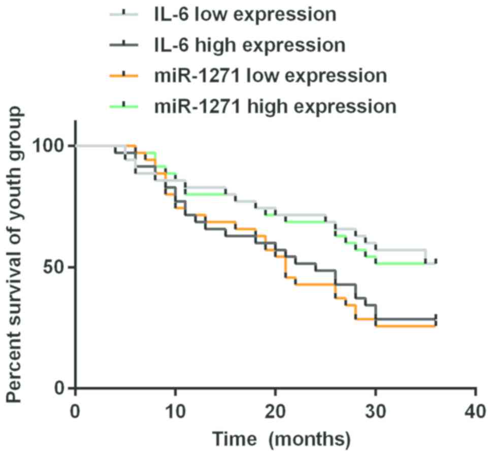
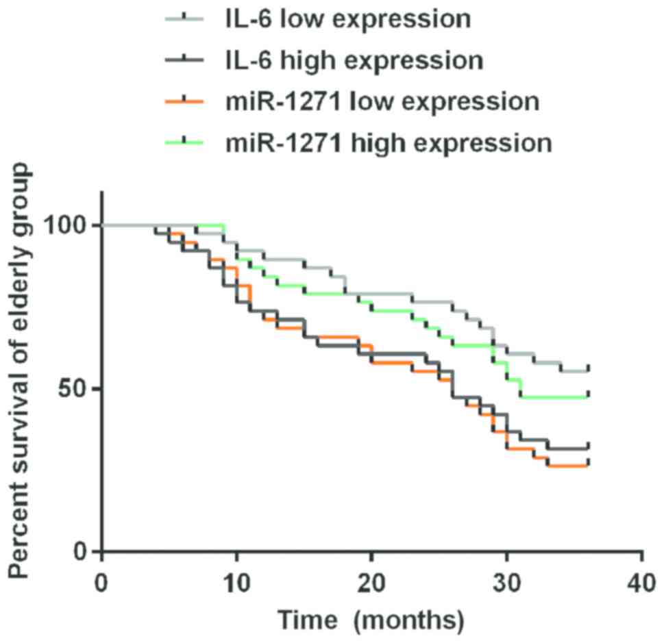
Correlation of expression levels of
IL-6 and miR-1271 with young and elderly gastric cancer
The median expression levels of IL-6 and miR-1271 in
young gastric cancer patients were 3.11±0.21 and 1.84±0.14,
respectively (data not shown). Based on this, the high and low
expression levels of the two indicators IL-6 and miR-1271 were
established, respectively, each with 35 cases. The 3-year overall
survival rate of patients with a low expression of IL-6 was better
than that of patients with a high expression in young gastric
cancer (P<0.05). Survival of patients with a high expression of
miR-1271 was better than that of patients with a low expression in
young gastric cancer (P<0.05). The median expression levels of
IL-6 and miR-1271 in elderly gastric cancer patients were 4.63±0.24
and 1.05±0.06, respectively (data not shown). Based on this, the
high and low expression levels of the indicators IL-6 and miR-1271
were established, respectively, each with 38 cases. The 3-year
overall survival rate of patients with low expression of IL-6 was
better than that of patients with high expression in elderly
gastric cancer patients (P<0.05). That of patients with the high
expression of miR-1271 was better than that of patients with the
low expression in elderly gastric cancer patients (P<0.05) as
per the Kaplan Meier analysis and log-rank test (Figs. 1 and 2, and Table
V).
| Table V.Correlation of expression levels of
IL-6 and miR-1271 with young and elderly gastric cancer. |
Table V.
Correlation of expression levels of
IL-6 and miR-1271 with young and elderly gastric cancer.
|
| Indicator |
| Survival rate
[(n)%] |
|
|---|
|
|
|
|
|
|
|---|
| Group |
|
| n | 1 year | 2 years | 3 years | P-value |
|---|
| Youth group | IL-6 | Low expression | 35 | 29 (82.86) | 25 (71.43) | 18 (51.43) | 0.043 |
|
|
| High
expression | 35 | 24 (68.57) | 17 (48.57) | 10 (28.57) |
|
|
| miR-1271 | Low expression | 35 | 25 (71.43) | 15 (42.86) | 9
(25.71) | 0.025 |
|
|
| High
expression | 35 | 28 (80.00) | 24 (68.57) | 16 (45.71) |
|
| Elderly group | IL-6 | Low expression | 38 | 34 (89.47) | 29 (76.32) | 21 (55.26) | 0.021 |
|
|
| High
expression | 38 | 28 (73.68) | 23 (60.53) | 12 (31.58) |
|
|
| miR-1271 | Low expression | 38 | 27 (71.05) | 21 (55.26) | 10 (26.32) | 0.047 |
|
|
| High
expression | 38 | 32 (84.21) | 26 (68.42) | 18 (47.37) |
|
Discussion
Gastric cancer accounts for 10.4% of the total
number of cancer deaths in Western countries (19). Currently, it is a comprehensive
treatment based on surgery, but its local recurrence rate following
surgery is still as high as 50%, with poor postoperative survival
rate (20). The formation of tumors
mainly depends on the activation of proto-oncogenes and mutation of
tumor suppressor genes. The development mechanism of gastric cancer
has not yet been clarified, owing to the abnormal lack of
specificity of the genes (21).
Nurul-Syakima et al (22) found that miR-1271 is highly expressed
in head and neck tumors, but Maurel et al (23) reported a low expression in hepatoma
cells, exerting a tumor suppressor effect. In this study, the
expression level of miR-1271 was significantly lower in young and
elderly gastric cancer tissues than that in adjacent tissues, with
a statistically significant difference (P<0.001). That of
miR-1271 was significantly higher in young gastric cancer tissues
than that in elderly gastric cancer tissues, with a statistically
significant difference (P<0.001). In addition, miR-1271 was not
significantly correlated with sex (P>0.05), but was correlated
with age, tumor size, lymph node metastasis, distant metastasis,
clinical stage and differentiation degree of gastric cancer
patients (P<0.05). Taken together, the above results indicate
that the expression levels and roles of miR-1271 are different in
different tumors, tissues and cells. The study of Xiang et
al (24) shows that miR-1271
expression is low in gastric cancer tissues of most patients, and
is negatively correlated with tumor size and clinical stage. Those
findings are consistent with our results. In this study, data of
the correlation analysis of miR-1271 with the clinical pathology of
gastric cancer were presented as mean standard deviation, but as
the number of cases in the studies were different, this may result
in statistically significant differences due to different
statistical calculation methods.
Our study showed that the expression level of IL-6
was significantly higher in young and elderly gastric cancer
tissues than that in adjacent tissues, with a statistically
significant difference (P<0.001). That of IL-6 was significantly
lower in young gastric cancer tissues than that in elderly gastric
cancer tissues, with a statistically significant difference
(P<0.001). That of IL-6 was not significantly correlated with
sex (P>0.05), but correlated with age, tumor size, lymph node
metastasis, distant metastasis, clinical stage and differentiation
degree of gastric cancer patients (P<0.05). The expression level
of IL-6 was higher in elderly gastric cancer than that in young
gastric cancer, probably because the elderly usually have low
immune function, susceptible to inflammatory infections (25). Sampaio et al (26) found that IL-6, highly expressed in
gastric cancer tissues, is correlated with tumor size and the T
stage of gastric cancer. The results of this study are consistent
with those of Sampaio et al (26). In this study, the data of the
correlation analysis of IL-6 with the clinical pathology of gastric
cancer were presented as mean standard deviation. According to
reports in the literature, IL-6 affects the growth of tumor cells
and the development of the disease (27). It also promotes angiogenesis, and the
adhesion and invasion of tumor cells (28).
The 3-year overall survival rate of patients with
the low expression of IL-6 was better than that of patients with
the high expression in young and elderly gastric cancer
(P<0.05). Survival of patients with the high expression of
miR-1271 was better than that of patients with the low expression
in young and elderly gastric cancer (P<0.05). Since no study of
the correlation of IL-6 and miR-1271 with the prognosis of young
and elderly gastric cancer was found, the results of this study
cannot be compared with those of other studies.
In summary, IL-6 is highly expressed but miR-1271 is
lowly expressed in gastric cancer tissues, and the expression
levels of both are correlated with age, tumor size, lymph node
metastasis, distant metastasis, clinical stage and differentiation
degree of gastric cancer patients. The 3-year overall survival rate
of patients with the low expression of IL-6 is better than that of
patients with the high expression in young and elderly gastric
cancer. Survival of patients with high expression of miR-1271 is
better than that of patients with low expression in young and
elderly gastric cancer.
Acknowledgements
Not applicable.
Funding
No funding was received.
Availability of data and materials
The datasets used and/or analyzed during the current
study are available from the corresponding author on reasonable
request.
Authors' contributions
SL was involved in writing the manuscript. SL and YZ
performed PCR. LZ and JWa were responsible for total RNA extraction
and reverse transcription reaction. RJ collected and analyzed the
general data of patients. YW, JWu and XY helped with ELISA. All
authors read and approved the final manuscript.
Ethics approval and consent to
participate
This study was approved by the Ethics Committee of
The First Hospital of Lanzhou University (Lanzhou, China). Patients
who participated in this research had complete clinical data.
Signed informed consents were obtained from the patients or the
guardians.
Patient consent for publication
Not applicable.
Competing interests
The authors declare that they have no competing
interests.
References
|
1
|
Karim S, Mirza Z, Naseer MI, Al-Qahtani MH
and Ali A: Clinicopathological characteristics and chronology of
p53 expression in the development of gastric cancer.
Hepatogastroenterology. 60:2113–2118. 2013.PubMed/NCBI
|
|
2
|
Krejs GJ: Gastric cancer: Epidemiology and
risk factors. Dig Dis. 28:600–603. 2010. View Article : Google Scholar : PubMed/NCBI
|
|
3
|
Dhobi MA, Wani KA, Parray FQ, Wani RA,
Wani ML, Peer GQ, Abdullah S, Wani IA, Wani MA, Shah MA, et al:
Gastric cancer in young patients. Int J Surg Oncol.
2013:9816542013.PubMed/NCBI
|
|
4
|
Kim DY, Joo JK, Ryu SY, Park YK, Kim YJ
and Kim SK: Clinicopathologic characteristics of gastric carcinoma
in elderly patients: A comparison with young patients. World J
Gastroenterol. 11:22–26. 2005. View Article : Google Scholar : PubMed/NCBI
|
|
5
|
Hsieh FJ, Wang YC, Hsu JT, Liu KH, Yeh CN,
Yeh T-S, Hwang T-L and Jan Y-Y: Clinicopathological features and
prognostic factors of gastric cancer patients aged 40 years or
younger. J Surg Oncol. 105:304–309. 2012. View Article : Google Scholar : PubMed/NCBI
|
|
6
|
Park HJ, Ahn JY, Jung HY, Lim H, Lee JH,
Choi KS, Kim DH, Choi KD, Song HJ, Lee GH, et al: Clinical
characteristics and outcomes for gastric cancer patients aged 18–30
years. Gastric Cancer. 17:649–660. 2014. View Article : Google Scholar : PubMed/NCBI
|
|
7
|
Kashiwagi Y, Kato N, Sassa T, Nishitsuka
K, Yamamoto T, Takamura H and Yamashita H: Cotylenin A inhibits
cell proliferation and induces apoptosis and PAX6 mRNA transcripts
in retinoblastoma cell lines. Mol Vis. 16:970–982. 2010.PubMed/NCBI
|
|
8
|
Tchernitsa O, Kasajima A, Schäfer R, Kuban
RJ, Ungethüm U, Györffy B, Neumann U, Simon E, Weichert W, Ebert
MP, et al: Systematic evaluation of the miRNA-ome and its
downstream effects on mRNA expression identifies gastric cancer
progression. J Pathol. 222:310–319. 2010. View Article : Google Scholar : PubMed/NCBI
|
|
9
|
Wang Y, Xu L and Jiang L: miR-1271
promotes non-small-cell lung cancer cell proliferation and invasion
via targeting HOXA5. Biochem Biophys Res Commun. 458:714–719. 2015.
View Article : Google Scholar : PubMed/NCBI
|
|
10
|
Naugler WE and Karin M: The wolf in
sheep's clothing: The role of interleukin-6 in immunity,
inflammation and cancer. Trends Mol Med. 14:109–119. 2008.
View Article : Google Scholar : PubMed/NCBI
|
|
11
|
Ashizawa T, Okada R, Suzuki Y, Takagi M,
Yamazaki T, Sumi T and Aoki T, Ohnuma S and Aoki T: Clinical
significance of interleukin-6 (IL-6) in the spread of gastric
cancer: Role of IL-6 as a prognostic factor. Gastric Cancer.
8:124–131. 2005. View Article : Google Scholar : PubMed/NCBI
|
|
12
|
Dey AB: World report on ageing and health.
Indian J Med Res. 145:150–151. 2017. View Article : Google Scholar
|
|
13
|
Sasaki M, Ikeda H, Sato Y and Nakanuma Y:
Proinflammatory cytokine-induced cellular senescence of biliary
epithelial cells is mediated via oxidative stress and activation of
ATM pathway: A culture study. Free Radic Res. 42:625–632. 2008.
View Article : Google Scholar : PubMed/NCBI
|
|
14
|
Bartek J, Hodny Z and Lukas J: Cytokine
loops driving senescence. Nat Cell Biol. 10:887–889. 2008.
View Article : Google Scholar : PubMed/NCBI
|
|
15
|
Akbaraly TN, Hamer M, Ferrie JE, Lowe G,
Batty GD, Hagger-Johnson G, Singh-Manoux A, Shipley MJ and Kivimäki
M: Chronic inflammation as a determinant of future aging
phenotypes. CMAJ. 185:E763–E770. 2013. View Article : Google Scholar : PubMed/NCBI
|
|
16
|
Gorelick PB: Role of inflammation in
cognitive impairment: Results of observational epidemiological
studies and clinical trials. Ann NY Acad Sci. 1207:155–162. 2010.
View Article : Google Scholar : PubMed/NCBI
|
|
17
|
Satizabal CL, Zhu YC, Mazoyer B, Dufouil C
and Tzourio C: Circulating IL-6 and CRP are associated with MRI
findings in the elderly: The 3C-Dijon Study. Neurology. 78:720–727.
2012. View Article : Google Scholar : PubMed/NCBI
|
|
18
|
Livak KJ and Schmittgen TD: Analysis of
relative gene expression data using real-time quantitative PCR and
the 2(-Delta Delta C(T)) method. Methods. 25:402–408. 2001.
View Article : Google Scholar : PubMed/NCBI
|
|
19
|
Guggenheim DE and Shah MA: Gastric cancer
epidemiology and risk factors. J Surg Oncol. 107:230–236. 2013.
View Article : Google Scholar : PubMed/NCBI
|
|
20
|
Paoletti X, Oba K, Burzykowski T, Michiels
S, Ohashi Y, Pignon JP, Rougier P, Sakamoto J, Sargent D, Sasako M,
et al GASTRIC (Global Advanced/Adjuvant Stomach Tumor Research
International Collaboration) Group, : Benefit of adjuvant
chemotherapy for resectable gastric cancer: A meta-analysis. JAMA.
303:1729–1737. 2010. View Article : Google Scholar : PubMed/NCBI
|
|
21
|
Skinnider BF and Mak TW: The role of
cytokines in classical Hodgkin lymphoma. Blood. 99:4283–4297. 2002.
View Article : Google Scholar : PubMed/NCBI
|
|
22
|
Nurul-Syakima AM, Yoke-Kqueen C, Sabariah
AR, Shiran MS, Singh A and Learn-Han L: Differential microRNA
expression and identification of putative miRNA targets and
pathways in head and neck cancers. Int J Mol Med. 28:327–336.
2011.PubMed/NCBI
|
|
23
|
Maurel M, Jalvy S, Ladeiro Y, Combe C,
Vachet L, Sagliocco F, Bioulac-Sage P, Pitard V, Jacquemin-Sablon
H, Zucman-Rossi J, et al: A functional screening identifies five
microRNAs controlling glypican-3: Role of miR-1271 down-regulation
in hepatocellular carcinoma. Hepatology. 57:195–204. 2013.
View Article : Google Scholar : PubMed/NCBI
|
|
24
|
Xiang XJ, Deng J, Liu YW, Wan LY, Feng M,
Chen J and Xiong JP: MiR-1271 inhibits cell proliferation, invasion
and EMT in gastric cancer by targeting FOXQ1. Cell Physiol Biochem.
36:1382–1394. 2015. View Article : Google Scholar : PubMed/NCBI
|
|
25
|
Judd LM, Alderman BM, Howlett M, Shulkes
A, Dow C, Moverley J, Grail D, Jenkins BJ, Ernst M and Giraud AS:
Gastric cancer development in mice lacking the SHP2 binding site on
the IL-6 family co-receptor gp130. Gastroenterology. 126:196–207.
2004. View Article : Google Scholar : PubMed/NCBI
|
|
26
|
Sampaio AM, Balseiro SC, Silva MR, Alarcão
A, d'Aguiar MJ, Ferreira T and Carvalho L: Association between IL-4
and IL-6 expression variants and gastric cancer among Portuguese
population. GE Port J Gastroenterol. 22:143–152. 2015. View Article : Google Scholar : PubMed/NCBI
|
|
27
|
Ohi S, Hashimoto H, Tachibana T, Tabei I,
Nakajima M, Sato K, Yanaga K and Ishikawa H: Establishment and
characterization of EB virus-free normal B-lymphocyte and
interleukin-6-producing poorly differentiated adenocarcinoma cell
lines derived from gastric tumor tissue. Hum Cell. 18:35–44. 2005.
View Article : Google Scholar : PubMed/NCBI
|
|
28
|
Huang SP, Wu MS, Wang HP, Yang CS, Kuo ML
and Lin JT: Correlation between serum levels of interleukin-6 and
vascular endothelial growth factor in gastric carcinoma. J
Gastroenterol Hepatol. 17:1165–1169. 2002. View Article : Google Scholar : PubMed/NCBI
|