Introduction
Lung cancer is the leading cause of cancer death
globally, accounting for 18.20% of all cancer deaths. The incidence
rate is ranked first in the global incidence of cancers, accounting
for 12.70% of all cancer cases (1).
According to statistics, among the number of lung cancer cases in
Serbia of southeastern Europe, the incidence rates in men were
higher than those of women and the mortality rates of male patients
were four times higher than those of female patients. This may be
due to the smoking habit in males (2). According to the histological type of
lung cancer, it can be divided into small cell lung cancer and
non-small cell lung cancer (NSCLC). NSCLC accounts for most causes
of lung cancer, according to statistics, NSCLC accounts for 80% of
all patients with lung cancers (3).
NSCLC has no obvious symptoms in the early stage, normally patients
are already in the advanced stage when obvious clinical symptoms or
physiological reactions occur. Some statistics have shown that most
NSCLC patients are already in the advanced stage at the time of
diagnosis (4). According to the
pathological type of NSCLC, it is mainly classified into large cell
carcinoma, squamous cell carcinoma and adenocarcinoma. Among them,
adenocarcinoma and squamous cell lung cancer are the main types of
cancers (5). Surgical resection is
the main treatment method for NSCLC, however, patients with NSCLC
have a higher recurrence rate (6).
With the development of medical technology, chemotherapy has become
more effective in the treatment of NSCLC. Although the prognosis of
patients with NSCLC has improved it is affected by drug resistance.
Moreover, the five-year survival rate of NSCLC patients is still
less than 20% and seriously affects people's health (7). Therefore, we have to continue to find
effective tumor markers to judge the prognosis of NSCLC
clinically.
microRNA (miRNA) is an endogenous
non-protein-encoded small RNA, located in eukaryotic cells and
approximately 22 nt in length. Studies have shown that its
expression has a certain impact on the occurrence, development,
invasion, metastasis and prognosis of many malignant tumors, which
has an important effect in inhibiting tumors or promoting tumor
development (8). miRNA has a great
significance in the diagnosis and treatment evaluation of NSCLC and
can be used as a non-invasive biomarker for prognosis (9).
miR-21 is one of the miRNAs located on human
chromosome 17q23.2 and with an abnormally high expression in many
malignant tumors, also is a type of oncogene (10). Studies have shown that the growth and
invasion of NSCLC cancer cells can be promoted by inhibiting the
tumor suppressor phosphatase and tensin homolog deleted from
chromosome 10 (PTEN). The expression levels of miR-21 were highly
expressed in NSCLC patients and the expression of miR-21 was
associated with TNM stages and distant metastasis in NSCLC patients
(11). miR-155 is an important
member of miRNA, the gene is located on chromosome 21 and it was
processed by non-protein-encoded transcripts of B cell fusion
cluster genes (12). miR-155
promotes the growth and development of tumor cells by regulating a
variety of cancer-promoting pathways and has a high expression in a
variety of malignancies (13).
Studies have shown that miR-155 was highly expressed in NSCLC and
induces the proliferation and invasion of NSCLC cancer cells by
directly inhibiting the programmed cell death gene 4 (14).
There are few studies on the high expression of
miR-155 and miR-21 and the recurrence or metastasis of NSCLC
patients. This research investigated the expression levels of serum
miR-155 and miR-21 in patients with recurrence or metastasis of
NSCLC. We also explored the relationship between the high
expression of miR-155 and miR-21 and the recurrence or metastasis
of NSCLC in order to provide clinical references.
Materials and methods
General information
Retrospective analysis of the clinical information
of 180 patients with NSCLC admitted to Daqing Oil Field General
Hospital (Daqing, China) from February 2012 to March 2015 were
carried out. These patients were the research group. There were 124
cases of males and 56 cases of females, with an average age
66.37±7.21 years. Physical examination information of 88 normal
medical examinees was selected during the same period of time, and
set as the control group. There were 63 cases of males and 25 cases
of females, average age 64.98±7.23 years. Sixty-eight patients in
the research group were newly diagnosed with NSCLC, and were set as
the newly diagnosed group and there were 46 cases of males and 22
cases of females. In addition, 112 patients with recurrence or
metastasis of NSCLC were the recurrence group, there were 78 cases
of males and 34 cases of females.
This investigation was approved by the Ethics
Committee of Daqing Oil Field General Hospital, patients or their
family members signed the informed consent form.
Inclusion and exclusion criteria
Inclusion criteria: Age ≥18 years; no other
hereditary diseases; patients with a complete clinical medical
records or physical examination information; included were only
patients who were treated in Daqing Oil Field General Hospital; no
deaths occurred during the treatment and the patients have been
discharged. Exclusion criteria: Patients with cardiovascular
disease, patients with second primary tumors; patients who had
taken antibiotics in the last three months; combined with severe
liver and kidney dysfunction; combined with other connective tissue
diseases, endocrine and metabolic diseases; and patients with a
history of mental illness or family history of mental illness.
Main reagents and instruments
PCR amplifier (manufacturer: Tianjin Jinside
Biotechnology Co., Ltd., Tianjin, China, model: ABI 2700); TRIzol
extractant (manufacturer: Beijing Suo Laibao Technology Co., Ltd.,
Beijing, China, batch number: 15596-026); high-speed centrifuge
(manufacturer: Anhui Zhongke Zhongjia Scientific Instrument Co.,
Ltd., Hefei, China, model: HC-2518R); reverse transcription RT kit
(manufacturer: Shanghai Jiemei Gene Pharmaceutical Technology Co.,
Ltd., Shanghai, China, batch number: GMS20020.4); RT-qPCR kit
(manufacturer: Shanghai Chuangye Biotechnology Co., Ltd., Shanghai,
China, article number: ZY131042) were used in the present study.
The miR-155 and miR-21 and the internal reference β-actin primer
sequences were designed and produced by Suzhou Yuxun Biotechnology
Co., Ltd. Suzhou, China (Table
I).
 | Table I.miR-155 and miR-21 and internal
reference β-actin primer sequences. |
Table I.
miR-155 and miR-21 and internal
reference β-actin primer sequences.
| Gene names | Upstream primer | Downstream
primer |
|---|
| miR-21 |
5′-TGCGCTAGCTTATCAGACTGAT-3′ |
5′-CCAGTGCAGGGTCCGAGGTATT-3′ |
| miR-155 |
5′-CTGTAT-CAAAAGGCCAACTGAA-3′ |
5′-GTGTCTATCCT-TATGAATCGCCA-3′ |
| β-actin |
5′-AGCGGGAAATCGTGCGTGACA-3′ |
5′-GTG-GACTTGGGAGAGGACTGG-3′ |
Collection of serum
The operation steps were all carried out in a
sterile environment, 6 ml of the external elbow venous blood was
extracted in the morning after all the experimental subjects had
fasted for 8 h. Blood was then placed in a vacuum blood collection
tube for 25 min, the serum was separated in a centrifuge at 3,000 ×
g at 22°C for approximately 15–20 min. After separation, 350 µl of
the supernatant was placed in an EP tube. After being placed in
liquid nitrogen for quick freezing, it was stored at −80°C.
Extraction of total RNA and synthesis
of cDNA
The RNA was extracted using TRIzol, serum was taken
from the refrigerator, 1.05 ml TRIzol was added and agitated for 5
min. The mixture was kept at room temperature for 10 min and 600 µl
of chloroform was added after the cells were completely lysed. The
mixture was mixed until the solution was emulsified and milky white
after which it was left to stand for 5 min, following
centrifugation at 800 × g for 15 min at 4°C. The top colorless
supernatant was carefully absorbed into the centrifuge tube using a
pipette, an equal volume of isopropanol was added and centrifuged
for further 10 min. After centrifugation, the precipitate was
washed by adding 1 ml of 75% ethanol to obtain RNA. The RNA
integrity was tested by using 1% agarose gel electrophoresis. Then
a qualified RNA sample was selected for cDNA synthesis, and reverse
transcribed cDNA using 1 µl of total RNA, the reaction was carried
out under reaction conditions of 42°C for 55 min.
Detection of the expression levels of
miR-155 and miR-21
The expression levels of miR-155 and miR-21 were
detected by RT-qPCR and the reaction system of 20 µl volume. First,
pre-denaturation at 95°C for 15 min, then denaturation at 94°C for
15 sec, annealing at 56°C for 30 sec and extension at 70°C for 30
sec, and repetition of the 40 cycles. The experiment was repeated 3
times and completed within 30 days. The standard curve was based on
the standard concentration and the average value of the cycle
depreciation corresponding to each concentration. According to the
standard curve, the instrument automatically calculates the copy
number/ml of miR-155, miR-21 and β-actin in serum. In addition, the
ratio of miR-155, miR-21 and β-actin was used as the expression
level of miR-155 and miR-21.
Follow up for the research group
After the treatment, all NSCLC patients discharged
from the hospital were followed up once every month for 36 months,
mainly by telephone. Home visit survey or consultation with
relatives were conducted for patients who had poor telephone
follow-ups and elderly patients or those with poor physical
fitness. This ensured the accurate recording of deaths in the
research group.
Statistical analysis
Statistical analysis was performed using SPSS19.0
[Yiyun (Shanghai) Information Technology Co., Ltd., Shanghai,
China] and measurement data was expressed as mean ± standard
deviation (mean ± SD). The t-test was used to compare the
measurement data between all the groups and the Chi-square test was
used to compare the countable data between the groups. Survival
analysis was performed using Kaplan-Meier and the log-rank test.
P<0.05 was considered to indicate a statistically significant
difference.
Results
General baseline information
Factors including age, sex, body mass index, heart
rate, smoking status, fasting blood glucose level, hemoglobin (Hb)
value, red blood cell (RBC) value, and platelet (PLT) value between
the research and control groups were compared. The difference
between the groups was not statistically significant (P>0.05)
(Table II).
| Table II.Comparison of baseline information
between the research group and the control group [n (%)] (mean ±
SD). |
Table II.
Comparison of baseline information
between the research group and the control group [n (%)] (mean ±
SD).
| Characteristics | Research group
(n=180) | Control group
(n=88) | χ2/t
value | P-value |
|---|
| Age (years) | 66.37±7.21 | 64.98±7.23 | 1.481 | 0.140 |
| Sex |
|
| 0.205 | 0.674 |
| Male | 124 (68.89) | 63 (71.59) |
|
|
|
Female | 56
(31.11) | 25 (28.41) |
|
|
| BMI
(kg/m2) |
|
| 0.164 | 0.755 |
|
<24 | 141 (78.33) | 67 (76.14) |
|
|
| ≥24 | 39
(21.67) | 21 (23.86) |
|
|
| Smoking status |
|
| 1.636 | 0.240 |
|
Smoker | 136 (75.56) | 60 (68.18) |
|
|
|
Non-smoker | 44
(24.44) | 28 (31.82) |
|
|
| Heart rate
(times/min) |
|
| 0.642 | 0.503 |
|
<60 | 15 (8.33) | 10 (11.36) |
|
|
| ≥60 | 165 (91.67) | 78 (88.64) |
|
|
| Fasting blood glucose
(mmol/l) | 4.48±0.62 | 4.53±0.56 | 0.640 | 0.523 |
| Hb (g/l) | 115.24±14.26 | 117.25±13.63 | 1.099 | 0.273 |
| RBC
(×1012/l) | 4.51±0.26 | 4.55±0.35 | 1.051 | 0.294 |
| PLT
(×109/l) | 224.24±36.73 | 218.99±55.36 | 0.924 | 0.357 |
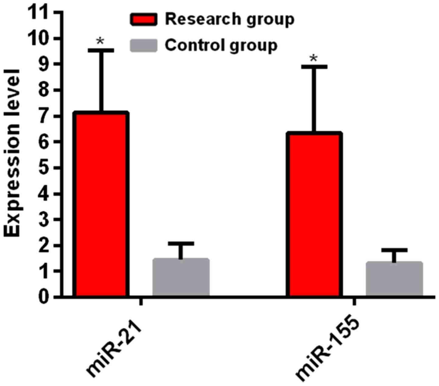
Expression levels of serum miR-155 and miR-21
between the research and control groups. The experiment results
showed that the expression level of miR-21 in the research group
was 7.13±3.76, which was higher than that in the control group
(1.45±0.62) and the difference between two groups was statistically
significant (t=14.060, P<0.01). The expression level of miR-155
in the research group was 6.34±3.93, which was higher than that in
the control group (1.32±0.52). The difference between two groups
was statistically significant (t=11.920, P<0.01) (Fig. 1).
Clinical information in the subgroup
of the research group
Factors including age, sex, body mass index, smoking
status, tumor diameter, differentiation, histological type, and TNM
stage between the recurrence group and the newly diagnosed group
were compared. The difference between the groups was not
statistically significant (P>0.05) (Table III).
| Table III.Comparison of clinical information
between the recurrence group and the newly diagnosed group [n
(%)]. |
Table III.
Comparison of clinical information
between the recurrence group and the newly diagnosed group [n
(%)].
| Characteristics | Recurrence group
(n=112) | Newly diagnosed group
(n=68) | χ2
value | P-value |
|---|
| Age (years) |
|
| 0.342 | 0.631 |
|
<65 | 38 (33.93) | 26 (38.24) |
|
|
| ≥65 | 74 (66.07) | 42 (61.76) |
|
|
| Sex |
|
| 0.079 | 0.868 |
|
Male | 78 (69.64) | 46 (67.65) |
|
|
|
Female | 34 (30.36) | 22 (32.35) |
|
|
| BMI
(kg/m2) |
|
| 0.418 | 0.579 |
|
<24 | 86 (76.79) | 55 (80.88) |
|
|
|
≥24 | 26 (23.21) | 13 (19.12) |
|
|
| Smoking status |
|
| 0.880 | 0.377 |
|
Smoker | 82 (73.21) | 54 (79.41) |
|
|
|
Non-smoker | 30 (26.79) | 14 (20.59) |
|
|
| Tumor diameter |
|
| 0.116 | 0.752 |
| <5
cm | 40 (35.71) | 26 (38.24) |
|
|
| ≥5
cm | 72 (64.29) | 42 (61.76) |
|
|
| Histological
type |
|
| 0.095 | 0.878 |
|
Squamous cell carcinoma | 57 (50.89) | 33 (48.53) |
|
|
|
Adenocarcinoma | 55 (49.11) | 35 (51.47) |
|
|
|
Differentiation |
|
| 0.702 | 0.482 |
| Mid and
low | 81 (72.32) | 53 (77.94) |
|
|
|
High | 31 (27.68) | 15 (22.06) |
|
|
| TNM stages |
|
| 0.142 | 0.755 |
|
I–II | 46 (41.07) | 26 (38.24) |
|
|
|
III–IV | 66 (58.93) | 42 (61.76) |
|
|
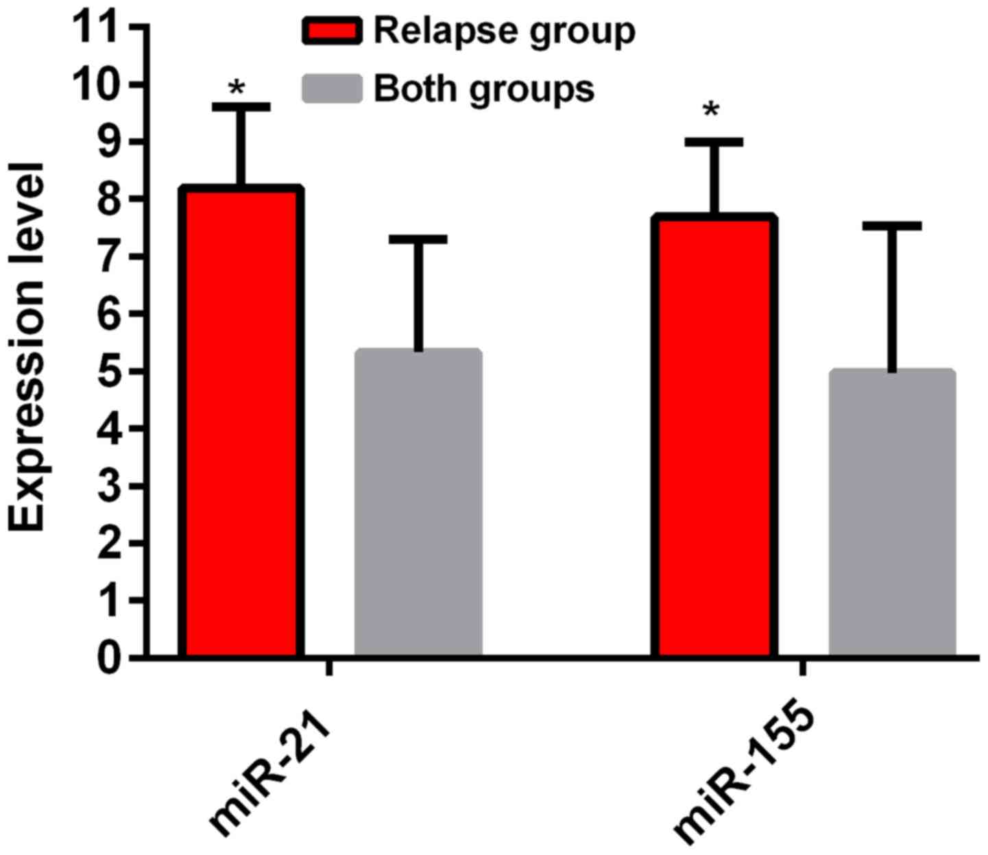
Expression of serum miR-155 and miR-21
in the subgroup of the research group
The results of the experiment showed that the
expression level of miR-21 in the recurrence group was 8.18±2.71,
which was higher than that in the newly diagnosed group
(5.33±1.96). The difference between the groups was statistically
significant (t=7.552, P<0.01). The expression level of miR-155
in the recurrence group was 7.69±2.58, which was higher than that
in the newly diagnosed group (4.97±2.56). The difference between
the groups was statistically significant (P<0.05) (Fig. 2).
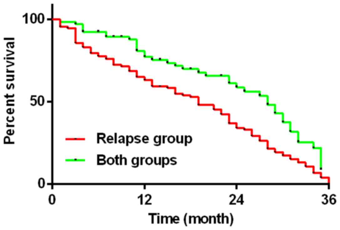
Subgroup of the research group
A 36-month fllow-up was conducted to both the
recurrence group and the newly diagnosed group. At follow-up, 103
cases of patients died in the recurrence group, with a median
survival time of 19 months and a mortality rate of 91.96%. In
addition, 39 patients died in the newly diagnosed group, the median
survival time was 28 months and the mortality rate was 57.35%. The
difference between two groups was statistically significant
(χ2=9.705, P<0.01) (Fig.
3).
Discussion
Lung cancer is the most malignant tumor with the
highest morbidity and mortality rate in the world (15). Assessment of the prognosis of NSCLC
is currently carried out using tissue cytological examination.
However, these are intrusive inspection methods and often require
obtaining tissues or cells, via lung puncture, biopsy or
bronchoscopy. These methods are not readily accepted by patients
due to inconvenience and trauma (16). In addition, a non-invasive,
easy-to-use and highly accurate biomarker is required to assess the
prognosis of NSCLC clinically.
miR-21 and miR-155 both have an important effect in
the occurrence and development of lung cancer. Studies have shown
that miR-155 has considerable regulatory effect in the
proliferation, differentiation and growth of T cells (17). miR-21 causes signal transduction and
activation on transcriptional activator 3, which increases the
vascular endothelial growth factor levels in recipient cells. This
indirectly leads to malignant transformation of human bronchial
epithelioid cells (18). The
expression levels of miR-21 and miR-155 are elevated in various
solid malignant tumors such as breast cancer, B cell lymphoma, lung
cancer and colon cancer. Moreover, decreased expression levels of
miR-21 and miR-155 can induce apoptosis and inhibit the
proliferation and invasion of cells (19).
Xue et al (20) reported that the expression levels of
miR-21 and miR-155 are increased in patients with NSCLC. In
addition, silencing miR-21 and miR-155 can effectively inhibit the
progression of NSCLC in the animals of xenotransplantation.
Furthermore, combined inhibition of miR-21 and miR-155 can
effectively improve the therapeutic effect of NSCLC, which is
similar to our research results. We studied the expression levels
of miR-21 and miR-155 in the serum between normal and NSCLC
patients. It was found that miR-21 and miR-155 were highly
expressed in the serum of NSCLC patients, which were higher than
those in the normal patients (P<0.05). We further investigated
the expression levels of miR-21 and miR-155 of the serum in
patients with recurrence of NSCLC or metastasis and found that the
expression levels of miR-21 and miR-155 with recurrence or
metastatic NSCLC were higher than those in the newly diagnosed
patients with NSCLC (P<0.05). The results of Yang et al
(21) were similar to our results,
they found that the high expression levels of miR-21 and miR-155 in
the post-operative recurrence and metastasis of patients with NSCLC
were higher than those of patients with low expression levels.
However, the survival rate was lower than that of patients with a
low expression level. We conducted a 36-month follow up in the
newly diagnosed patients and NSCLC patients with recurrence or
metastasis. The mortality rate of the NSCLC with recurrence or
metastasis was 91.96%, the median survival time was 19 months and
the initial diagnosis of NSCLC patients had a mortality rate of
57.35% and the median survival time was 28 months. The difference
in survival rate between the two groups was statistically
significant (P<0.05). Moreover, patients with recurrence or
metastasis of NSCLC had a poor survival rate. The results of this
investigation are similar to those of Lashiotaki et al
(22) who considered that the high
expression of miR-21 and miR-155 may reduce the overall survival
and recurrence-free survival rate in patients with NSCLC. This can
lead to poor prognosis in patients with NSCLC and reduces patient
survival time. Donnem et al (23) reported that highly expressed miR-155
has a significant negative impact on the survival rates and
prognosis of NSCLC and it is an independent prognostic factor for
NSCLC. Gironella et al (24)
considered that miR-155 is abnormally highly expressed in
pancreatic cancer. It may be caused by inhibition of the mRNA
function of TP53INP1 and resulted in a decrease in its expression
level. Therefore, it inhibits tumor cell apoptosis and promotes
tumor cell proliferation. In addition, Meng et al (25) believe that the expression level of
PTEN can be increased by inhibiting the expression of miR-21 in
liver cancer cells and therefore the proliferation and metastasis
of tumors were inhibited. The PTEN gene may be impacted by miR-21
and could affect the metastasis and recurrence in tumors. This
indicates that high expression of miR-21 and miR-155 is associated
with recurrence and metastasis of tumors.
Retrospective case analysis was used during this
study and the pathology was strictly selected according to the
inclusion and exclusion criteria. The basic patient information
investigation, clinical examination, and special inspection were
strictly recorded and organized. Moreover, a 36-month follow-up was
conducted in the NSCLC patients, this accurately recorded the
patient death rate and obtained a reliable result.
miR-21 and miR-155 were highly expressed in the
serum of patients with NSCLC, and the expression levels of serum in
NSCLC patients with recurrence and metastasis was even higher.
Highly expressed miR-21 and miR-155 were associated with recurrence
and metastasis in patients with NSCLC reducing survival rate and
affecting prognosis of patients.
Acknowledgements
Not applicable.
Funding
No funding was received.
Availability of data and materials
The datasets used and/or analyzed during the present
study are available from the corresponding author on reasonable
request.
Authors' contributions
SX drafted the manuscript and was responsible for
the extraction of total RNA and synthesis of cDNA. SX and LS
performed PCR. Both authors read and approved the final
manuscript.
Ethics approval and consent to
participate
The study was approved by the Ethics Committee of
Daqing Oil Field General Hospital (Daqing, China). Signed informed
consents were obtained from the patients or the guardians.
Patient consent for publication
Not applicable.
Competing interests
The authors declare that they have no competing
interests.
References
|
1
|
Scagliotti GV, Szczesna A, Ramlau R,
Cardenal F, Mattson K, Van Zandwijk N, Price A, Lebeau B, Debus J
and Manegold C: Docetaxel-based induction therapy prior to
radiotherapy with or without docetaxel for non-small-cell lung
cancer. Br J Cancer. 94:1375–1382. 2006. View Article : Google Scholar : PubMed/NCBI
|
|
2
|
Kocić B, Petrović B, Rancić N and Ilić M:
Lung cancer trends in Southeastern Serbia. Cent Eur J Public
Health. 21:17–21. 2013. View Article : Google Scholar : PubMed/NCBI
|
|
3
|
Meza R, Meernik C, Jeon J and Cote ML:
Lung cancer incidence trends by gender, race and histology in the
United States, 1973–2010. PLoS One. 10:e01213232015. View Article : Google Scholar : PubMed/NCBI
|
|
4
|
Pathak AK, Bhutani M, Mohan A, Guleria R,
Bal S and Kochupillai V: Non small cell lung cancer (NSCLC):
Current status and future prospects. Indian J Chest Dis Allied Sci.
46:191–203. 2004.PubMed/NCBI
|
|
5
|
Haroun RA, Zakhary NI, Mohamed MR,
Abdelrahman AM, Kandil EI and Shalaby KA: Assessment of the
prognostic value of methylation status and expression levels of
FHIT, GSTP1 and p16 in non-small cell lung cancer in Egyptian
patients. Asian Pac J Cancer Prev. 15:4281–4287. 2014. View Article : Google Scholar : PubMed/NCBI
|
|
6
|
Kuo CH, Wu CY, Lee KY, Lin SM, Chung FT,
Lo YL, Liu CY, Hsiung TC, Yang CT and Wu YC: Chronic obstructive
pulmonary disease in stage I non-small cell lung cancer that
underwent anatomic resection: The role of a recurrence promoter.
COPD. 11:407–413. 2014. View Article : Google Scholar : PubMed/NCBI
|
|
7
|
Kumpiro S, Sriuranpong V and Areepium N:
Impact of the copper transporter protein 1 (CTR1) polymorphism on
adverse events among advanced nonsmall cell lung cancer patients
treated with a carboplatin/gemcitabine regimen. Asian Pac J Cancer
Prev. 17:4391–4394. 2016.PubMed/NCBI
|
|
8
|
Merino MJ, Gil S, Macias CG and Lara K:
The unknown microRNA expression of male breast cancer. Similarities
and differences with female ductal carcinoma. Their role as tumor
biomarker. J Cancer. 9:450–459. 2018. View Article : Google Scholar : PubMed/NCBI
|
|
9
|
Feng B, Zhang K, Wang R and Chen L:
Non-small-cell lung cancer and miRNAs: Novel biomarkers and
promising tools for treatment. Clin Sci (Lond). 128:619–634. 2015.
View Article : Google Scholar : PubMed/NCBI
|
|
10
|
Ribas J, Ni X, Castanares M, Liu MM, Esopi
D, Yegnasubramanian S, Rodriguez R, Mendell JT and Lupold SE: A
novel source for miR-21 expression through the alternative
polyadenylation of VMP1 gene transcripts. Nucleic Acids Res.
40:6821–6833. 2012. View Article : Google Scholar : PubMed/NCBI
|
|
11
|
Zhang JG, Wang JJ, Zhao F, Liu Q, Jiang K
and Yang GH: MicroRNA-21 (miR-21) represses tumor suppressor PTEN
and promotes growth and invasion in non-small cell lung cancer
(NSCLC). Clin Chim Acta. 411:846–852. 2010. View Article : Google Scholar : PubMed/NCBI
|
|
12
|
Xie Q, Chen X, Lu F, Zhang T, Hao M, Wang
Y, Zhao J, McCrae MA and Zhuang H: Aberrant expression of microRNA
155 may accelerate cell proliferation by targeting sex-determining
region Y box 6 in hepatocellular carcinoma. Cancer. 118:2431–2442.
2012. View Article : Google Scholar : PubMed/NCBI
|
|
13
|
Gao Y, Fu S, Jiang W, Li B, Tian Y and Fu
X: Association of MiR-155 expression with prognosis in resected
stage III non-small cell lung cancer. Zhongguo Fei Ai Za Zhi.
17:417–423. 2014.(In Chinese). PubMed/NCBI
|
|
15
|
Liu F, Song D, Wu Y, Liu X, Zhu J and Tang
Y: MiR-155 inhibits proliferation and invasion by directly
targeting PDCD4 in non-small cell lung cancer. Thorac Cancer.
8:613–619. 2017. View Article : Google Scholar : PubMed/NCBI
|
|
16
|
Gyoba J, Shan S, Roa W and Bédard EL:
Diagnosing lung cancers through examination of micro-RNA biomarkers
in blood, plasma, serum and sputum: A review and summary of current
literature. Int J Mol Sci. 17:4942016. View Article : Google Scholar : PubMed/NCBI
|
|
17
|
Roesel C, Terjung S, Weinreich G, Hager T,
Chalvatzoulis E, Metzenmacher M and Welter S: Sarcomatoid carcinoma
of the lung: A rare histological subtype of non-small cell lung
cancer with a poor prognosis even at earlier tumour stages.
Interact Cardiovasc Thorac Surg. 24:407–413. 2017.PubMed/NCBI
|
|
18
|
Liu J, Shi K, Chen M, Xu L, Hong J, Hu B,
Yang X and Sun R: Elevated miR-155 expression induces
immunosuppression via CD39(+) regulatory T-cells in sepsis patient.
Int J Infect Dis. 40:135–141. 2015. View Article : Google Scholar : PubMed/NCBI
|
|
19
|
Liu Y, Luo F, Wang B, Li H, Xu Y, Liu X,
Shi L, Lu X, Xu W, Lu L, et al: STAT3-regulated exosomal miR-21
promotes angiogenesis and is involved in neoplastic processes of
transformed human bronchial epithelial cells. Cancer Lett.
370:125–135. 2016. View Article : Google Scholar : PubMed/NCBI
|
|
20
|
Wang Y, Li J, Tong L, Zhang J, Zhai A, Xu
K, Wei L and Chu M: The prognostic value of miR-21 and miR-155 in
non-small-cell lung cancer: A meta-analysis. Jpn J Clin Oncol.
43:813–820. 2013. View Article : Google Scholar : PubMed/NCBI
|
|
21
|
Xue X, Liu Y, Wang Y, Meng M, Wang K, Zang
X, Zhao S, Sun X, Cui L, Pan L, et al: MiR-21 and MiR-155 promote
non-small cell lung cancer progression by downregulating SOCS1,
SOCS6, and PTEN. Oncotarget. 7:84508–84519. 2016. View Article : Google Scholar : PubMed/NCBI
|
|
22
|
Yang M, Shen H, Qiu C, Ni Y, Wang L, Dong
W, Liao Y and Du J: High expression of miR-21 and miR-155 predicts
recurrence and unfavourable survival in non-small cell lung cancer.
Eur J Cancer. 49:604–615. 2013. View Article : Google Scholar : PubMed/NCBI
|
|
23
|
Lasithiotaki I, Tsitoura E, Koutsopoulos
A, Lagoudaki E, Koutoulaki C, Pitsidianakis G, Spandidos DA,
Siafakas NM, Sourvinos G and Antoniou KM: Aberrant expression of
miR-21, miR-376c and miR-145 and their target host genes in Merkel
cell polyomavirus-positive non-small cell lung cancer. Oncotarget.
8:112371–112383. 2016.PubMed/NCBI
|
|
24
|
Donnem T, Eklo K, Berg T, Sorbye SW,
Lonvik K, Al-Saad S, Al-Shibli K, Andersen S, Stenvold H, Bremnes
RM, et al: Prognostic impact of MiR-155 in non-small cell lung
cancer evaluated by in situ hybridization. J Transl Med. 9:62011.
View Article : Google Scholar : PubMed/NCBI
|
|
25
|
Gironella M, Seux M, Xie MJ, Cano C,
Tomasini R, Gommeaux J, Garcia S, Nowak J, Yeung ML, Jeang KT, et
al: Tumor protein 53-induced nuclear protein 1 expression is
repressed by miR-155, and its restoration inhibits pancreatic tumor
development. Proc Natl Acad Sci USA. 104:16170–16175. 2007.
View Article : Google Scholar : PubMed/NCBI
|
|
26
|
Meng F, Henson R, Wehbe-Janek H, Ghoshal
K, Jacob ST and Patel T: MicroRNA-21 regulates expression of the
PTEN tumor suppressor gene in human hepatocellular cancer.
Gastroenterology. 133:647–658. 2007. View Article : Google Scholar : PubMed/NCBI
|