Introduction
Gastric cancer is considered to be the fourth
leading cause of cancer-associated mortality worldwide (1,2), this is
due partially to late diagnosis coupled with disease recurrence and
chemoresistance. Therefore, the discovery of predictive markers for
early detection and prognostic markers to optimize treatment is
necessary (3,4). MicroRNAs (miRNAs/miRs) are endogenous,
non-coding RNA molecules in eukaryotes, 21–25 nucleotides in length
(5,6). Being present in various animal and
plant cells, miRNAs serve important roles in a number of biological
processes in humans, including development, cell proliferation and
differentiation, apoptosis, angiogenesis and tumor growth. miRNAs
act primarily by regulating signaling-associated molecules,
including proliferation factors, pro-apoptotic and anti-apoptotic
genes (7–9).
miRNAs are highly evolutionarily conserved, and
their expression is time-dependent and tissue-specific (10,11).
miRNA mutations may potentially result in gene mutations and
functional disorder, thus potentially inducing disease, including
numerous types of cancer (12,13). In
oncology, the abnormal expression of miRNAs serves a noteworthy
role in the proliferation, differentiation, apoptosis, migration
and invasion of tumor cells, and miRNAs serve dual roles in tumor
suppression and carcinogenesis (14,15). In
addition, miRNAs have been associated with the occurrence and
progression of numerous tumor types (16,17).
The convenient detection of early diagnostic markers
has been a long-term aspiration for tumor screening and diagnosis.
In previous decades, miRNAs have been widely studied as potential
tumor markers due to their key roles in carcinogenesis. However,
miRNA detection has primarily relied on tumor tissues obtained by
surgery and biopsy, which has restricted their application as tumor
markers. Previous studies have indicated that mature miRNAs are
secreted from tumor tissues and exist stably in the peripheral
blood (18,19), which provides a convenient and
potential source for the screening of miRNAs.
miR-141 is a member of the miR-200 family, which is
associated with the formation of cancer stem cells and the
regulation of epithelial-mesenchymal transition (20). Although miR-141 has been closely
associated with the development of numerous types of cancer,
including colorectal (21), ovarian
(22) and gastric cancer (23), the association between miR-141 and
gastric cancer lacks in-depth study. miR-141 modulates cisplatin
sensitivity in gastric cancer cells by attenuating the functions of
programmed cell death protein 4; therefore, the upregulation of
miR-141 may be a novel therapeutic strategy for reducing the
chemoresistance of gastric cancer cells (24). Inhibiting the functions of miR-141
suppresses gastric cell proliferation and increases caspase-3
activity in H1299 and docetaxel-treated H2009 cells (25). However, these studies focused on
miR-141 expression from resected tumor tissue or cancer cell lines,
and did not investigate expression levels in patient plasma
(26). In the present study, miR-141
expression levels were detected in the peripheral plasma of
patients with gastric cancer. The potential roles of miR-141 in
gastric cancer pathogenesis, including grade, metastasis, prognosis
and survival were also investigated.
Patients and methods
Study subjects
A total of 164 patients with gastric cancer who
underwent tumor resection between January 2010 and December 2017 at
the Tianjin Nankai Hospital (Nankai, China) were enrolled. The
patient age range was 18–70 years with 93 male and 71 female
subjects, and a median age of 56 years. The inclusion criteria were
as follows: i) Confirmation of gastric cancer by pathological
identification; Exclusion criteria were as follows: i)
Gastrointestinal tract complications; ii) hemolysis; and iii) high
blood lipid concentration (a plasma triglyceride concentration
>150 mg/dl or hypercholesterolemia >200 mg/dl). Peripheral
blood plasma and tissue samples were obtained from each patient.
The tissue specimens collected were fixed in 10% formaldehyde at
4°C overnight, embedded in paraffin and cut into 1 cm sections for
pathological diagnosis. All diagnoses were confirmed based on
histopathological examination, and histological grade was
determined according to the criteria formulated by the World Health
Organization (WHO; 2007) (27).
Patient follow-up was conducted at 3-month intervals until December
31, 2017. A total of 109 healthy patients with a similar age and
sex distribution were enrolled as controls. The present study was
approved by the Tianjin Nankai Hospital Ethics Committee, and
written informed consent was obtained from each subject.
RNA isolation and miR-141
quantification
Total plasma RNA was isolated from patients and
healthy subjects using TRIzol® reagent (Invitrogen;
Thermo Fisher Scientific, Inc., Waltham, MA, USA) according to the
manufacturer's protocol. Reverse transcription-quantitative
polymerase chain reaction (RT-qPCR) was used to assess the
expression levels of miR-141 in each group. Total RNA (250 ng) from
each sample was reverse transcribed using single strand reverse
transcription (SuperScript III First-Strand Synthesis SuperMix;
Invitrogen; Thermo Fisher Scientific, Inc.) according to the
manufacturer's protocol. TaqMan PCR assay kits were purchased from
Applied Biosystems (Thermo Fisher Scientific, Inc.), and the primer
sequences were as follows: miR-141, forward,
5′-GTCCATCTTCCAGTACAGTGTTG-3′ and reverse,
5′-AGCCATCTTTACCAGACAGTGT-3′; and RNU6, forward,
5′-GCTTGCTTCAGCAGCACATA-3′ and reverse,
5′-AAAAACATGGAACTCTTCACG-3′. The relative expression levels of
miR-141 were determined using the 2−ΔΔCq method
(28). The thermocycling conditions
were as follows: One cycle at 95°C for 30 sec, followed by 40
cycles at 95°C for 15 sec, 60°C for 30 sec and 72°C for 30 sec; the
final extension was conducted for 5 min at 72°C, prior to a
temperature decrease to 4°C.
Cell culture and miR-141
transfection
The gastric cancer cell line BGC-823 was purchased
from the American Type Culture Collection (Manassas, VA, USA).
Cells were cultured in RPMI-1640 medium (Gibco; Thermo Fisher
Scientific, Inc.) supplemented with 10% fetal bovine serum (Gibco;
Thermo Fisher Scientific, Inc.) and incubated at 37°C in an
atmosphere containing 5% CO2. miR-141 mimics were
synthesized by Gene Company, Ltd. (Hong Kong, China), and the
negative control (NC) RNA duplex was non-homologous to any human
genome sequence. The miR-141 mimic sequence was
5′-ACAAAGUUCUGUGAUGCACUGA-3′ a 2′-O-methyl-modified
oligoribonucleotide, and the NC sequence was
5′-CAGUACUUUUGUGUAGUACAA-3′. Cells (1×10⁶ per well in a
six-well plate) were transfected with RNA mimics (50 nM) and NCs,
using Lipofectamine® 2000 reagent (Invitrogen; Thermo
Fisher Scientific, Inc.) according to the manufacturer's protocol.
Transfection efficiency was determined using western blotting and
RT-qPCR.
Cell proliferation assay
Cell proliferation was assessed using an MTT assay.
A total of 24 h post-transfection, ~8×10³ cells/well
were seeded into 96-well culture plates and incubated for a further
24, 48, 72, 96, 122 and 146 h. Subsequently, 20 µl MTT (5 mg/ml)
was added to the cells for 4 h at 37°C, and the formazan crystals
were solubilized using 150 µl DMSO for 20 min at room temperature.
The optical density was measured using a spectrophotometer
(Multiskan MK3; Thermo Fisher Scientific, Inc.) at a wavelength of
490 nm (29).
Wound-healing assay
The migratory ability of cells was determined using
a wound-healing assay. Briefly, 2×10⁴ cells were
inoculated in 6-cm tissue culture dishes and cultured overnight.
The cell monolayer was scratched when the cells reached >90%
confluence. Migration images were captured at 0, 8, 16 and 24 h,
and the migration rate (gap closure, mm) of cells was
calculated.
Transwell migration assay
A 100-µl cell suspension (5×10⁵/ml in
RPMI-1640 without FBS) was added to the upper chamber (pore size, 8
µm) of a Transwell system, and 600 µl medium containing 10% FBS was
added to the lower chamber. After 6 h, the medium in the chamber
was disposed of and unmigrated cells were removed. Cells were fixed
with 4% paraformaldehyde for 10 min, and subsequently stained with
crystal violet for a further 10 min at 4°C. The filter membrane was
sealed with neutral gum, and images were captured using an inverted
light microscope (magnification, ×200). Cells were counted using
Image-Pro Plus Version 6 (Media Cybernetics, Inc., Rockville, MD,
USA); three wells in each group and five visual fields of each well
were randomly selected, and the average cell number was
determined.
Tumorigenicity assays in nude
mice
A total of 21 female nude mice (BALB/C, nu/nu, 15–18
g) were purchased from the Institute of Laboratory Animal Sciences,
Chinese Academy of Medical Sciences (Beijing, China) and housed in
a laminar air-flow cabinet under specific pathogen free conditions;
the mice were maintained at 20–22°C with 40–60% relative humidity
and a 12-h light/dark cycle. Furthermore, the animals were provided
with food and water ad libitum. Female BALB/c athymic nude mice
(age, 6 weeks) were subcutaneously injected with 1.5×10⁶
cells in 0.2 ml PBS into the right armpit region. The following
were administered to three separate groups of mice (n=7/group); i)
Group 1, BGC-823 cells transfected with miR-141 mimics; ii) group
2, BGC-823 cells transfected with NC RNA; and iii) group 3,
untransfected BGC-823 cells. Tumor sizes were measured every 5 days
using calipers, and tumor volumes were calculated using the
following formula: [length (mm) × width (mm)2]/2. When
the tumor with the largest diameter reached 15 mm in 4 weeks, the
mice were euthanized with an intraperitoneal injection of
pentobarbital sodium (250 mg/kg), and the tumor weights were
determined. All animal experiments were approved by the Ethics
Committee of Laboratory Animals of Tianjin Nankai Hospital
(approval no. 00000531). The Guide for the Care and Use of
Laboratory Animals (NIH publication no. 80-23, revised 1996)
(30) and the institutional ethical
guidelines for animal experiments were used as guides for all
animal experiments. Animal health and behavior were monitored
daily. To confirm that xenograft tumors expressed higher levels of
miR-141 following the administration of miR-141 mimics, total RNA
was extracted from tumor tissues and miR-141 levels were quantified
using RT-qPCR.
Statistical analysis
Data are presented as the mean ± standard deviation,
and all experiments were repeated ≥3 times. Statistical analyses
were conducted using SPSS 19.0 software (IBM Corp., Armonk, NY,
USA). The Wilcoxon-Mann-Whitney test was used to compare the
miR-141 expression levels between patients with gastric cancer and
healthy controls. To investigate the association between miR-141
expression status and clinical-pathological characteristics,
χ2 test was used. Kaplan-Meier analysis was used to
measure survival analysis, and comparison of overall survival
between two groups was performed using the log-rank test. The
hazard ratio (HR) of each covariate was measured using multivariate
Cox regression analysis. For the comparison of biological
characteristics among untransfected, NC and miR-141 transfected
cells, the data were analyzed using one-way analysis of variance,
followed by the Bonferroni test. P<0.05 was considered to
indicate a statistically significant difference.
Results
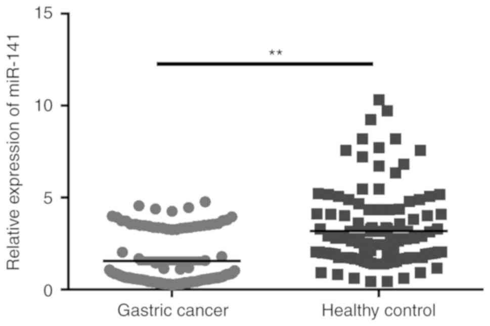
Plasma miR-141 expression level is
decreased in patients with gastric cancer
The plasma expression levels of miR-141 were
compared between 109 healthy controls and patients with gastric
cancer. As shown in Fig. 1, plasma
miR-141 levels were significantly higher in the blood of healthy
subjects compared with in patients with gastric cancer
(P<0.01).
Association between miR-141 expression
and clinicopathological characteristics
Patients were categorized into low- and high miR-141
expression groups, with the median value set as the cutoff. A total
of 115 patients were categorized as having low expression, whereas
49 patients were identified as having high expression. It was
demonstrated that miR-141 expression was not associated with age,
tumor size, tumor location, resection range or adjuvant therapy.
However, a significant association was observed between miR-141
expression and WHO grade (grade I+II vs. grade III+IV), recurrence
time and overall survival time (Table
I). As grades III and IV refer to lymph node and distant
metastasis, respectively, the significant association for WHO grade
also suggests that a lower miR-141 expression level may increase
the risk of lymph node and distant metastasis in gastric
cancer.
 | Table I.Association between miR-141
expression and clinicopathological characteristics. |
Table I.
Association between miR-141
expression and clinicopathological characteristics.
|
|
| miR-141 expression
level, n |
|
|---|
|
|
|
|
|
|---|
| Clinicopthological
characteristics | Patients, n | Low | High | P-value |
|---|
| Age (years) |
|
|
| 0.058 |
|
<60 | 94 | 58 | 36 |
|
|
≥60 | 70 | 53 | 17 |
|
| Tumor size |
|
|
| 0.349 |
| <3
cm | 89 | 58 | 31 |
|
| ≥3
cm | 75 | 54 | 21 |
|
| World Health
Organization grade |
|
|
| 0.075 |
| I | 17 | 7 | 10 |
|
| II | 76 | 47 | 29 |
|
|
III | 52 | 39 | 13 |
|
| IV | 19 | 13 | 6 |
|
|
I+II |
| 54 | 39 | 0.049 |
|
III+IV |
| 52 | 19 |
|
| Resection
range |
|
|
| 0.644 |
| Total
resection | 71 | 49 | 22 |
|
| Local
resection | 93 | 61 | 32 |
|
| Adjuvant
therapy |
|
|
| 0.971 |
|
Radiotherapy | 13 | 9 | 4 |
|
|
Chemotherapy | 35 | 23 | 12 |
|
|
Radiotherapy and
chemotherapy | 46 | 31 | 15 |
|
| Recurrence
time |
|
|
| <0.0001 |
| <3
months | 96 | 77 | 19 |
|
| ≥3
months | 68 | 33 | 35 |
|
| Survival
duration |
| <15
months | 96 | 72 | 24 | 0.002 |
| ≥15
months | 68 | 35 | 33 |
|
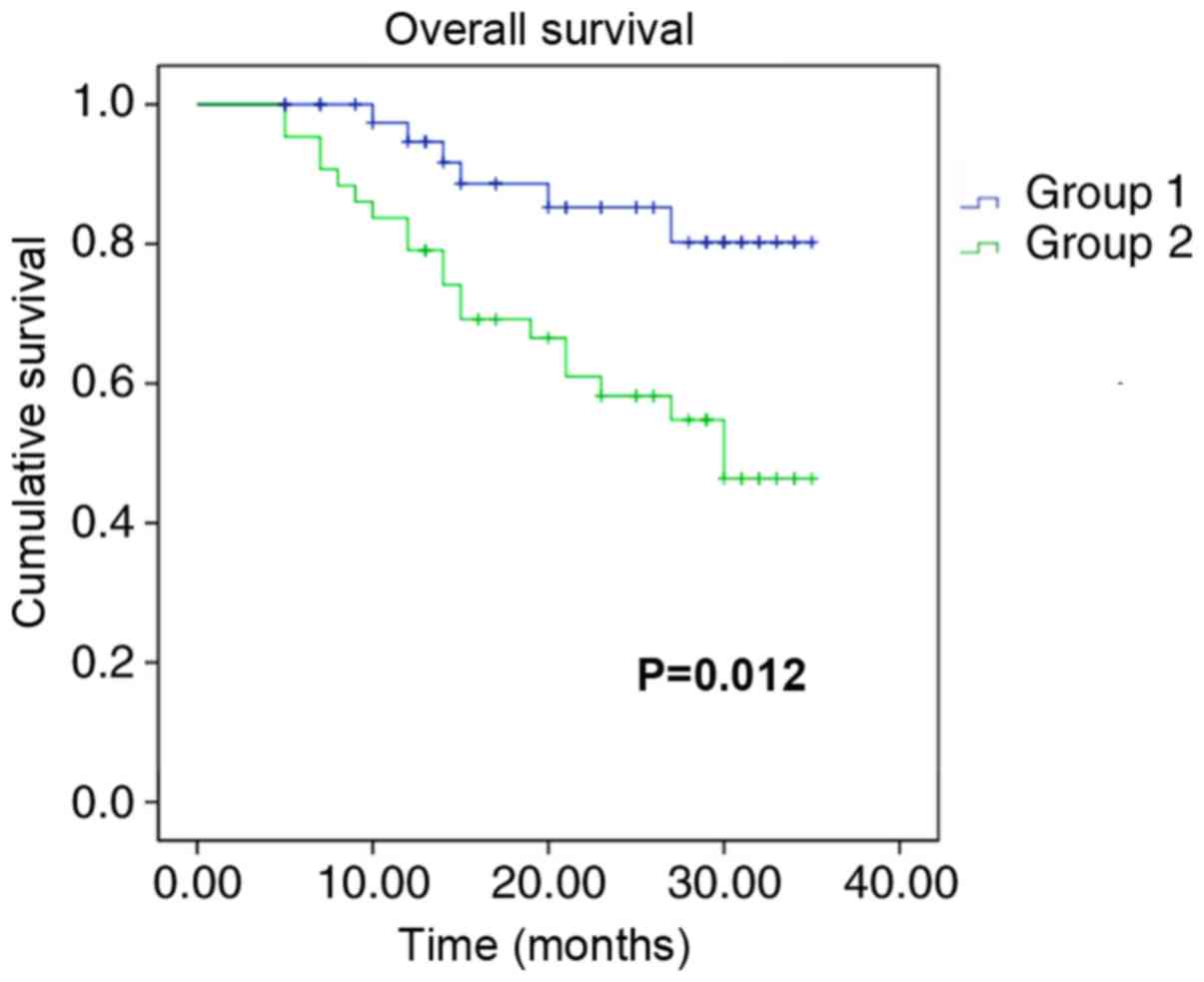
miR-141 expression level is associated
with the overall survival of patients with gastric cancer
Kaplan-Meier survival analysis was performed to
determine whether miR-141 expression was associated with the
survival of patients with gastric cancer. Due to the loss of
patients, follow-up was not conducted for seven of the original
recruits. Patients with high expression levels of miR-141 exhibited
greater overall survival times compared with patients with low
miR-141 expression levels (Fig. 2).
The HR associated with miR-141 low expression was 2.352 (Table II). These data indicated that the
aggressiveness of gastric cancer was associated with decreased
expression levels of miR-141.
| Table II.Multivariate Cox regression analysis
of risk factors associated with the overall survival of patients
with cancer gastric. |
Table II.
Multivariate Cox regression analysis
of risk factors associated with the overall survival of patients
with cancer gastric.
| Clinical
characteristics | Hazard ratio | 95% confidence
interval | P-value |
|---|
| Age | 1.005 | 0.978–1.032 | 0.719 |
| Tumor size | 0.678 | 0.422–1.088 | 0.014 |
| World Health
Organization grade | 0.709 | 0.435–1.158 | 0.001 |
| Resection
range | 0.980 | 0.620–1.547 | 0.930 |
| Adjuvant
therapy | 0.934 | 0.576–1.516 | 0.784 |
| Low microRNA-141
expression | 2.352 | 1.379–4.012 | 0.002 |
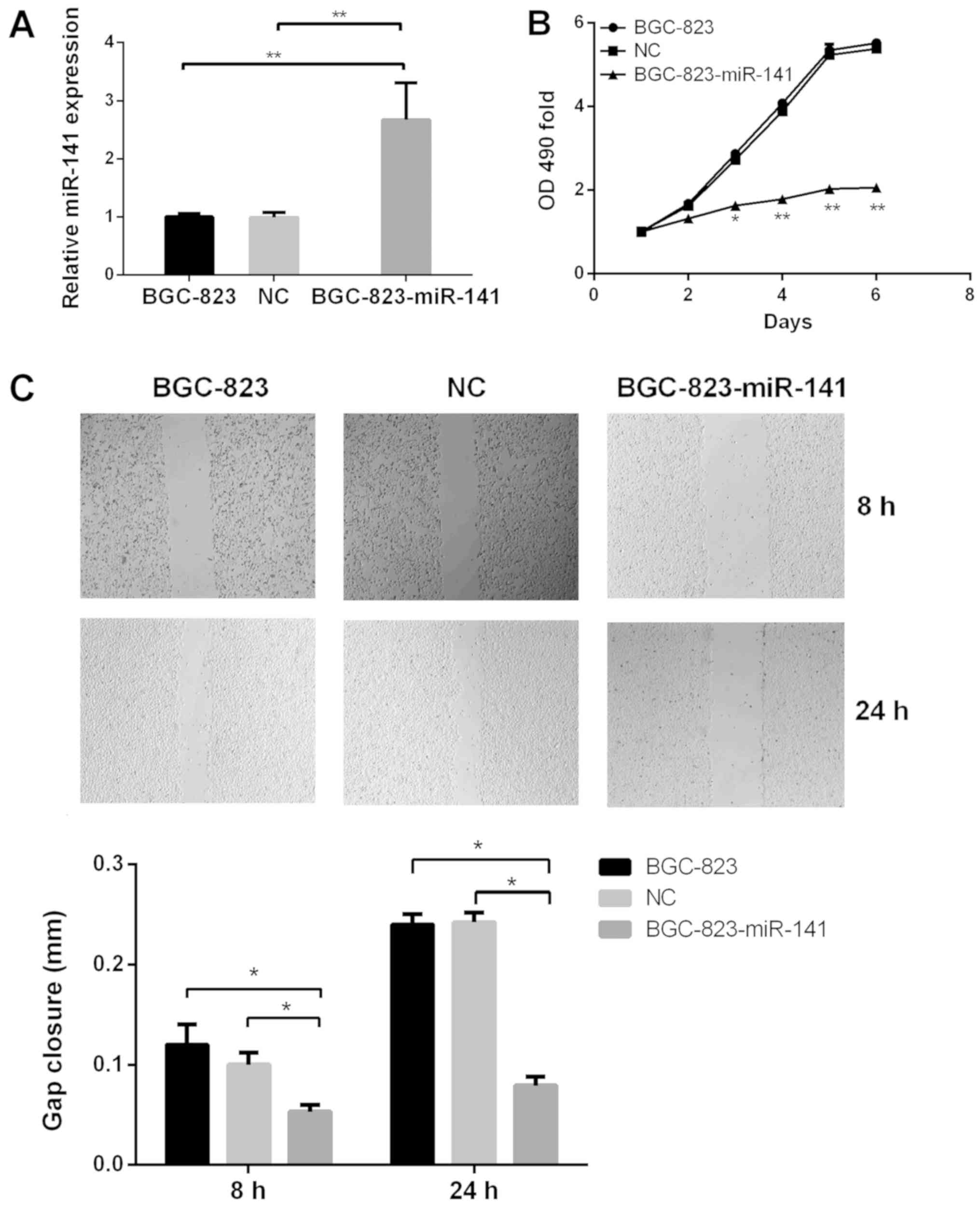
miR-141 inhibits cell proliferation
and migration in vitro
Following the observation that miR-141 expression
was significantly downregulated in gastric cancer samples, the
functional role of miR-141 in the development of gastric cancer was
investigated. Exogenous expression of miR-141 was significantly
increased in miR-141-transfected BGC-823 cells compared with in
untransfected and BGC-823-NC cells (Fig.
3A). The MTT assay revelaed that the proliferative ability of
BGC-823-miR-141 cells was significantly decreased, compared with
that of the control cell groups (Fig.
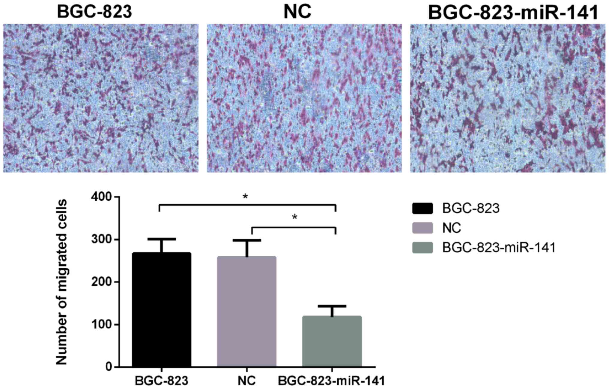
3B). Furthermore, the migratory ability of BGC-823-miR-141
cells, as determined using wound-healing (Fig. 3C) and Transwell assays(Fig. 4), was significantly decreased
(P<0.05) compared with in the control cell groups. These results
suggested that miR-141 may serve a key role in the proliferation
and migration of gastric cancer cells.
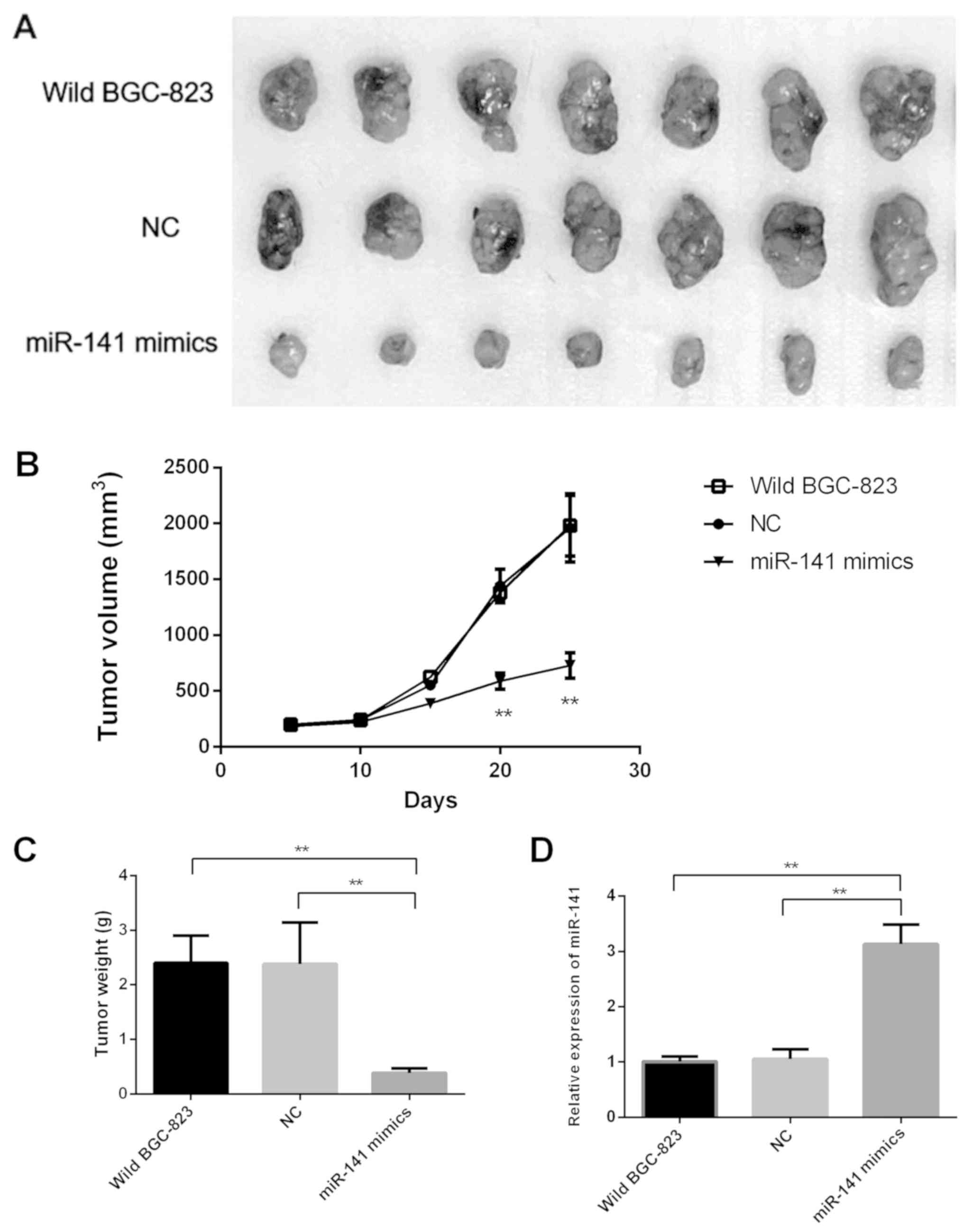
miR-141 suppresses gastric cancer
tumorigenicity in vivo
In order to validate the aforementioned results,
in vivo experimentation was performed; untransfected BGC-823
cells, and those transfected with miR-141 mimics or NC were
inoculated into three separate groups of mice. After 4 weeks, the
BGC-823-miR-141 mimic group exhibited significantly decreased tumor
growth and weight compared with the control groups (Fig. 5A-C). Furthmore, RT-qPCR analysis
confirmed that miR-141 expression levels in tumor tissues derived
from BGC-823-miR-141 mimics were significantly higher compared with
in the other two groups (Fig.
5D).
Discussion
In the present study, the prognostic value of
miR-141 in patients with gastric cancer was evaluated. It was
revealed that plasma miR-141 expression levels in patients with
gastric cancer were significantly lower compared with those in
healthy control subjects. This is in contrast to miR-141 expression
in gastric cancer tissues, in which a previous study suggested
higher miR-141 expression levels in gastric cancer tissues compared
with in healthy subjects (26).
Patients with lower expression levels of miR-141 were revealed to
have poor clinical outcomes, which supports the hypothesis that
miR-141 serves a key role in the survival of patients with gastric
cancer. Therefore, miR-141 expression may be used as an independent
prognostic indicator of gastric cancer.
miRNAs are implicated as key regulators of normal
cell function (31,32), and their aberrant expression is
associated with various diseases, including gastric cancer. A
number of miRNAs have been associated with multiple regulatory
networks in gastric cancer pathogenesis. It has previously been
reported that miR-218 serves as a tumor suppressor regulating cell
invasion, migration, proliferation and the maintenance of cancer
cell stemness (33,34), which provides clues for the
investigation of other miRs, including miR-141. Other miRNAs have
also been associated with clinical endpoints and diagnostic
potential (35–38). Regardless, there are still a large
number of miRNAs which have not been characterized, and which may
serve prominent roles in the progression of gastric cancer.
As numerous circulating miRNAs have emerged as
biomarkers for cancer diagnosis, miR-141 has also attracted
attention. The present study investigated whether circulating
miR-141 expression levels were associated with the prognosis and
overall survival of patients. It was demonstrated that plasma
miR-141 expression levels were significantly lower in patients with
gastric cancer compared with in healthy controls, and that this was
significantly associated with the survival duration and recurrence
rate of patients with gastric cancer. The results suggested that
miR-141 may be considered an independent prognostic marker for
predicting patient survival, which may also aid the personalized
treatment of patients with cancer.
Furthermore, in vitro and in vivo
methods were used to investigate the role of miR-141 in the
progression and pathophysiology of gastric cancer. miR-141 mimics
were successfully transfected into BGC-823 cells, and the results
revealed that overexpression of miR-141 significantly inhibited the
proliferation and migration of gastric cancer cells. These results
were consistent with the clinical association results of the
present study, which are displayed in Tables I and II.
There were a number of limitations to the present
study. Firstly, the patient population was small, thus the
conclusions regarding the association of miR-141 with
clinicopathological characteristics require further verification
using a larger patient cohort. Secondly, only one cell line was
used for the in vitro study, thus the use of additional
gastric cancer cell lines may further support the role of miR-141
in gastric cancer cells. Thirdly, a mechanistic study to determine
the target of miR-141, or how miR-141 regulates tumor growth, was
not conducted.
In conclusion, to the best of our knowledge, the
present study was the first to evaluate the plasma expression
levels of miR-141 in patients with gastric cancer from a Chinese
population. Although the population size was small, a significant
association between the expression of miR-141 and patient survival
time was observed. A larger population is required to further
confirm the prognostic significance of miR-141 in gastric cancer.
Furthermore, the possible molecular targets of miR-141, in addition
to its potential interference with signaling pathways, require
further investigation.
Acknowledgements
Not applicable.
Funding
No funding was received.
Availability of data and materials
The datasets used and/or analyzed during the current
study are available from the corresponding author on reasonable
request.
Authors' contributions
TW and JZ carried out the laboratory experiments. JT
and SH collected the clinical information from patients. RW
conducted the statistical analysis. LC designed the study and
drafted the manuscript.
Ethics approval and consent to
participate
The present study was approved by the Tianjin Nankai
Hospital Ethics Committee, and written informed consent was
obtained from each subject. All animal experiments were approved by
the Ethics Committee of Laboratory Animals of Tianjin Nankai
Hospital (approval no. 00000531).
Patient consent for publication
Written informed consent was obtained from all
patients.
Competing interests
The authors declare that they have no competing
interests.
References
|
1
|
Crew KD and Neugut AI: Epidemiology of
gastric cancer. World J Gastroenterol. 12:354–362. 2006. View Article : Google Scholar : PubMed/NCBI
|
|
2
|
Song YX, Yue ZY, Wang ZN, Xu YY, Luo Y, Xu
HM, Zhang X, Jiang L, Xing CZ and Zhang Y: MicroRNA-148b is
frequently down-regulated in gastric cancer and acts as a tumor
suppressor by inhibiting cell proliferation. Mol Cancer. 10:12011.
View Article : Google Scholar : PubMed/NCBI
|
|
3
|
Zuberi M, Khan I, Mir R, Gandhi G, Ray PC
and Saxena A: Utility of Serum miR-125b as a diagnostic and
prognostic indicator and its alliance with a panel of tumor
suppressor genes in epithelial ovarian cancer. PLoS One.
11:e01539022016. View Article : Google Scholar : PubMed/NCBI
|
|
4
|
Kinose Y, Sawada K, Nakamura K and Kimura
T: The role of microRNAs in ovarian cancer. Biomed Res Int.
2014:2493932014. View Article : Google Scholar : PubMed/NCBI
|
|
5
|
Tilghman SL, Rhodes LV, Bratton MR,
Carriere P, Preyan LC, Boue SM, Vasaitis TS, McLachlan JA and Burow
ME: Phytoalexins, miRNAs and breast cancer: A review of
phytochemical-mediated miRNA regulation in breast cancer. J Health
Care Poor Underserved. 24 (Suppl 1):S36–S46. 2013. View Article : Google Scholar
|
|
6
|
Chen J, Deng S, Zhang S, Chen Z, Wu S, Cai
X, Yang X, Guo B and Peng Q: The role of miRNAs in the
differentiation of adipose-derived stem cells. Curr Stem Cell Res
Ther. 9:268–279. 2014. View Article : Google Scholar : PubMed/NCBI
|
|
7
|
Du L, Borkowski R, Zhao Z, Ma X, Yu X, Xie
XJ and Pertsemlidis A: A high-throughput screen identifies miRNA
inhibitors regulating lung cancer cell survival and response to
paclitaxel. RNA Biol. 10:1700–1713. 2013. View Article : Google Scholar : PubMed/NCBI
|
|
8
|
He J, Zhang JF, Yi C, Lv Q, Xie WD, Li JN,
Wan G, Cui K, Kung HF, Yang J, et al: miRNA-mediated functional
changes through co-regulating function related genes. PLoS One.
5:e135582010. View Article : Google Scholar : PubMed/NCBI
|
|
9
|
Recchiuti A and Serhan CN: Pro-resolving
lipid mediators (SPMs) and their actions in regulating miRNA in
novel resolution circuits in inflammation. Front Immunol.
3:2982012. View Article : Google Scholar : PubMed/NCBI
|
|
10
|
Wolff LJ, Wolff JA and Sebestyén MG:
Effect of tissue-specific promoters and microRNA recognition
elements on stability of transgene expression after hydrodynamic
naked plasmid DNA delivery. Hum Gene Ther. 20:374–388. 2009.
View Article : Google Scholar : PubMed/NCBI
|
|
11
|
Xia JH, He XP, Bai ZY and Yue GH:
Identification and characterization of 63 MicroRNAs in the Asian
seabass Lates calcarifer. PLoS One. 6:e175372011. View Article : Google Scholar : PubMed/NCBI
|
|
12
|
Vos S, Vesuna F, Raman V, van Diest PJ and
van der Groep P: miRNA expression patterns in normal breast tissue
and invasive breast cancers of BRCA1 and BRCA2 germ-line mutation
carriers. Oncotarget. 6:32115–32137. 2015. View Article : Google Scholar : PubMed/NCBI
|
|
13
|
Mushtaq G, Greig NH, Anwar F, Zamzami MA,
Choudhry H, Shaik MM, Tamargo IA and Kamal MA: miRNAs as
circulating biomarkers for Alzheimer's disease and Parkinson's
disease. Med Chem. 12:217–225. 2016. View Article : Google Scholar : PubMed/NCBI
|
|
14
|
Ren X, Bai X, Zhang X, Li Z, Tang L, Zhao
X, Li Z, Ren Y, Wei S, Wang Q, et al: Quantitative nuclear
proteomics identifies that miR-137-mediated EZH2 reduction
regulates resveratrol-induced apoptosis of neuroblastoma cells. Mol
Cell Proteomics. 14:316–328. 2015. View Article : Google Scholar : PubMed/NCBI
|
|
15
|
Wang LG, Ni Y, Su BH, Mu XR, Shen HC and
Du JJ: MicroRNA-34b functions as a tumor suppressor and acts as a
nodal point in the feedback loop with Met. Int J Oncol. 42:957–962.
2013. View Article : Google Scholar : PubMed/NCBI
|
|
16
|
Aure MR, Leivonen SK, Fleischer T, Zhu Q,
Overgaard J, Alsner J, Tramm T, Louhimo R, Alnæs GI, Perälä M, et
al: Individual and combined effects of DNA methylation and copy
number alterations on miRNA expression in breast tumors. Genome
Biol. 14:R1262013. View Article : Google Scholar : PubMed/NCBI
|
|
17
|
Qin W, Zhang K, Clarke K, Weiland T and
Sauter ER: Methylation and miRNA effects of resveratrol on mammary
tumors vs. Nutr Cancer. 66:270–277. 2014. View Article : Google Scholar : PubMed/NCBI
|
|
18
|
Schwarzenbach H, Nishida N, Calin GA and
Pantel K: Clinical relevance of circulating cell-free microRNAs in
cancer. Nat Rev Clin Oncol. 11:145–156. 2014. View Article : Google Scholar : PubMed/NCBI
|
|
19
|
Xu J, Cao Z, Liu W, You L, Zhou L, Wang C,
Lou W, Sun B, Miao Y, Liu X, et al: Plasma miRNAs effectively
distinguish patients with pancreatic cancer from controls: A
multicenter study. Ann Surg. 263:1173–1179. 2016. View Article : Google Scholar : PubMed/NCBI
|
|
20
|
Park SM, Gaur AB, Lengyel E and Peter ME:
The miR-200 family determines the epithelial phenotype of cancer
cells by targeting the E-cadherin repressors ZEB1 and ZEB2. Genes
Dev. 22:894–907. 2008. View Article : Google Scholar : PubMed/NCBI
|
|
21
|
Long ZH, Bai ZG, Song JN, Zheng Z, Li J,
Zhang J, Cai J, Yao HW, Wang J, Yang YC, et al: miR-141 inhibits
proliferation and migration of colorectal cancer SW480 Cells.
Anticancer Res. 37:4345–4352. 2017.PubMed/NCBI
|
|
22
|
van Jaarsveld MT, Helleman J, Boersma AW,
van Kuijk PF, van Ijcken WF, Despierre E, Vergote I, Mathijssen RH,
Berns EM, Verweij J, et al: miR-141 regulates KEAP1 and modulates
cisplatin sensitivity in ovarian cancer cells. Oncogene.
32:4284–4293. 2013. View Article : Google Scholar : PubMed/NCBI
|
|
23
|
Zhou X, Su J, Zhu L and Zhang G:
Helicobacter pylori modulates cisplatin sensitivity in gastric
cancer by down-regulating miR-141 expression. Helicobacter.
19:174–181. 2014. View Article : Google Scholar : PubMed/NCBI
|
|
24
|
Fu WF, Chen WB, Dai L, Yang GP, Jiang ZY,
Pan L, Zhao J and Chen G: Inhibition of miR-141 reverses cisplatin
resistance in non-small cell lung cancer cells via upregulation of
programmed cell death protein 4. Eur Rev Med Pharmacol Sci.
20:2565–2572. 2016.PubMed/NCBI
|
|
25
|
Wang D, Ma J, Ji X, Xu F and Wei Y:
miR-141 regulation of EIF4E expression affects docetaxel
chemoresistance of non-small cell lung cancer. Oncol Rep.
37:608–616. 2017. View Article : Google Scholar : PubMed/NCBI
|
|
26
|
Tejero R, Navarro A, Campayo M, Viñolas N,
Marrades RM, Cordeiro A, Ruíz-Martínez M, Santasusagna S, Molins L,
Ramirez J and Monzó M: miR-141 and miR-200c as markers of overall
survival in early stage non-small cell lung cancer adenocarcinoma.
PLoS One. 9:e1018992014. View Article : Google Scholar : PubMed/NCBI
|
|
27
|
Kim YH, Kim JH, Kim H, Kim H, Lee YC, Lee
SK, Shin SK, Park JC, Chung HS, Park JJ, et al: Is the recent WHO
histological classification for gastric cancer helpful for
application to endoscopic resection? Gastric Cancer. 19:869–875.
2016. View Article : Google Scholar : PubMed/NCBI
|
|
28
|
Livak KJ and Schmittgen TD: Analysis of
relative gene expression data using real-time quantitative PCR and
the 2(-Delta Delta C(T)) method. Methods. 25:402–408. 2001.
View Article : Google Scholar : PubMed/NCBI
|
|
29
|
Sen Z, Zhan XK, Jing J, Yi Z and Wanqi Z:
Chemosensitizing activities of cyclotides from Clitoria ternatea in
paclitaxel-resistant lung cancer cells. Oncol Lett. 5:641–644.
2013. View Article : Google Scholar : PubMed/NCBI
|
|
30
|
Zhang S, Xin H, Li Y, Zhang D, Shi J, Yang
J and Chen X: Skimmin, a coumarin from hydrangea paniculata, slows
down the progression of membranous glomerulonephritis by
Anti-inflammatory effects and inhibiting immune complex deposition.
Evid Based Complement Alternat Med. 2013:8192962013.PubMed/NCBI
|
|
31
|
Kontomanolis EN and Koukourakis MI:
MicroRNA: The potential regulator of endometrial carcinogenesis.
Microrna. 4:18–25. 2015. View Article : Google Scholar : PubMed/NCBI
|
|
32
|
Fan Y, Zhou Y, Zhou X, Sun F, Gao B, Wan
M, Zhou X, Sun J, Xu X, Cheng L, et al: MicroRNA 224 regulates ion
transporter expression in ameloblasts to coordinate enamel
mineralization. Mol Cell Biol. 35:2875–2890. 2015. View Article : Google Scholar : PubMed/NCBI
|
|
33
|
Hu Y, Xu K and Yagüe E: miR-218 targets
survivin and regulates resistance to chemotherapeutics in breast
cancer. Breast Cancer Res Treat. 151:269–280. 2015. View Article : Google Scholar : PubMed/NCBI
|
|
34
|
Li Q, Zhu F and Chen P: miR-7 and miR-218
epigenetically control tumor suppressor genes RASSF1A and Claudin-6
by targeting HoxB3 in breast cancer. Biochem Biophys Res Commun.
424:28–33. 2012. View Article : Google Scholar : PubMed/NCBI
|
|
35
|
Yang L, Xu Q, Xie H, Gu G and Jiang J:
Expression of serum miR-218 in hepatocellular carcinoma and its
prognostic significance. Clin Transl Oncol. 18:841–847. 2016.
View Article : Google Scholar : PubMed/NCBI
|
|
36
|
Jiang Z, Song Q, Yang S, Zeng R, Li X,
Jiang C, Ding W, Zhang J and Zheng Y: Serum microRNA-218 is a
potential biomarker for esophageal cancer. Cancer Biomark.
15:381–389. 2015. View Article : Google Scholar : PubMed/NCBI
|
|
37
|
Xin SY, Feng XS, Zhou LQ, Sun JJ, Gao XL
and Yao GL: Reduced expression of circulating microRNA-218 in
gastric cancer and correlation with tumor invasion and prognosis.
World J Gastroenterol. 20:6906–6911. 2014. View Article : Google Scholar : PubMed/NCBI
|
|
38
|
Zhang XL, Shi HJ, Wang JP, Tang HS, Wu YB,
Fang ZY, Cui SZ and Wang LT: MicroRNA-218 is upregulated in gastric
cancer after cytoreductive surgery and hyperthermic intraperitoneal
chemotherapy and increases chemosensitivity to cisplatin. World J
Gastroenterol. 20:11347–11355. 2014. View Article : Google Scholar : PubMed/NCBI
|