Introduction
Lung cancer, a complex disease involving both
epigenetic and genetic changes, is the leading cause of
cancer-associated mortality worldwide (1,2). Lung
cancer has had a high incidence rate and a poor 5-year survival
rate of <19% in the United States between 2006 and 2012
(2). One cause of the high mortality
rate is the lack of specific early detection methods and the
majority of patients are diagnosed with middle- or late-stage
disease (3). Therefore, early
detection and treatment strategies for lung cancer are urgently
required.
Several imaging and cytology-based strategies have
been utilized for early lung cancer detection. However, none have
been demonstrated to completely reduce lung cancer mortality
(3–5). Previous studies have reported that
aberrant epigenetic changes are one of the most frequent
cancer-associated events and are regarded as important mechanisms
in carcinogenesis (6). Investigation
of the associated molecular mechanisms can be exploited to diagnose
early-stage lung cancer (3–5). Furthermore, methylation profiles may be
potential biomarkers for early cancer diagnosis and they have been
demonstrated to exhibit good prognostic value (4,7–9). Previously, accumulating evidence has
confirmed that tumor tissues can be characterized by
hypermethylation at promoter-associated CpG islands (CGIs) or
global hypomethylation of the genome compared with normal tissues
(9–11). Furthermore, certain studies have
suggested that methylation of DNA CpG sites is an epigenetic
regulator of gene expression that usually results in gene silencing
(12,13). Hao et al (12) reported that methylation patterns can
predict prognosis and survival, and identified an association
between differential methylation of CpG sites and the expression of
cancer-associated genes. Their findings demonstrate the utility of
methylation biomarkers for cancer molecular characterization,
diagnosis and prognosis determination. Therefore, a number of
specific tumor targets can be developed for use as DNA
methylation-based biomarkers (4,7,9).
At present, numerous useful cancer biomarkers have
been identified. Pituitary tumor transforming gene 1 binding factor
(PTTG1IP; also termed PBF) is a ubiquitously expressed
proto-oncogene. PTTG1IP was first identified through its ability to
bind to human securin, also termed pituitary tumor transforming
gene (PTTG) (14,15). Thus far, PTTG1IP has been
reported to be highly expressed in thyroid, breast, colorectal, and
liver cancer (16–19). However, to the best of our knowledge,
its expression levels in lung cancer have not been reported. The
present study investigated PTTG1IP expression in early
non-small cell lung cancer (NSCLC) and examined the correlation
between the PTTG1IP promoter region methylation level and
the gene expression level.
Materials and methods
Tissue samples
In total, 18 pairs of early-stage (stage I or II)
NSCLC tissues and adjacent tissues were obtained from the South
Hospital of Renji Hospital Shanghai Jiao Tong University School of
Medicine (Shanghai, China) between January 2014 and March 2015
(Table I). A total of 12 male and 6
female patients aged between 45 and 75 years were included in the
present study. During excision surgery, 50 mg fresh cancer tissue
and 50 mg adjacent normal tissue (<2 cm from cancer margin) was
obtained from each patient. Tissue samples were immediately frozen
in liquid nitrogen following resection and stored at −80°C until
use. All included samples were histologically confirmed primary
NSCLC and pathological stage I or II according to the
Tumor-Node-Metastasis staging system (20). Written informed consent was obtained
from all patients and the study was approved by the Ethics
Committee of South Hospital of Renji Hospital Shanghai Jiao Tong
University School of Medicine.
 | Table I.Basic information of the paired lung
cancer tissue and adjacent tissue samples. |
Table I.
Basic information of the paired lung
cancer tissue and adjacent tissue samples.
| Sample no. | Sex | Diagnosis | Staged |
|---|
| Pair 1c | Female | Lung
adenocarcinoma | II |
| Pair 2a,c | Male | Lung squamous cell
carcinoma | II |
| Pair 3a,c | Female | Lung
adenocarcinoma | II |
| Pair 4a,c | Male | Lung squamous cell
carcinoma | II |
| Pair 5a,c | Male | Lung squamous cell
carcinoma | II |
| Pair 6a,c | Female | Lung
adenocarcinoma | I |
| Pair 7a,c | Male | Lung
adenocarcinoma | II |
| Pair 8a,c | Female | Lung
adenocarcinoma | II |
| Pair 9a,c | Male | Lung
adenocarcinoma | II |
| Pair
10a,c | Male | Lung
adenocarcinoma | II |
| Pair
11a,c | Male | Lung
adenocarcinoma | II |
| Pair
12c | Male | Lung
adenocarcinoma | I |
| Pair
13b | Male | Lung
adenocarcinoma | I |
| Pair
14b | Male | Lung
adenocarcinoma | II |
| Pair
15b | Male | Lung squamous cell
carcinoma | II |
| Pair
16b | Female | Lung
adenocarcinoma | I |
| Pair
17b | Male | Lung
adenocarcinoma | I |
| Pair
18b | Female | Lung
adenocarcinoma | II |
Cell culture and treatments
A549 cells of the human lung adenocarcinoma cell
line were cultured in Roswell Park Memorial Institute-1640 medium
(Thermo Fisher Scientific, Inc., Waltham, MA, USA) with 10% fetal
bovine serum (FBS; Thermo Fisher Scientific, Inc., Waltham, MA,
USA) and 1% penicillin/streptomycin. MRC5 cells of the human
embryonic lung fibroblast cell line were cultured in minimum
essential medium (Thermo Fisher Scientific, Inc., Waltham, MA, USA)
with 10% FBS and 1% penicillin/streptomycin. Cell cultures were
incubated at 37°C in a humidified atmosphere with 5%
CO2. Following A549 cell culture to ~90% confluence, 1
µM 5-aza-2′-deoxycytidine (5-aza-dC, Sigma-Aldrich; Merck KGaA,
Darmstadt, Germany) was added to the culture medium. DMSO
(Sigma-Aldrich; Merck KGaA) treatment was used as a control. The
cells were harvested following 48-h treatment, and total RNA and
genomic DNA were obtained according to standard protocols. In
brief, TRIzol® reagent (Sigma-Aldrich; Merck KGaA,
Darmstadt, Germany) was used for RNA extraction. Precipitated RNA
was washed to remove impurities, and then resuspended for use in
downstream applications. DNA extractions were performed using the
High Pure PCR Template Preparation kit (Roche Diagnostics, Basel,
Switzerland). Cells were lysed with Lysis Buffer and Proteinase K,
and released DNA was bound on a glass fiber filter and washed prior
to elution.
Plasmid construction and cell
transfection
To generate a vector expressing myc-PTTG1IP,
the corresponding sequences were subcloned into a pcDNA3.1 vector
(Invitrogen; Thermo Fisher Scientific, Inc., Waltham, MA, USA) that
was subsequently termed pcDNA3.1/3Xmyc-PTTG1IP. Cells were
plated the day prior to transfection and cultured to 70%
confluence. Transfection was performed with
Lipofectamine® 2000 (Invitrogen; Thermo Fisher
Scientific, Inc., Waltham, MA, USA) with 1 µg plasmid/well,
according to the manufacturer's protocol. Cells transfected with
empty pcDNA3.1 vector was used as a control. The medium was
replaced with new culture medium 6 h post-transfection. Cells were
harvested 48 h following transfection and then prepared for
subsequent assays.
Cell proliferation assay
Cell proliferation was assessed by Cell Counting
Kit-8 (Dojindo Molecular Technologies, Inc., Kumamoto, Japan)
assays. Cells were seeded at 1,000 cells/well into 96-well plates
with 100 µl culture medium. Subsequently, 10 µl CCK-8 solution was
added to the cells at every 24 h for 5 days and the cells were
incubated for 2 h at 37°C. The reaction product was quantified
according to the manufacturer's protocol by measuring the
absorbance at 450 nm.
RNA and DNA extraction
Total RNA was extracted from cultured cells and
tissue samples using TRIzol® reagent (Sigma-Aldrich;
Merck KGaA, Darmstadt, Germany), according to the manufacturer's
protocol. Genomic DNA was extracted from cultured cells and tissue
samples using a High Pure PCR Template Preparation kit (Roche
Diagnostics, Basel, Switzerland), according to the manufacturer's
protocol.
Reverse transcription-quantitative
polymerase chain reaction (RT-qPCR)
RT was performed with a mix of oligo dT primer and
random primers for mRNA using a PrimeScript RT Reagent kit (Takara
Biotechnology Co., Ltd., Dalian, China). qPCR was performed using a
CFX96 Real-Time PCR detector (Bio-Rad Laboratories, Inc., Hercules,
CA, USA) and SYBR Premix Ex Taq™ (Takara Biotechnology Co., Ltd.,
Dalian, China). The thermocycling conditions were: 95°C for 2 min;
40 cycles of 95°C for 20 sec, 60°C for 20 sec and 72°C for 20 sec.
The comparative 2−ΔΔCq method was used to calculate fold
changes (21). GAPDH was used
as an endogenous reference. The primers used for qPCR were as
follows: PTTG1IP forward, 5′-GTCTGGACTACCCAGTTACAAGC-3′ and
reverse, 5′-CGCCTCAAAGTTCACCCAA-3′; GAPDH forward,
5′-GGAGTCCACTGGCGTCTTC-3′ and reverse,
5′-GCTGATGATCTTGAGGCTGTTG-3′. The experiment was performed in
triplicate.
Reduced representation bisulfite
sequencing (RRBS)
Genomic DNA was used to perform RRBS. RRBS library
construction was performed as described previously (22). The library was sequenced on a
next-generation sequencing (NGS) HiSeq platform (Illumina, Inc.,
San Diego, CA, USA). The sequencing data were aligned to a
reference genome (UCSC hg19) using Bismark (a flexible aligner and
methylation caller for Bisulfite-Seq applications) with default
parameters. The methylation level of each cytosine was calculated
using the R package MethylKit (version 1.0.0; http://code.google.com/p/methylkit), which is a
comprehensive R package for analysis of genome-wide DNA methylation
profiles (23).
Bisulfite amplicon sequencing
The DNA methylation level of the PTTG1IP
promoter was analyzed in cells or tissue samples via bisulfite
amplification followed by NGS. A total of 500 ng DNA was bisulfite
treated using an EZ DNA Methylation Gold-kit (Zymo Research Corp.,
Irvine, CA, USA). The bisulfite-converted DNA was used to amplify
the candidate fragment with a Takara EX Taq Hot Start Version kit
(Takara Biotechnology Co., Ltd., Dalian, China). The PCR products
were loaded on a 1.5% agarose gel for analysis and recovered for
library construction and NGS using a MiSeq platform (Illumina,
Inc., San Diego, CA, USA). The DNA methylation level of candidate
fragments was determined by analyzing the NGS data. The primers for
amplification were as follows: PTTG1IP forward,
5′-GTATTGTTGAAGGGTGTAGAGATG-3′ and PTTG1IP reverse,
5′-CCACCCACCAAAACTTAATAATTA-3′.
Statistical analysis
The statistical significance of mean values in a
two-sample comparison was determined with Student's t-test.
P<0.05 was considered to indicate a statistically significant
difference. Data are presented as the mean ± standard error. The
lung adenocarcinoma and lung squamous cell carcinoma data sets from
The Cancer Genome Atlas (TCGA) were used to further validate the
relationship between promoter methylation and gene expression of
PTTG1IP. Gene expression data (RNASeq) and DNA methylation
data (Illumina methylation beadchip HM450 K) from 456 lung
adenocarcinoma samples and 370 squamous cell carcinoma samples were
downloaded from TCGA database on the cBioportal website (www.cbioportal.org). Spearman's non-parametric
correlation test was performed to evaluate the correlation between
gene methylation and expression using R software (version 3.3.2;
http://www.R-project.org) (24).
Results
Decreased PTTG1IP expression in
early-stage non-small cell lung cancer
Although PTTG1IP has been reported to be
abnormally expressed in a variety of tumor types (17–19,25), to
the best of our knowledge, its association with lung cancer remains
to be reported. The present study analyzed PTTG1IP
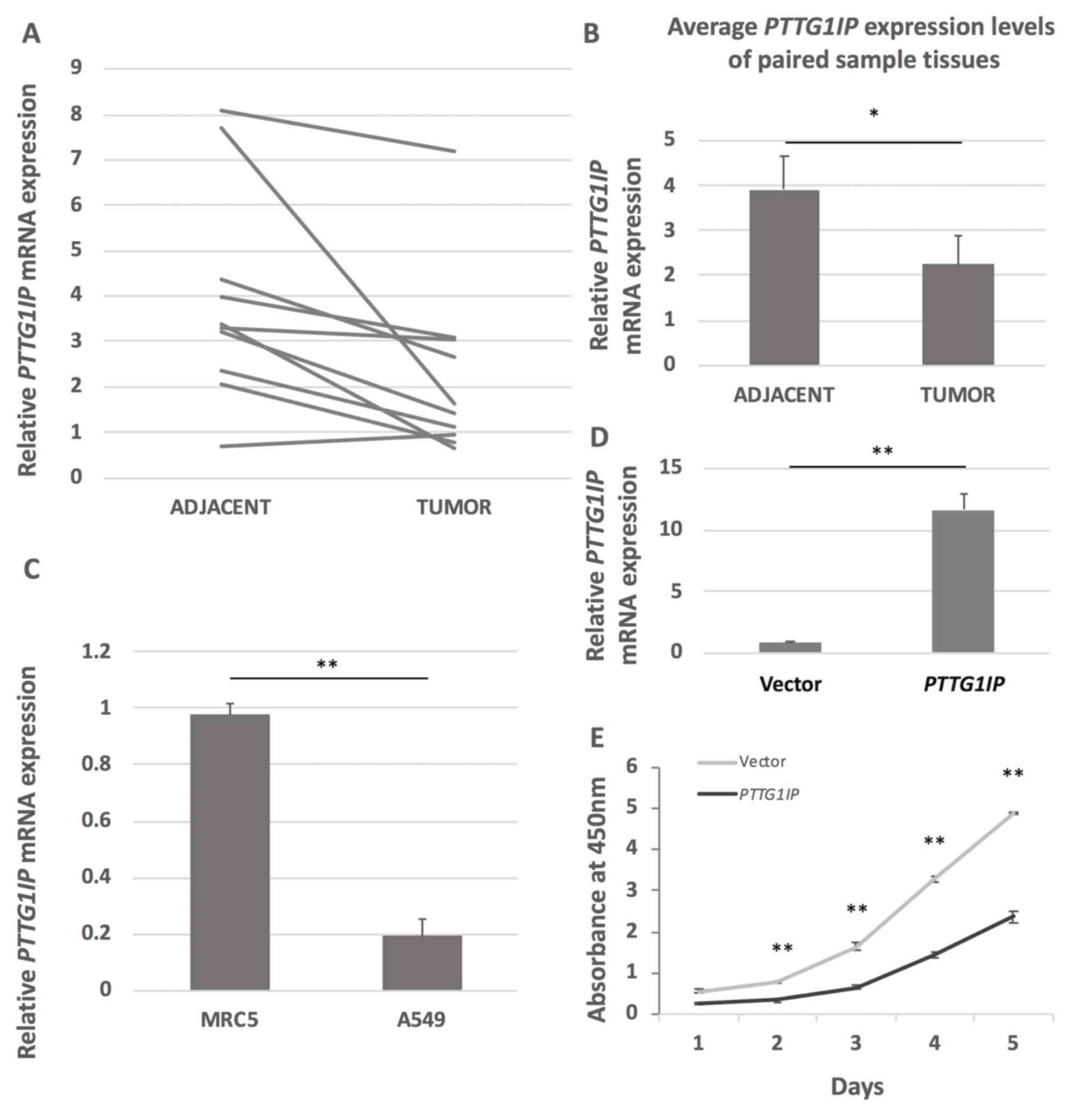
expression in 10 paired early-stage NSCLC tissue samples (Table I). The RT-qPCR results revealed that
PTTG1IP expression was decreased in all the cancer tissues
except sample pair 10 (Fig. 1A). The
mean mRNA level in the lung cancer tissues was significantly lower
compared with that in the adjacent tissues (Fig. 1B). PTTG1IP expression was
reduced by 80.1% in the lung cancer cell line A549 compared with
the normal lung cell line MRC5 (Fig.
1C). To evaluate whether the expression level of PTTG1IP
is associated with the proliferation capacity of lung cancer cells,
PTTG1IP was overexpressed in A549 cells. The expression
level of PTTG1IP was ~11 times higher in cells transfected
with pcDNA3.1/3Xmyc-PTTG1IP compared with those transfected
with empty pcDNA3.1 vector 2 days after transfection (Fig. 1D). A cell proliferation assay
revealed that the proliferation of
pcDNA3.1/3Xmyc-PTTG1IP-transfected cells was significantly
inhibited compared with the control cells. By day five, the number
of transfected cells was <50% of that in the control group
(Fig. 1E).
DNA methylation analysis of the
PTTG1IP promoter
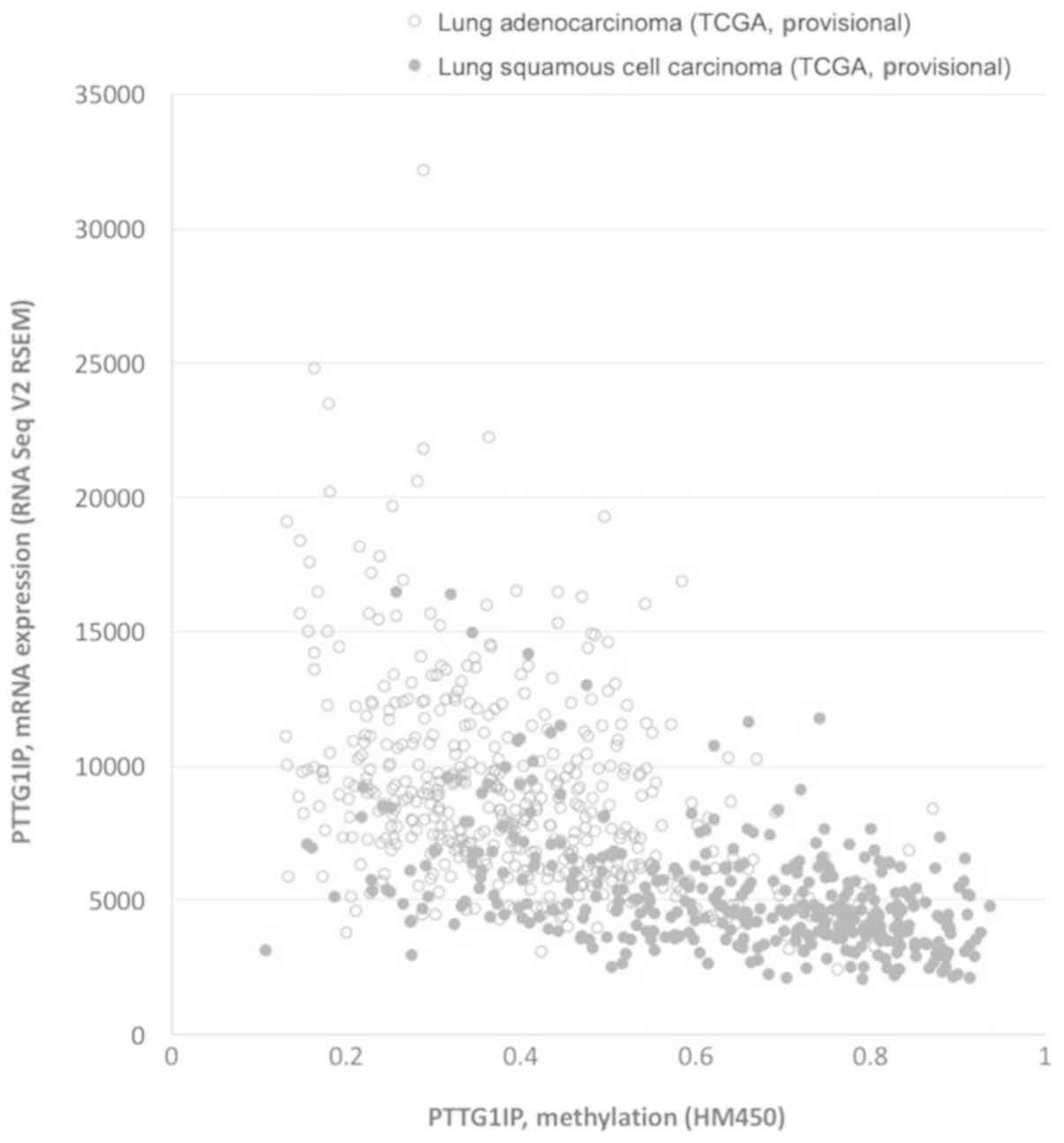
To investigate the regulatory mechanism driving the
decreased expression of PTTG1IP in lung cancer, the present
study first downloaded RNAseq data and DNA methylation chip 450k
data of lung adenocarcinoma and lung squamous cell carcinoma from
The Cancer Genome Atlas (TCGA) database on the cBioportal website
(www.cbioportal.org/). Correlation
analysis revealed a significant negative correlation between the
PTTG1IP gene methylation level and mRNA level in both lung
adenocarcinoma and lung squamous cell carcinoma, with Spearman
correlation coefficients of −0.415 and −0.457, respectively
(Fig. 2).
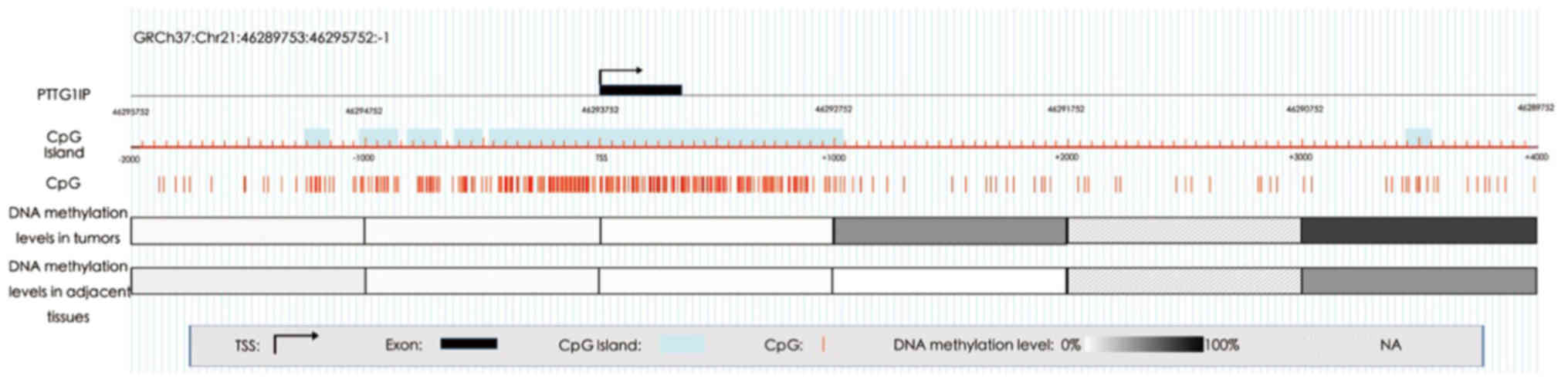
To further determine the association between
PTTG1IP promoter methylation and gene expression, an RRBS
study was conducted with six pairs of early-stage NSCLC tissue
samples (Table I). As presented in
Fig. 3, a plurality of CGIs were
distributed among the 2,000 bp upstream and the 2,000 bp downstream
of the PTTG1IP transcription start site (TSS). However,
regional DNA methylation analysis demonstrated that the region from
2,000 bp upstream to 1,000 bp downstream of the TSS was
hypomethylated both in tumor tissues and adjacent tissues, although
CpG loci were very concentrated in this region. However, a
difference was identified in the CGI shore region of 1,000-2,000 bp
downstream of the TSS between lung cancer tissues and adjacent
tissues, with an mean DNA methylation difference of 50%. In the
region 5,000-6,000 bp downstream of the TSS, DNA was
hypermethylated and the methylation level in cancer tissues was
higher compared with that in adjacent tissues. Therefore,
hypermethylation of the CGI shore region within the PTTG1IP
gene promoter might be associated with its low expression.
DNA methylation level of the CGI shore
region within the PTTG1IP gene promoter is associated with PTTG1IP
expression
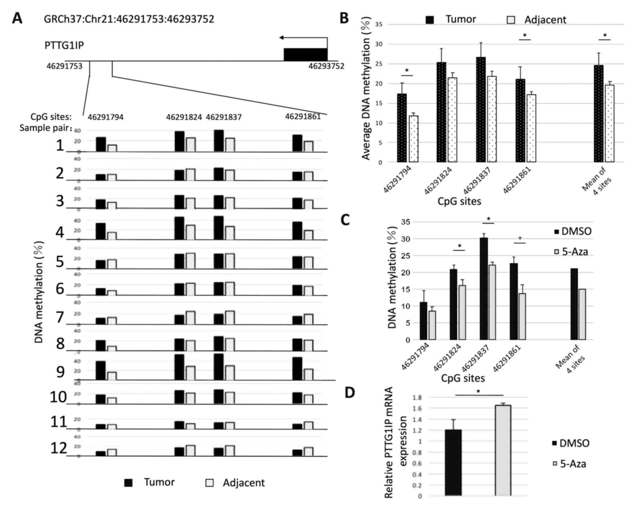
Subsequently, the methylation level of a fragment
composed of four CG sites in the CGI shore region within the
PTTG1IP promoter was measured in 12 pairs of early-stage
NSCLC samples using bisulfite amplicon sequencing (Fig. 4A). Hypermethylation was identified in
>50% of the cancer tissues in the sample pairs. As presented in
Fig. 4B, the mean methylation level
of the four CG loci in tumor tissues was higher compared with that
in adjacent cancerous tissues. The mean methylation level of the
four CG loci was 22.6 and 18.0%, respectively, and the difference
was significant. The trend of these results was consistent with
that observed in the RRBS analysis. To verify the association
between DNA methylation and gene expression in this region, A549
cells were treated with 5-aza-2′-deoxycytidine (1 µM) to reduce DNA
methylation levels. Following 48 h of treatment, a significant
decrease in methylation of the three CG sites (except for site
46291794) was observed (Fig. 4C).
The mean methylation level of the fragment was reduced from 21 to
15%. Furthermore, RT-qPCR revealed that PTTG1IP gene
expression was significantly increased following treatment with
5-aza-2′-deoxycytidine (Fig. 4D).
These results suggest that hypermethylation in the CGI shore within
the PTTG1IP promoter is essential for silencing of
PTTG1IP.
Discussion
The present study reported a negative correlation
between PTTG1IP gene expression and the methylation level of
its promoter region in lung cancer. In addition, it was identified
that PTTG1IP was highly methylated in the early stage of
lung cancer and exhibited a low expression level. Cytological
experiments indicated that PTTG1IP overexpression may
inhibit lung cancer cell proliferation. The present study provides
a possible new mechanism for lung cancer development and a
potential novel marker for early diagnosis of lung cancer.
The National Lung Screening Trial demonstrated a 20%
reduction in lung cancer mortality using low-dose computed
tomography (CT) screening (19).
This survival benefit comes at the cost of testing numerous
indeterminate pulmonary nodules, with an overall false-positive
rate of 96.4% (26,27). One possible way to improve CT
screening specificity is to use cancer-specific biomarkers from
sputum and plasma. Previous studies have examined DNA methylation
as a biomarker of cancer risk; however, the current low sensitivity
and/or specificity of lung cancer screening is not sufficient
(28–31). Epigenetic biomarkers, particularly
DNA methylation, have become one of the most promising options for
improving cancer diagnosis and have several advantages compared
with other markers, including gene expression or genetic markers
(32).
One surprising finding in cancer biology that has
emerged from TCGA sequencing projects is the wide diversity of
mutations that promote cancer development (33). DNA methylation changes are covalent
modifications that are very stable and usually occur early in
carcinogenesis. In addition, DNA methylation can be detected by a
variety of sensitive and low-cost techniques, even in samples with
low tumor cell purity (32). This
epigenetic modification can also be detected in different
biological fluids and is one of the most promising noninvasive
cancer detection tools (32).
Previously, different epigenetic candidates have
been proposed but have not yet reached clinical requirements, which
is predominantly due to the fact that the majority of studies are
based on a single candidate gene (34–38). For
example, methylated CDKN2A, commonly referred to as p16, was
an early focus in the search for diagnostic biomarkers in lung
cancer plasma; however, although earlier studies identified
CDKN2A promoter methylation in the plasma of patients with
lung cancer (39–42), subsequent studies have described low
sensitivity and specificity of this method (32,43,44).
Methylated plasma CDKN2A may be used to detect lung cancer;
however, it is more likely to be used as one part of a biomarker
panel rather than as a single gene diagnostic marker. Other
candidate genes include adenomatous polyposis coli (45,46), ras
association domain family 1A gene (34,43,44,46,47),
retinoic acid receptor β (43,44,46,48) and
cadherin 13 (43,44,46);
however, the sensitivity of these genes is generally low. The
diagnostic firm Theracode identified short stature homeobox protein
2 as a potential biomarker (49);
however, only 60% sensitivity (95% confidence interval, 53–67%) and
90% specificity (95% confidence interval, 84–94%) were identified
(49). A multigene panel is a viable
solution to the sensitivity and specificity concerns; however, more
candidate genes need to be identified. Another consideration is
that if early diagnosis of lung cancer requires a panel approach to
assess plasma circulating tumor DNA, a panel with tumor type
specificity is required, which requires a single gene methylation
change in the panel or a combination of gene methylation changes
indicating lung cancer. The present study demonstrated that
PTTG1IP may be a new and specific gene that is aberrantly
methylated in lung cancer.
PTTG1IP, also termed PBF, was originally reported to
bind and promote the nuclear translocation of PTTG1 (50). PTTG1 is a marker of invasive
colorectal cancer (51) and is a key
signature gene associated with tumor metastasis (52). The functional interaction between
PTTG1 and p53 has been demonstrated in transformed cells (53,54).
A number of studies have suggested that the
subcellular localization of PTTG1IP and PTTG1 is crucial for
progression of mitosis through the metaphase-anaphase transition
(14,15,18).
PTTG1IP promotes PTTG1 activation by promoting transfer of PTTG1
from the cytoplasm to the nucleus, thereby allowing the interaction
between separase and PTTG1 (50). In
addition to its role in metaphase/anaphase transition, PTTG1IP is
also involved in transactivation of fibroblast growth factor 2
(50) and regulation of the human
symporter in thyroid cells through its interaction with PTTG1
(55). However, to date, the full
functionality of PTTG1IP has not been revealed.
PTTG1IP overexpression has been previously
observed in certain types of malignancy, including thyroid
(25), breast (53) and colorectal (52) cancer. However, to the best of our
knowledge, PTTG1IP expression in other cancer types,
including lung cancer, has not been reported. Expression data for
all genes in lung adenocarcinoma, breast cancer, colorectal cancer,
kidney cancer, melanoma, liver cancer and ovarian cancer (GSE1007,
GSE20347, GSE32323, GSE6344, GSE3189, GSE14520 and GSE14407) were
downloaded from the Gene Expression Omnibus database in NCBI. The
ID_REF for PTTG1IP is 200677_at. The results of the analysis
demonstrated that expression changes were not consistent among the
tumor types, suggesting that PTTG1IP may perform different
roles in different tumors (data not shown). Furthermore, it was
revealed that the expression of PTTG1IP was regulated by the
DNA methylation level. Further investigation demonstrated that DNA
methylation at the shore of the CGI in the promoter region was
negatively associated with PTTG1IP expression. More
importantly, this region was hypermethylated in early-stage NSCLC.
An appropriate gene methylation marker for early diagnosis of lung
cancer may be a lung cancer-specific hypermethylated DNA site.
Therefore, the unique performance of PTTG1IP in early-stage
NSCLC suggests it can be used as an early biomarker for lung cancer
diagnosis. Of course, prior to application in the clinic, further
investigations are required to verify whether hypermethylation of
the PTTG1IP promoter can be detected in body fluids,
including sputum and plasma, from patients with early-stage
NSCLC.
In conclusion, to the best of our knowledge, the
present study investigated the expression of PTTG1IP in
early-stage lung cancer for the first time. Low expression and
promoter hypermethylation were identified. Furthermore, a negative
correlation between PTTG1IP expression and methylation
levels was revealed. These findings indicate that the methylation
level of the PTTG1IP promoter region may be a candidate
biomarker for early diagnosis of lung cancer.
Acknowledgements
Not applicable.
Funding
The present study was supported by The National
Natural Science Foundation of China (grant no. 81872103 and no.
81372768), Natural Science Foundation of Minhang District (grant
no. 2015MHZ069), Training Program of Renji Hospital (grant no.
2017PYQA09) and the Science and Technology Climbing Fund of SIPPR
(grant nos. PD2017-2 and PD2017-4).
Availability of data and materials
The datasets used and/or analyzed during the present
study are available from the corresponding author on reasonable
request.
Authors' contributions
XT and HJ provided the samples. XT, SZ and HG
performed the experiments. WH, MX and QW analyzed the data. XT and
QW wrote the manuscript. XN and HJ designed and supervised the
study and wrote the manuscript.
Ethics approval and consent to
participate
All experimental protocols were approved by the
Ethics Committee of South Hospital of Renji Hospital Shanghai Jiao
Tong University School of Medicine (Shanghai, China). Written
informed consent was obtained from each patient prior to
participation.
Patient consent for publication
Not applicable.
Competing interests
The authors declare that they have no competing
interests.
Glossary
Abbreviations
Abbreviations:
|
CT
|
computed tomography
|
|
NGS
|
next-generation sequencing
|
|
PTTG1IP
|
pituitary tumor transforming gene 1
binding factor
|
|
RRBS
|
reduced representation bisulfite
sequencing
|
|
TCGA
|
The Cancer Genome Atlas
|
|
TSS
|
transcription start site
|
References
|
1
|
Torre LA, Bray F, Siegel RL, Ferlay J,
Lortet-Tieulent J and Jemal A: Global cancer statistics, 2012. CA
Cancer J Clin. 65:87–108. 2015. View Article : Google Scholar : PubMed/NCBI
|
|
2
|
Siegel RL, Miller KD and Jemal A: Cancer
statistics, 2017. CA Cancer J Clin. 67:7–30. 2017. View Article : Google Scholar : PubMed/NCBI
|
|
3
|
Anglim PP, Alonzo TA and Laird-Offringa
IA: DNA methylation-based biomarkers for early detection of
non-small cell lung cancer: An update. Mol Cancer. 7:812008.
View Article : Google Scholar : PubMed/NCBI
|
|
4
|
Guo S, Yan F, Xu J, Bao Y, Zhu J, Wang X,
Wu J, Li Y, Pu W, Liu Y, et al: Identification and validation of
the methylation biomarkers of non-small cell lung cancer (NSCLC).
Clin Epigenetics. 7:32015. View Article : Google Scholar : PubMed/NCBI
|
|
5
|
Sardi AH and Islam S: Early lung cancer
detection, mucosal, and alveolar imaging. Curr Opin Pulm Med.
22:271–280. 2016. View Article : Google Scholar : PubMed/NCBI
|
|
6
|
Yang IV and Schwartz DA: Epigenetic
control of gene expression in the lung. Am J Respir Crit Care Med.
183:1295–1301. 2011. View Article : Google Scholar : PubMed/NCBI
|
|
7
|
Belinsky SA: Gene-promoter
hypermethylation as a biomarker in lung cancer. Nat Rev Cancer.
4:707–717. 2004. View
Article : Google Scholar : PubMed/NCBI
|
|
8
|
Ooki A, Maleki Z, Tsay JJ, Goparaju C,
Brait M, Turaga N, Nam HS, Rom WN, Pass HI, Sidransky D, et al: A
panel of novel detection and prognostic methylated DNA markers in
primary non-small cell lung cancer and serum DNA. Clin Cancer Res.
23:7141–7152. 2017. View Article : Google Scholar : PubMed/NCBI
|
|
9
|
Walter K, Holcomb T, Januario T, Yauch RL,
Du P, Bourgon R, Seshagiri S, Amler LC, Hampton GM and S Shames D:
Discovery and development of DNA methylation-based biomarkers for
lung cancer. Epigenomics. 6:59–72. 2014. View Article : Google Scholar : PubMed/NCBI
|
|
10
|
Bedford MT and van Helden PD:
Hypomethylation of DNA in pathological conditions of the human
prostate. Cancer Res. 47:5274–5276. 1987.PubMed/NCBI
|
|
11
|
Portela A, Liz J, Nogales V, Setién F,
Villanueva A and Esteller M: DNA methylation determines nucleosome
occupancy in the 5′-CpG islands of tumor suppressor genes.
Oncogene. 32:5421–5428. 2013. View Article : Google Scholar : PubMed/NCBI
|
|
12
|
Hao X, Luo H, Krawczyk M, Wei W, Wang W,
Wang J, Flagg K, Hou J, Zhang H, Yi S, et al: DNA methylation
markers for diagnosis and prognosis of common cancers. Proc Natl
Acad Sci USA. 114:7414–7419. 2017. View Article : Google Scholar : PubMed/NCBI
|
|
13
|
Herman JG and Baylin SB: Gene silencing in
cancer in association with promoter hypermethylation. N Engl J Med.
349:2042–2054. 2003. View Article : Google Scholar : PubMed/NCBI
|
|
14
|
Read ML, Seed RI, Fong JC, Modasia B, Ryan
GA, Watkins RJ, Gagliano T, Smith VE, Stratford AL, Kwan PK, et al:
The PTTG1-binding factor (PBF/PTTG1IP) regulates p53 activity in
thyroid cells. Endocrinology. 155:1222–1234. 2014. View Article : Google Scholar : PubMed/NCBI
|
|
15
|
Imruetaicharoenchoke W, Fletcher A, Lu W,
Watkins RJ, Modasia B, Poole VL, Nieto HR, Thompson RJ, Boelaert K,
Read ML, et al: Functional consequences of the first reported
mutations of the proto-oncogene PTTG1IP/PBF. Endocr Relat Cancer.
24:459–474. 2017. View Article : Google Scholar : PubMed/NCBI
|
|
16
|
Stratford AL, Boelaert K, Tannahill LA,
Kim DS, Warfield A, Eggo MC, Gittoes NJ, Young LS, Franklyn JA and
McCabe CJ: Pituitary tumor transforming gene binding factor: A
novel transforming gene in thyroid tumorigenesis. J Clin Endocrinol
Metab. 90:4341–4349. 2005. View Article : Google Scholar : PubMed/NCBI
|
|
17
|
Watkins RJ, Read ML, Smith VE, Sharma N,
Reynolds GM, Buckley L, Doig C, Campbell MJ, Lewy G, Eggo MC, et
al: Pituitary tumor transforming gene binding factor: A new gene in
breast cancer. Cancer Res. 70:3739–3749. 2010. View Article : Google Scholar : PubMed/NCBI
|
|
18
|
Read ML, Seed RI, Modasia B, Kwan PP,
Sharma N, Smith VE, Watkins RJ, Bansal S, Gagliano T, Stratford AL,
et al: The proto-oncogene PBF binds p53 and is associated with
prognostic features in colorectal cancer. Mol Carcinog. 55:15–26.
2016. View Article : Google Scholar : PubMed/NCBI
|
|
19
|
Li C, Wang Y, Wang S, Wu B, Hao J, Fan H,
Ju Y, Ding Y, Chen L, Chu X, et al: Hepatitis B virus mRNA-mediated
miR-122 inhibition upregulates PTTG1-binding protein, which
promotes hepatocellular carcinoma tumor growth and cell invasion. J
Virol. 87:2193–2205. 2013. View Article : Google Scholar : PubMed/NCBI
|
|
20
|
Edge SB and Compton CC: The American Joint
Committee on Cancer: The 7th edition of the AJCC cancer staging
manual and the future of TNM. Ann Surg Oncol. 17:1471–1474. 2010.
View Article : Google Scholar : PubMed/NCBI
|
|
21
|
Livak KJ and Schmittgen TD: Analysis of
relative gene expression data using real-time quantitative PCR and
the 2(-Delta Delta C(T)) method. Methods. 25:402–408. 2001.
View Article : Google Scholar : PubMed/NCBI
|
|
22
|
Gu H, Bock C, Boyle P, Gnirke A and
Meissner A: Preparation of reduced representation bisulfite
sequencing libraries for genome-scale DNA methylation profiling.
Nat Protoc. 6:468–481. 2011. View Article : Google Scholar : PubMed/NCBI
|
|
23
|
Akalin A, Kormaksson M, Li S,
Garrett-Bakelman FE, Figueroa ME, Melnick A and Mason CE:
methylKit: A comprehensive R package for the analysis of
genome-wide DNA methylation profiles. Genome Biol. 13:R872012.
View Article : Google Scholar : PubMed/NCBI
|
|
24
|
R Core Team, . R: A language and
environment for statistical. computing. R Foundation for
Statistical Computing; Vienna, Austria: 2013
|
|
25
|
Hsueh C, Lin JD, Chang YS, Hsueh S, Chao
TC, Yu JS, Jung SM, Tseng NM, Sun JH, Kuo SY and Ueng SH:
Prognostic significance of pituitary tumour-transforming
gene-binding factor (PBF) expression in papillary thyroid
carcinoma. Clin Endocrinol (Oxf). 78:303–309. 2013. View Article : Google Scholar : PubMed/NCBI
|
|
26
|
National Lung Screening Trial Research
Team, ; Aberle DR, Adams AM, Berg CD, Black WC, Clapp JD,
Fagerstrom RM, Gareen IF, Gatsonis C, Marcus PM and Sicks JD:
Reduced lung-cancer mortality with low-dose computed tomographic
screening. N Engl J Med. 365:395–409. 2011. View Article : Google Scholar : PubMed/NCBI
|
|
27
|
Tammemägi MC, Katki HA, Hocking WG, Church
TR, Caporaso N, Kvale PA, Chaturvedi AK, Silvestri GA, Riley TL,
Commins J and Berg CD: Selection criteria for lung-cancer
screening. N Engl J Med. 368:728–736. 2013. View Article : Google Scholar : PubMed/NCBI
|
|
28
|
Brock MV, Hooker CM, Ota-Machida E, Han Y,
Guo M, Ames S, Glöckner S, Piantadosi S, Gabrielson E, Pridham G,
et al: DNA methylation markers and early recurrence in stage I lung
cancer. N Engl J Med. 358:1118–1128. 2008. View Article : Google Scholar : PubMed/NCBI
|
|
29
|
Leng S, Do K, Yingling CM, Picchi MA, Wolf
HJ, Kennedy TC, Feser WJ, Baron AE, Franklin WA, Brock MV, et al:
Defining a gene promoter methylation signature in sputum for lung
cancer risk assessment. Clin Cancer Res. 18:3387–3395. 2012.
View Article : Google Scholar : PubMed/NCBI
|
|
30
|
Sandoval J, Mendez-Gonzalez J, Nadal E,
Chen G, Carmona FJ, Sayols S, Moran S, Heyn H, Vizoso M, Gomez A,
et al: A prognostic DNA methylation signature for stage I
non-small-cell lung cancer. J Clin Oncol. 31:4140–4147. 2013.
View Article : Google Scholar : PubMed/NCBI
|
|
31
|
Yang X, Dai W, Kwong DL, Szeto CY, Wong
EH, Ng WT, Lee AW, Ngan RK, Yau CC, Tung SY, et al: Epigenetic
markers for noninvasive early detection of nasopharyngeal carcinoma
by methylation-sensitive high resolution melting. Int J Cancer.
136:E127–E135. 2015. View Article : Google Scholar : PubMed/NCBI
|
|
32
|
Heyn H and Esteller M: DNA methylation
profiling in the clinic: Applications and challenges. Nat Rev
Genet. 13:679–692. 2012. View Article : Google Scholar : PubMed/NCBI
|
|
33
|
Vogelstein B, Papadopoulos N, Velculescu
VE, Zhou S, Diaz LA Jr and Kinzler KW: Cancer genome landscapes.
Science. 339:1546–1558. 2013. View Article : Google Scholar : PubMed/NCBI
|
|
34
|
Belinsky SA, Klinge DM, Dekker JD, Smith
MW, Bocklage TJ, Gilliland FD, Crowell RE, Karp DD, Stidley CA and
Picchi MA: Gene promoter methylation in plasma and sputum increases
with lung cancer risk. Clin Cancer Res. 11:6505–6511. 2005.
View Article : Google Scholar : PubMed/NCBI
|
|
35
|
Topaloglu O, Hoque MO, Tokumaru Y, Lee J,
Ratovitski E, Sidransky D and Moon CS: Detection of promoter
hypermethylation of multiple genes in the tumor and bronchoalveolar
lavage of patients with lung cancer. Clin Cancer Res. 10:2284–2288.
2004. View Article : Google Scholar : PubMed/NCBI
|
|
36
|
Geng J, Sun J, Lin Q, Gu J, Zhao Y, Zhang
H, Feng X, He Y, Wang W, Zhou X and Yu J: Methylation status of
NEUROG2 and NID2 improves the diagnosis of stage I NSCLC. Oncol
Lett. 3:901–906. 2012.PubMed/NCBI
|
|
37
|
Schmidt B, Liebenberg V, Dietrich D,
Schlegel T, Kneip C, Seegebarth A, Flemming N, Seemann S, Distler
J, Lewin J, et al: SHOX2 DNA methylation is a biomarker for the
diagnosis of lung cancer based on bronchial aspirates. BMC Cancer.
10:6002010. View Article : Google Scholar : PubMed/NCBI
|
|
38
|
Nikolaidis G, Raji OY, Markopoulou S,
Gosney JR, Bryan J, Warburton C, Walshaw M, Sheard J, Field JK and
Liloglou T: DNA methylation biomarkers offer improved diagnostic
efficiency in lung cancer. Cancer Res. 72:5692–5701. 2012.
View Article : Google Scholar : PubMed/NCBI
|
|
39
|
Bearzatto A, Conte D, Frattini M,
Zaffaroni N, Andriani F, Balestra D, Tavecchio L, Daidone MG and
Sozzi G: p16(INK4A) Hypermethylation detected by fluorescent
methylation-specific PCR in plasmas from non-small cell lung
cancer. Clin Cancer Res. 8:3782–3787. 2002.PubMed/NCBI
|
|
40
|
Kurakawa E, Shimamoto T, Utsumi K, Hirano
T, Kato H and Ohyashiki K: Hypermethylation of p16(INK4a) and
p15(INK4b) genes in non-small cell lung cancer. Int J Oncol.
19:277–281. 2001.PubMed/NCBI
|
|
41
|
An Q, Liu Y, Gao Y, Huang J, Fong X, Li L,
Zhang D and Cheng S: Detection of p16 hypermethylation in
circulating plasma DNA of non-small cell lung cancer patients.
Cancer Lett. 188:109–114. 2002. View Article : Google Scholar : PubMed/NCBI
|
|
42
|
Ng CS, Zhang J, Wan S, Lee TW, Arifi AA,
Mok T, Lo DY and Yim AP: Tumor p16M is a possible marker of
advanced stage in non-small cell lung cancer. J Surg Oncol.
79:101–106. 2002. View Article : Google Scholar : PubMed/NCBI
|
|
43
|
Hsu HS, Chen TP, Hung CH, Wen CK, Lin RK,
Lee HC and Wang YC: Characterization of a multiple epigenetic
marker panel for lung cancer detection and risk assessment in
plasma. Cancer. 110:2019–2026. 2007. View Article : Google Scholar : PubMed/NCBI
|
|
44
|
Wang YC, Hsu HS, Chen TP and Chen JT:
Molecular diagnostic markers for lung cancer in sputum and plasma.
Ann N Y Acad Sci 1075. 179–184. 2006. View Article : Google Scholar
|
|
45
|
Usadel H, Brabender J, Danenberg KD,
Jerónimo C, Harden S, Engles J, Danenberg PV, Yang S and Sidransky
D: Quantitative adenomatous polyposis coli promoter methylation
analysis in tumor tissue, serum and plasma DNA of patients with
lung cancer. Cancer Res. 62:371–375. 2002.PubMed/NCBI
|
|
46
|
Rykova EY, Skvortsova TE, Laktionov PP,
Tamkovich SN, Bryzgunova OE, Starikov AV, Kuznetsova NP, Kolomiets
SA, Sevostianova NV and Vlassov VV: Investigation of tumor-derived
extracellular DNA in blood of cancer patients by
methylation-specific PCR. Nucleosides Nucleotides Nucleic Acids.
23:855–859. 2004. View Article : Google Scholar : PubMed/NCBI
|
|
47
|
Ponomaryova AA, Rykova EY, Cherdyntseva
NV, Skvortsova TE, Dobrodeev AY, Zav'yalov AA, Tuzikov SA, Vlassov
VV and Laktionov PP: RARβ2 gene methylation level in the
circulating DNA from blood of patients with lung cancer. Eur J
Cancer Prev. 20:453–455. 2011. View Article : Google Scholar : PubMed/NCBI
|
|
48
|
Ponomaryova AA, Rykova EY, Cherdyntseva
NV, Skvortsova TE, Dobrodeev AY, Zav'yalov AA, Bryzgalov LO,
Tuzikov SA, Vlassov VV and Laktionov PP: Potentialities of
aberrantly methylated circulating DNA for diagnostics and
post-treatment follow-up of lung cancer patients. Lung Cancer.
81:397–403. 2013. View Article : Google Scholar : PubMed/NCBI
|
|
49
|
Kneip C, Schmidt B, Seegebarth A,
Weickmann S, Fleischhacker M, Liebenberg V, Field JK and Dietrich
D: SHOX2 DNA methylation is a biomarker for the diagnosis of lung
cancer in plasma. J Thorac Oncol. 6:1632–1638. 2011. View Article : Google Scholar : PubMed/NCBI
|
|
50
|
Chien W and Pei L: A novel binding factor
facilitates nuclear translocation and transcriptional activation
function of the pituitary tumor-transforming gene product. J Biol
Chem. 275:19422–19427. 2000. View Article : Google Scholar : PubMed/NCBI
|
|
51
|
Heaney AP, Singson R, McCabe CJ, Nelson V,
Nakashima M and Melmed S: Expression of pituitary-tumour
transforming gene in colorectal tumours. Lancet. 355:716–719. 2000.
View Article : Google Scholar : PubMed/NCBI
|
|
52
|
Carvalho L, Yu J, Schwartsmann G, McLeod
HL and Fleshman JW: RNA expression of the molecular signature genes
for metastasis in colorectal cancer. Oncol Rep. 25:1321–1327.
2011.PubMed/NCBI
|
|
53
|
Bernal JA, Luna R, Espina A, Lázaro I,
Ramos-Morales F, Romero F, Arias C, Silva A, Tortolero M and
Pintor-Toro JA: Human securin interacts with p53 and modulates
p53-mediated transcriptional activity and apoptosis. Nat Genet.
32:306–311. 2002. View
Article : Google Scholar : PubMed/NCBI
|
|
54
|
Kim DS, Fong J, Read ML and McCabe CJ: The
emerging role of pituitary tumour transforming gene (PTTG) in
endocrine tumourigenesis. Mol Cell Endocrinol. 278:1–6. 2007.
View Article : Google Scholar : PubMed/NCBI
|
|
55
|
Boelaert K, Smith VE, Stratford AL, Kogai
T, Tannahill LA, Watkinson JC, Eggo MC, Franklyn JA and McCabe CJ:
PTTG and PBF repress the human sodium iodide symporter. Oncogene.
26:4344–4356. 2007. View Article : Google Scholar : PubMed/NCBI
|