Introduction
Gastric cancer (GC) is the third leading cause of
cancer-associated mortality worldwide (1). According to the National Central Cancer
Registry of China, GC is the second most common cause of
cancer-associated mortality in China, accounting for ~498,000
deaths in 2015 (2). The majority of
patients with GC (>80%) are diagnosed at an advanced stage,
therefore leading to 5-year survival rates of <25% (3).
MicroRNAs (miRNAs/miRs) are short non-coding RNA
molecules of 18–25 nucleotides in length. They serve important
roles in the initiation and progression of human cancer and maybe
used as promising therapeutic targets (4) It has been reported that several miRNAs
are involved in cancer progression and diagnosis, and are
associated with patient survival or drug-resistance in GC (5–9). For
example, circulating levels of miR-627, miR-629 and miR-652 may be
used as promising classifiers for GC, and greatly enhance the
feasibility of circulating miRNAs as non-invasive diagnostic
markers (10). Downregulation of
miR-491-5p is associated with larger tumor size, and promotes GC
metastasis by regulating SNAIL family transcriptional repressor and
fibroblast growth factor receptor 4 (11). A three-miRNA signature (miR-145-3p,
miR-125b-5p and miR-99a-5p) is significantly associated with the
survival of patients with GC (12).
miR-17-5p may induce cisplatin-resistance of GC cells by modulating
apoptosis via targeting p21 (13).
Therefore, examining the expression profile of miRNAs in GC is
important.
In the present study, expression of the
miR-125a/let-7e/miR-99b cluster was analyzed using The Cancer
Genome Atlas (TCGA) database; it was shown that miR-125a expression
was decreased in GC tissues compared with in adjacent normal
tissues. Additionally, the results showed that reduced miR-125a-5p
expression was associated with high tumor diameter and poor
survival of patients with GC. However, inhibitory effects of
miR-125a-5p on cancer cell proliferation, migration and invasion
were not identified in vitro.
Materials and methods
TCGA database
Stomach adenocarcinoma (STAD) miRNA expression data
was downloaded. A total of 43 pairs of matched tumor and adjacent
normal tissues were included within the TCGA-STAD dataset and were
obtained using SPSS (version 18.0.0; SPSS, Inc.) software based on
the sample IDs [criteria for each pair of matched samples: i) The
first 13 characters of each sample ID were the same; and ii) the
last two characters of each sample ID represented either primary
tumor (code: 01) or solid tissue normal (code: 11)] from the TCGA
database (https://cancergenome.nih.gov/). Then, the normalized
expression data (level 3) of the miR-125a/let-7e/miR-99b cluster
according to miRNA-seq calculated using the log2
algorithm (tumor/normal) in the selected samples were obtained, and
the statistical significance between tumor and adjacent normal
tissues was evaluated using a paired t-test.
Tissue specimens
A total of 150 GC tissue specimens and 97 matched
adjacent normal tissues were obtained from patients (mean age,
58.96±11.76 years; age range, 32–82 years; 109 males and 41
females) that underwent surgery at Peking University Cancer
Hospital and Institute (Beijing, China) between December 2004 and
October 2008. The following inclusion criteria were applied: i)
Stomach adenocarcinoma; ii) without preoperative radiation or
chemotherapy; and iii) with records of overall survival (OS)
information. The present study was approved by the Ethics Committee
of Peking University Cancer Hospital and Institute (no. 2017KT79).
Written informed consent was obtained from all participants.
Cell culture
The NCI-N87, NUGC-3, SGC-7901, AGS, MGC-803 and
BGC-823 human GC cell lines were obtained from the Cell Center of
Peking Union Medical University (Beijing, China). All cells were
cultured in RPMI-1640 medium (Gibco; Thermo Fisher Scientific,
Inc.) supplemented with 10% FBS (Gibco; Thermo Fisher Scientific,
Inc.), 100 U/ml penicillin and 100 mg/ml streptomycin at 37°C with
5% CO2.
Cell transfection with miR-125a-5p
mimics or inhibitors
miRNA mimics or inhibitors for miRabse accession no.
MIMAT0000443 were synthesized by Guangzhou RiboBio Co., Ltd., and
transiently transfected into 1×10⁵ cells at a 50 nmol/l
concentration using Lipofectamine® 2000 (Invitrogen;
Thermo Fisher Scientific, Inc.), according to the manufacturer's
protocol for 24 h prior to subsequent experimentation. The mimics
sequence (5′-UCCCUGAGACCCUUUAACCUGUGA-3′), inhibitors sequence
(5′-TCACAGGUUAAAGGGTCTCAGGGA-3′) and negative control sequence (NC;
5′-UUCUCCGAACGUGUCACGUU-3′) were all designed as chemically
modified double strands.
Reverse transcription-quantitative PCR
(RT-qPCR)
Total RNA was isolated from tissues and cells using
miRNeasy Mini kits (Qiagen, Inc.) according to the manufacturer's
protocols. Poly(A) tails were added to total RNA 3′-ends (14,15).
Subsequently, RNA was reverse transcribed with an oligodT-adaptor
primer (5′-GCGAGCACAGAATTAATACGACTCACTATAGGTTTTTTTTTTTTVN-3′) and
Moloney Murine Leukemia Virus Reverse Transcriptase (Invitrogen;
Thermo Fisher Scientific, Inc.) for 1 h at 37°C. The following
primers were used: miR-125a-5p, forward 5′-TGAGACCCTTTAACCTGTGA-3′
and reverse 5′-GCGAGCACAGAATTAATACGAC-3′; U6, forward
5′-CTCGCTTCGGCAGCACA-3′and reverse 5′-AACGCTTCACGAATTTGCGT-3′. The
expression levels of miRNA were evaluated using SYBR™ Select Master
Mix (Thermo Fisher Scientific, Inc.) and an ABI7500 fast System
(Applied Biosystems; Thermo Fisher Scientific, Inc.). The cycling
conditions were as follows: 2 min at 50°C and 10 min at 95°C
followed by 40 cycles of 95°C for 3 sec and 60°C for 30 sec. U6 was
used as the reference gene. The expression level of miR-125a-5p was
calculated using the 2−ΔΔCq method, whereby ΔCq=Cq
(miR-125a-5p)-Cq (U6) (16,17).
Cell viability assay
A total of 5×10³ cells/well were seeded
into 96-well plates and cultured for 24, 48, 72 and 96 h. Cell
growth was evaluated using a Cell Counting Kit-8 (CCK8; Dojindo
Molecular Technologies, Inc.), according to the manufacturer's
protocol. Absorbance was measured at a wavelength of 450 nm using a
microplate reader (iMark; Bio-Rad Laboratories, Inc.).
Transwell migration and invasion
assays
Cells (1×10⁴) were seeded into the upper
chambers of the Transwell plate, with or without Matrigel, in 100
µl RPMI-1640 medium containing 1% FBS. A total of 500 µl RPMI-1640
medium containing 10% FBS was placed in each lower chamber as a
chemoattractant. After a 24-h incubation at 37°C, migrated or
invaded cells were fixed with 4% formaldehyde at room temperature
for 5 min and stained with 1% crystal violet for 5 min at room
temperature. The number of migrated or invasive cells was counted
using a light microscope (magnification, ×20) in five
randomly-selected microscopic fields.
Statistical analysis
Data were analyzed using SPSS 13.0 software (SPSS,
Inc.). The relevant data are expressed as the mean ± standard
deviation from three independent experiments. Statistical
significance was determined for two groups using a two-tailed
Student's t-test, and for multiple comparisons using one-way ANOVA
with a Bonferroni post hoc test. A Mann Whitney U test was used for
the comparison of two independent groups as a nonparametric test on
ranked data. A two-tailed χ2 test was used to evaluate
the association between miR-125a-5p expression and
clinicopathological factors. OS was analyzed using the Kaplan-Meier
method and a log-rank test. The cut-off value for miR-125a-5p
expression was based on the receiver-operator curve (ROC)
corresponding to the maximum Youden index. A univariate Cox
analysis was performed followed by a multivariate Cox regression
analysis including only variables which were significant in the
univariate Cox analysis (18,19).
Pearson's correlation analysis was conducted to determine the
correlation between the level of miR-125a-5p expression and the
migration or invasion ability in GC cell lines. P<0.05 was
considered to indicate a statistically significant difference.
Results
Expression of miR-125a in GC
tissues
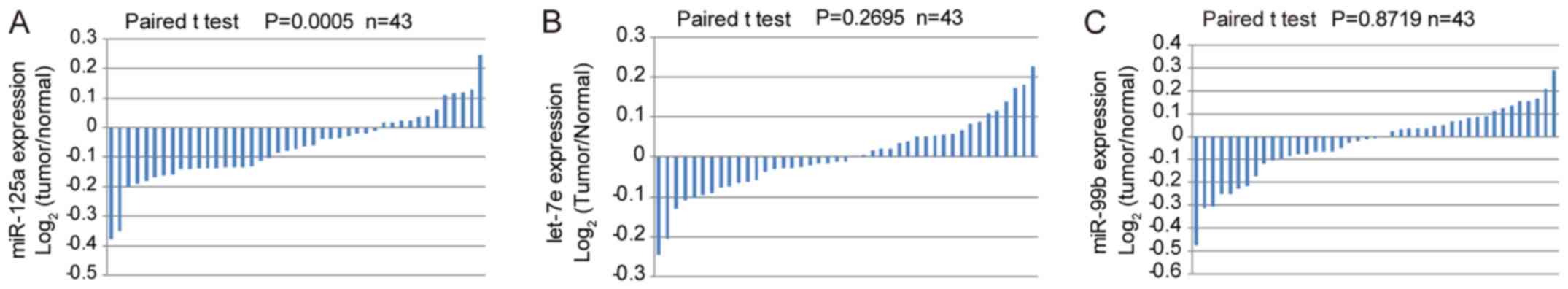
miR-125a/let-7e/miR-99b expression levels were
analyzed in GC tissues based on data derived from the TCGA
database. Even though the three miRNAs derive from the same
precursor, only miR-125a exhibited low expression in GC tissues
compared with adjacent normal tissues (Fig. 1A). There was no significant
difference in the expression of let-7e or miR-99b between GC and
matched adjacent normal tissues (Fig. 1B
and C).
Low expression of miR-125a-5p is
associated with larger tumor diameter and poor survival of patients
with GC
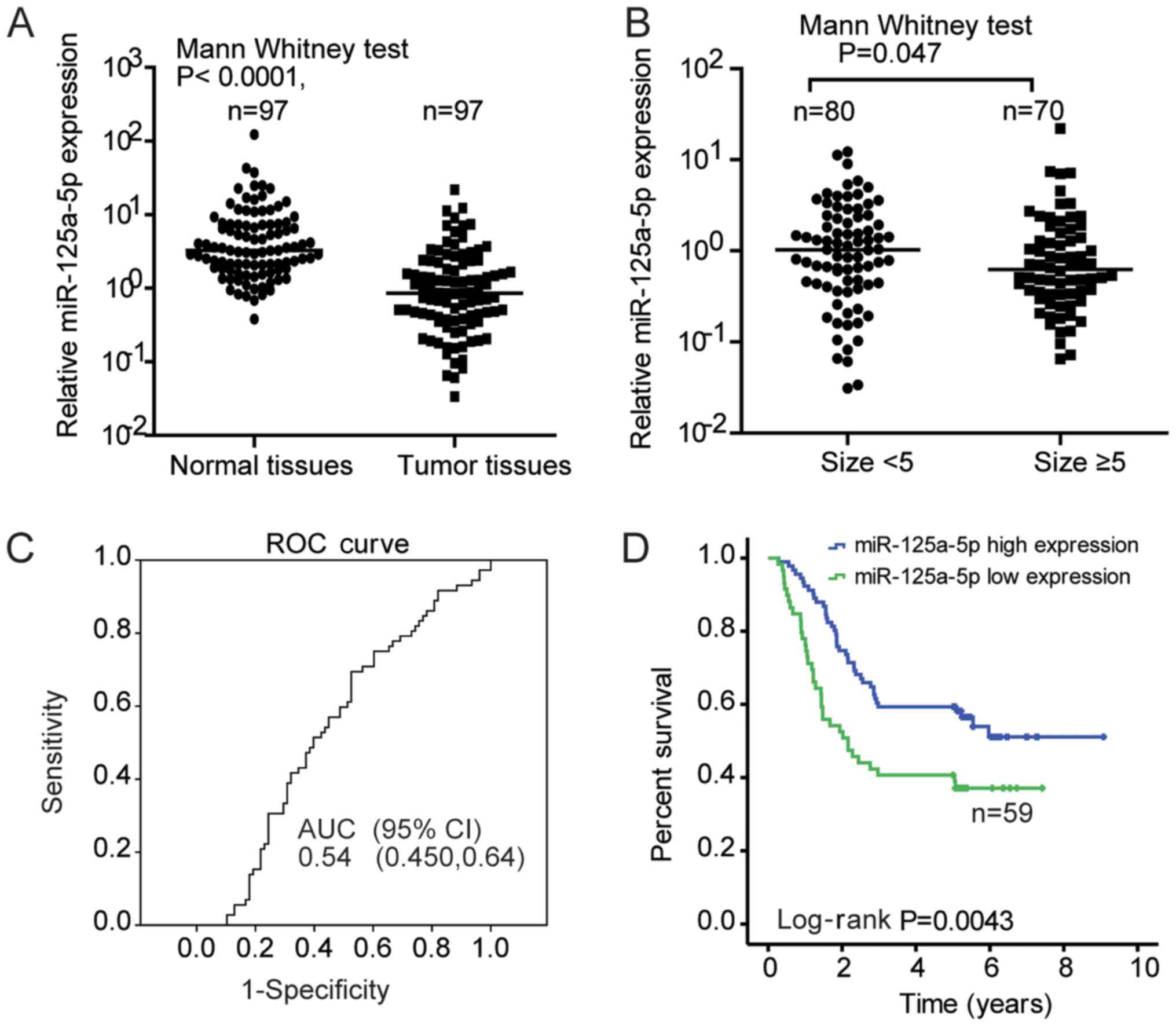
To further confirm the expression profile of
miR-125a-5p, RT-qPCR was performed using 97 pairs of matched tumor
and adjacent normal tissues. The results indicated significantly
decreased expression levels of miR-125a-5p in GC tumor tissues
compared with adjacent normal tissues (Mann Whitney U test;
P<0.001; Fig. 2A). The expression
level of miR-125a-5p was subsequently detected in 150 cases of
patients with GC, and the association between miR-125a-5p
expression and the clinicopathological parameters of patients with
GC was evaluated. The average expression level of miR-125a-5p was
decreased when the tumor diameter was ≥5 cm, compared with tumors
of <5 cm diameter (1.491±2.929 vs. 1.778±2.278; P=0.0474;
Fig. 2B). According to the cut-off
value of the ROC curve corresponding to the maximum Youden index
(Fig. 2C), patients were divided
into high or low miR-125a expression groups. As shown in Table I, the number of patients with low
miR-125a-5p expression was higher in if tumor size is ≥5 cm and
Ki67 expression is high, indicating that low miR-125a-5p expression
may be associated with tumor growth. However, there was no
association between miR-125a-5p expression and other
clinicopathological features, including sex, differentiation, TNM
stage, lymph node metastasis, tumor embolus and distant metastasis
(Table I). Nevertheless, the number
of patients with low miR-125a-5p expression was higher in patients
≥60 years of age (Table I). The
association between miR-125a-5p expression and aging requires
further investigation.
 | Table I.Association between miR-125a-5p
expression levels and clinical characteristics. |
Table I.
Association between miR-125a-5p
expression levels and clinical characteristics.
|
| microRNA-125a-5p |
|
|---|
| Characteristic | Low (%) | High
(%) | P-value |
|---|
| Sex |
|
| 0.855 |
|
Male | 55 (50.46) | 54 (49.54) |
|
|
Female | 20 (48.72) | 21 (51.22) |
|
| Age (years) |
|
| 0.003 |
|
<60 | 30 (38.46) | 48 (61.54) |
|
|
≥60 | 45 (62.50) | 27 (37.50) |
|
|
Differentiation |
|
| 0.497 |
| Poor,
undifferentiated | 62 (48.82) | 65 (51.18) |
|
| Well,
moderate | 13 (56.52) | 10 (43.48) |
|
| Tumor size
(cm) |
|
| 0.021 |
|
<5 | 34 (42.50) | 46 (57.50) |
|
| ≥5 | 43 (61.43) | 27 (38.57) |
|
| TNM stage |
|
| 0.249 |
| I,
II | 39 (45.88) | 46 (54.12) |
|
| III,
IV | 36 (55.38) | 29 (44.62) |
|
| Lymph node
metastasis |
|
| 0.402 |
|
Negative (N0) | 16 (57.14) | 12 (42.86) |
|
|
Positive (N1, N2, N3) | 59 (48.36) | 63 (51.64) |
|
| Tumor embolus |
|
| 0.570 |
|
Negative | 32 (52.46) | 29 (47.54) |
|
|
Positive | 42 (47.73) | 46 (52.27) |
|
| Metastasis |
|
| 0.206 |
|
Negative | 50 (46.73) | 57 (53.27) |
|
|
Positive | 25 (58.14) | 18 (41.86) |
|
| Ki67 |
|
| 0.032 |
|
Negative | 0
(0.00) | 2
(100) |
|
|
<25% | 3
(23.08) | 10 (76.92) |
|
|
25–50% | 9
(40.91) | 13 (59.09) |
|
|
>50% | 18 (66.67) | 9
(33.33) |
|
Furthermore, the association between the expression
level of miR-125a-5p and patient OS was assessed. Kaplan-Meier
survival analysis revealed that patients with low miR-125a-5p
expression exhibited a significantly shorter OS compared with those
with high miR-125a-5p expression (Fig.
2D). Similarly, from the univariate analysis results of Cox
proportional hazard model analysis, the higher tumor size, advanced
TNM stage, and low miR-125a-5p expression are prognostic factors
for survival (Table II). While
using multivariate Cox's proportional hazard model analysis, it was
identified that advanced TNM stage and low miR-125a-5p expression
were independent risk factors for survival (Table II). Univariate Cox analysis also
indicated an association between metastasis and survival for
patients with GC (Table II;
P<0.001). Considering that TNM contains metastasis as a
component in this staging system, it was excluded in the Cox
multivariate regression analysis. Patients with low miR-125a-5p
expression exhibited a 2-fold increased risk of fatal outcome
compared with those with high miR-125a-5p expression. Collectively,
the data suggested that miR-125a-5p may be a potential prognostic
biomarker for patients with GC.
| Table II.Univariate and multivariate Cox
regression analysis of risk factors for patients with gastric
carcinoma. |
Table II.
Univariate and multivariate Cox
regression analysis of risk factors for patients with gastric
carcinoma.
|
| Univariate Cox
analysis | Multivariate Cox
analysis |
|---|
|
|
|
|
|---|
| Variable | HR (95% CI) | P-value | HR (95% CI) | P-value |
|---|
| Sex (female vs.
male) | 0.920
(0.553–1.530) | 0.748 |
|
|
| Age (≥60 vs. <60
years) | 1.184
(0.760–1.847) | 0.455 |
|
|
| Differentiation
(poor vs. well) | 1.102
(0.607–2.000) | 0.749 |
|
|
| Ki67 (high vs.
low) | 1.502
(0.190–11.892) | 0.610 |
|
|
| Metastasis (yes vs.
no) | 3.200
(2.040–5.021) | <0.001 |
|
|
| Tumor size (≥5 vs.
<5 cm) | 1.704
(1.090–2.660) | 0.019 | 1.286
(0.808–1.237) | 0.288 |
| TNM stage (III and
IV vs. I and II) | 2.064
(1.655–4.098) | <0.001 | 2.550
(1.595–4.077) | <0.001 |
| microRNA-125a-5p
(high vs. low) | 0.528
(0.338–0.825) | 0.005 | 0.497
(0.314–0.789) | 0.003 |
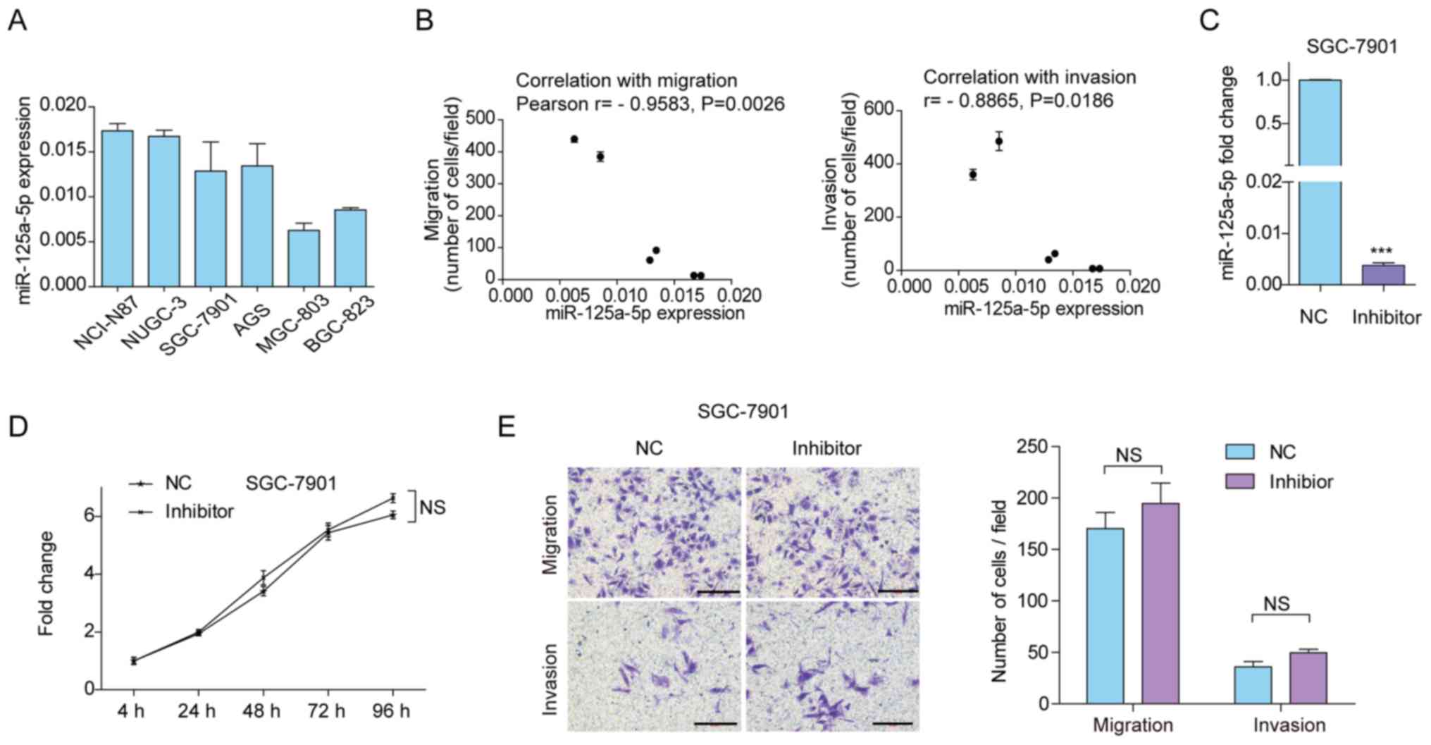
miR-125a-5p expression levels in GC
cell lines
In order to determine suitable cell lines to perform
functional assays to assess the effects of miR-125a-5p
overexpression and inhibition, its expression was evaluated in six
GC cell lines. Four GC cell lines, including NCI-N87, NUGC-3,
SGC-7901 and AGS, had relatively high expression, while MGC-803 and
BGC-823 had relatively low expression (Fig. 3A). Therefore, SGC-7901 cells were
selected to assess the effects of miR-125a-5p silencing, and
MGC-803 cells were selected to assess the effects of miR-125a-5p
overexpression. Previously, we evaluated the migration and invasion
ability of these gastric cancer cells by Transwell assays (20). The correlation between miR-125a-5p
expression levels and migration or invasion ability was
subsequently analyzed in GC cells. As indicated in Fig. 3B, the expression level of miR-125a-5p
was negatively correlated with cell migration (r=−0.9583, P=0.0026)
and invasion (r=−0.8865; P=0.0186) ability.
Inhibition of miR-125a-5p fails to
enhance the viability, migration and invasion of GC cells in
vitro
As it was hypothesized that miR-125a-5p may act as a
tumor suppressor in GC, the phenotypic changes were evaluated
following the downregulation of miR-125a-5p by miRNA inhibitors in
SGC-7901 cells with a relatively higher endogenous expression
(Fig. 3A). RT-qPCR analysis
demonstrated 99.6% transfection efficiency in response to treatment
using miRNA inhibitors (Fig. 3C).
However, there was no significant effect on cell viability in
response to treatment using miR-125a-5p inhibitors as assessed
using CCK8 assay (Fig. 3D). The
results of the Transwell assay demonstrated no significant changes
in the migratory or invasive ability of SGC-7901 cells transfected
with miR-125a-5p inhibitors and NC (Fig.
3E). These results suggest that miR-125a-5p silencing has no
effect on SGC-7901 cell proliferation, migration and invasion in
vitro.
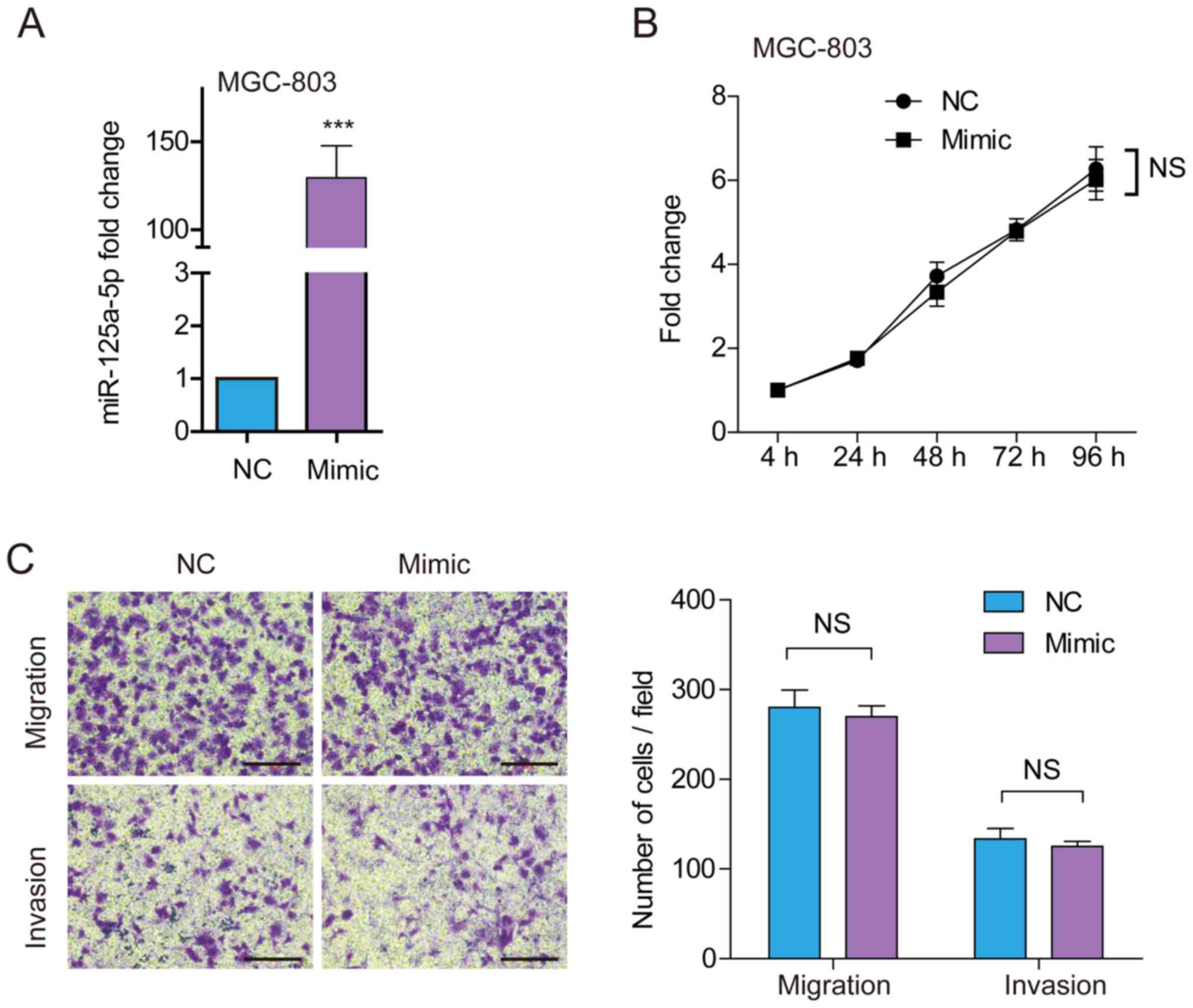
Ectopic overexpression of miR-125a-5p
fails to inhibit cell viability, migration and invasion in
vitro
To further confirm the aforementioned results on
viability and the migratory and invasive ability of GC cells,
MGC-803 cells, which showed relatively lower endogenous miR-125a-5p
expression among the six GC cell lines, were treated with
miR-125a-5p mimics. RT-qPCR analysis demonstrated that miR-125a-5p
expression was increased 129.4-fold in cells transfected with
miR-125a-5p mimics (Fig. 4A).
Consistently with the aforementioned results, there was no
significant difference in cell viability (Fig. 4B), cell migration or invasion
(Fig. 4C) in MGC-803 cells
transfected with miR-125a mimics and NC. Therefore, miR-125a-5p
overexpression has no effect on MGC-803 cell proliferation,
migration and invasion in vitro.
Discussion
The hsa-miR-125a/let-7e/miR-99b cluster is located
within the 19q13.2–19q13.4 region (21). Further understanding of
hsa-miR-125a/let-7e/miR-99b cluster biogenesis and function is
required. In physiological conditions, high levels of all cluster
members or miR-125a-5p are essential in maintaining the phenotype
of mouse hematopoietic stem/progenitor cells (22). Furthermore, a single cluster member,
miR-125a is sufficient to induce the amplification of hematopoietic
stem cells, suggesting that miR-125a/let-7e/miR-99b cluster members
differ in their expression patterns and functions, although they
are derived from a common RNA precursor (23). Increased expression of these miRNAs
is essential for osteoclast differentiation from primary human
monocytes (24). These studies
suggest the important role of miR-125a/let-7e/miR-99b cluster
members in normal cell differentiation.
The miR-125a/let-7e/miR-99b cluster or its members
may be involved in human pathological processes.
miR-99b/let-7e/miR-125a cluster may promote metastasis in
esophageal squamous cell carcinoma (ESCC) (25). Overexpression of this miRNA cluster
was demonstrated to induce ESCC cell migration and invasion in
vitro, and experimental metastasis in vivo (25). The miR-125a/let-7e/miR-99b cluster
members were also shown to be upregulated in synovial sarcomas,
however only two cluster members, let-7e and miR-99b, promoted
cancer cell proliferation (26).
Similarly, two cluster members, miR-99b and miR-125a, were
previously implicated in the pathogenesis of cystic fibrosis
(21). Decreased expression level of
miR-125a, but not of miR-99b or let-7e, was identified in
hepatocellular carcinoma biopsies compared with normal liver
tissues, and predicted poor prognosis of patients with
hepatocellular carcinoma (27).
miR-125a-5p has been reported to be a potential tumor suppressor in
lung carcinoma cells (28),
retinoblastoma (29) and cervical
carcinoma (30). All of these
studies support that these miRNAs may have diverse functions in
disease. In accordance with the aforementioned studies, the results
of the present study demonstrated that the expression of
miR-125a-5p was downregulated in GC tissues compared with adjacent
normal tissues.
Consistent with previous research (31–34), the
results of the present study identified decreased expression of
miR-125a-5p in GC tissues compared with adjacent normal tissues,
and decreased miR-125a-5p expression predicted poor OS in patients
with GC. From the univariate analysis results of Cox proportional
hazard model analysis, higher tumor size, advanced TNM stage and
low miR-125a-5p expression were associated with reduced survival of
patients with GC. Furthermore, advanced TNM stage and low
miR-125a-5p expression were found to be independent prognostic
factors associated with survival, as assessed using multivariate
Cox's analysis, which is in accordance with a previous study
(30).
Decreased expression levels of miR-125a-5p were
reported to be associated with enhanced malignant potential,
including tumor size, tumor invasion, liver metastasis and poor
prognosis (32). In the present
study, the association between low expression of miR-125a-5p and
tumor size was established, however miR-125a-5p was not associated
with distant metastasis. Differences in sample size or distribution
may contribute to the outcomes of the study. Consistent with a
previous study (27), the results of
the present study demonstrated that low expression level of
miR-125a-5p was associated with high Ki67 expression, a cellular
marker for proliferation in GC; however, evaluation of Ki67
expression in cells transfected with miR125a-5p mimics or
inhibitors remains to be investigated.
However, in vitro experiments demonstrated
that miR-125a-5p mimics and inhibitors had no effect on GC cell
proliferation, migration and invasion. These results differ from a
previous report (34), where
proliferation, migration and invasion of AGS and MGC-803 cells
treated with pre-miR-125a were evaluated. Since miR-125a-3p, a
known tumor suppressor in GC (35),
derives from the 3′-arm of the same precursor (pre-miR-125a), these
inhibitory effects on GC cell proliferation, migration and invasion
may be mediated by miR-125a-3p, miR-125a-5p or even both. In the
present study, MGC-803 cells were transfected with miR-125a-5p
mimics (specifically altering miR-125a-5p expression) and SGC-7901
cells were transfected with miR-125a-5p inhibitors. However, there
were no significant changes in cell proliferation, migration and
invasion. Previously, it had been reported that low expression of
miR-125a-5p was able to promote the paracrine vascular endothelial
growth factor A signaling pathway in GC, while the latter increased
Akt phosphorylation in endothelial cells and, thereby, promoted
tumor angiogenesis (36). Therefore,
even though there were no changes in GC cell proliferation,
migratory and invasive ability in vitro, it is possible that
miR-125a-5p functions as a tumor suppressor in vivo by
regulating the tumor microenvironment, including effects on tumor
angiogenesis. It is still possible that miR-125a-5p may regulate
stemness (37), a critical feature
of tumor formation, just like the tumor size. Therefore, in
vivo experiments assessing the effect of miR-125a-5p on tumor
growth and metastasis are required. TargetScan indicates that there
are 3,991 candidate target genes for miR-125a-3p; however, which
ones are real targets of miR-125a-5p requires further investigation
using expression correlation and dual-luciferase reporter
assays.
The probe sequences of miR-125a were checked in
TGCA, which were matched to the chromosome 19 plus strand from
56888319-56888404. This indicates that the miR-125a expression data
obtained from TCGA includes both miR-125a-3p and miR-125a-5p
expression. Only the miR-125a-5p expression was analyzed in the
cohort in the present study. The expression of miR-125a-3p
expression in the GC tissues should be investigated in future
studies.
Taken together, the results of the present study
support that reduced miR-125a-5p expression is associated with
larger tumor size, high Ki67 expression and poor prognosis in
patients with GC. Therefore, miR-125a-5p may have prognostic value
as a tumor biomarker for patients with GC.
Acknowledgements
Not applicable.
Funding
This work was supported by the grants from ‘San
Ming’ Project of Shenzhen city, China (no. SZSM 201612051).
Availability of data and materials
The datasets used and/or analyzed during the current
study are available from the corresponding author on reasonable
request.
Authors' contributions
GL performed most experiments, including the RNA
extraction, quantitative PCR analysis and cell transfection. SA and
JH participated in the in vitro study and acquired cell
viability data. GQL designed the experiments, coordinated the
project and wrote the manuscript. All authors have read and
approved the final version of this manuscript.
Ethics approval and consent to
participate
Written informed consents were obtained from all
patients participated in the present study and this work was
approved by the Peking University Cancer Hospital and Institute
Ethical Committee (approval no. 2017KT79).
Patient consent for publication
Written informed consent for publication was
obtained from all patients who participated in the present
study.
Competing interests
The authors declare that they have no competing
interests.
References
|
1
|
Torre LA, Bray F, Siegel RL, Ferlay J,
Lortet-Tieulent J and Jemal A: Global cancer statistics, 2012. CA
Cancer J Clin. 65:87–108. 2015. View Article : Google Scholar : PubMed/NCBI
|
|
2
|
Chen W, Zheng R, Baade PD, Zhang S, Zeng
H, Bray F, Jemal A, Yu XQ and He J: Cancer statistics in China,
2015. CA Cancer J Clin. 66:115–132. 2016. View Article : Google Scholar : PubMed/NCBI
|
|
3
|
Zong L, Abe M, Seto Y and Ji J: The
challenge of screening for early gastric cancer in China. Lancet.
388:26062016. View Article : Google Scholar : PubMed/NCBI
|
|
4
|
Svoronos AA, Engelman DM and Slack FJ:
OncomiR or Tumor suppressor? The duplicity of microRNAs in cancer.
Cancer Res. 76:3666–3670. 2016. View Article : Google Scholar : PubMed/NCBI
|
|
5
|
Tsai MM, Wang CS, Tsai CY, Huang HW, Chi
HC, Lin YH, Lu PH and Lin KH: Potential diagnostic, prognostic and
therapeutic targets of MicroRNAs in human gastric cancer. Int J Mol
Sci. 17(pii): E9452016. View Article : Google Scholar : PubMed/NCBI
|
|
6
|
da Silva Oliveira KC, Thomaz Araujo TM,
Albuquerque CI, Barata GA, Gigek CO, Leal MF, Wisnieski F,
Rodrigues Mello Junior FA, Khayat AS, de Assumpção PP, et al: Role
of miRNAs and their potential to be useful as diagnostic and
prognostic biomarkers in gastric cancer. World J Gastroenterol.
22:7951–7962. 2016. View Article : Google Scholar : PubMed/NCBI
|
|
7
|
Ishiguro H, Kimura M and Takeyama H: Role
of microRNAs in gastric cancer. World J Gastroenterol.
20:5694–5699. 2014. View Article : Google Scholar : PubMed/NCBI
|
|
8
|
Riquelme I, Letelier P, Riffo-Campos AL,
Brebi P and Roa JC: Emerging role of miRNAs in the drug resistance
of gastric cancer. Int J Mol Sci. 17:4242016. View Article : Google Scholar : PubMed/NCBI
|
|
9
|
Wu HH, Lin WC and Tsai KW: Advances in
molecular biomarkers for gastric cancer: miRNAs as emerging novel
cancer markers. Expert Rev Mol Med. 16:e12014. View Article : Google Scholar : PubMed/NCBI
|
|
10
|
Shin VY, Ng EK, Chan VW, Kwong A and Chu
KM: A three-miRNA signature as promising non-invasive diagnostic
marker for gastric cancer. Mol Cancer. 14:2022015. View Article : Google Scholar : PubMed/NCBI
|
|
11
|
Yu T, Wang LN, Li W, Zuo QF, Li MM, Zou QM
and Xiao B: Downregulation of miR-491-5p promotes gastric cancer
metastasis by regulating SNAIL and FGFR4. Cancer Sci.
109:1393–1403. 2018. View Article : Google Scholar : PubMed/NCBI
|
|
12
|
Zhang C, Zhang CD, Ma MH and Dai DQ:
Three-microRNA signature identified by bioinformatics analysis
predicts prognosis of gastric cancer patients. World J
Gastroenterol. 24:1206–1215. 2018. View Article : Google Scholar : PubMed/NCBI
|
|
13
|
Wang Z and Ji F: Downregulation of
microRNA-17-5p inhibits drug resistance of gastric cancer cells
partially through targeting p21. Oncol Lett. 15:4585–4591.
2018.PubMed/NCBI
|
|
14
|
Shi R and Chiang VL: Facile means for
quantifying microRNA expression by real-time PCR. Biotechniques.
39:519–525. 2005. View Article : Google Scholar : PubMed/NCBI
|
|
15
|
Wang S, Han H, Hu Y, Yang W, Lv Y, Wang L,
Zhang L and Ji J: MicroRNA-130a-3p suppresses cell migration and
invasion by inhibition of TBL1XR1-mediated EMT in human gastric
carcinoma. Mol Carcinog. 57:383–392. 2018. View Article : Google Scholar : PubMed/NCBI
|
|
16
|
Pfaffl MW: A new mathematical model for
relative quantification in real-time RT-PCR. Nucleic Acids Res.
29:e452001. View Article : Google Scholar : PubMed/NCBI
|
|
17
|
Livak KJ and Schmittgen TD: Analysis of
relative gene expression data using real-time quantitative PCR and
the 2(-Delta Delta C(T)) method. Methods. 25:402–408. 2001.
View Article : Google Scholar : PubMed/NCBI
|
|
18
|
Yang D, Li R, Wang H, Wang J, Li Y, Wang
H, Wang W and Liu Z: Clinical significance of tumor necrosis factor
receptor 2 in middle and lower thoracic esophageal squamous cell
carcinoma. Oncol Lett. 16:2971–2978. 2018.PubMed/NCBI
|
|
19
|
Yoon J, Chung YE, Lim JS and Kim MJ:
Quantitative assessment of mesorectal fat: New prognostic biomarker
in patients with mid-to-lower rectal cancer. Eur Radiol.
29:1240–1247. 2019. View Article : Google Scholar : PubMed/NCBI
|
|
20
|
Wang S, Han H, Hu Y, Yang W, Lv Y, Wang L,
Zhang L and Ji J: SLC3A2, antigen of mAb 3G9, promotes migration
and invasion by upregulating of mucins in gastric cancer.
Oncotarget. 8:88586–88598. 2017.PubMed/NCBI
|
|
21
|
Endale Ahanda ML, Bienvenu T,
Sermet-Gaudelus I, Mazzolini L, Edelman A, Zoorob R and Davezac N:
The hsa-miR-125a/hsa-let-7e/hsa-miR-99b cluster is potentially
implicated in Cystic Fibrosis pathogenesis. J Cyst Fibros.
14:571–579. 2015. View Article : Google Scholar : PubMed/NCBI
|
|
22
|
Gerrits A, Walasek MA, Olthof S, Weersing
E, Ritsema M, Zwart E, van Os R, Bystrykh LV and de Haan G: Genetic
screen identifies microRNA cluster 99b/let-7e/125a as a regulator
of primitive hematopoietic cells. Blood. 119:377–387. 2018.
View Article : Google Scholar
|
|
23
|
Guo S, Lu J, Schlanger R, Zhang H, Wang
JY, Fox MC, Purton LE, Fleming HH, Cobb B, Merkenschlager M, et al:
MicroRNA miR-125a controls hematopoietic stem cell number. Proc
Natl Acad Sci USA. 107:14229–14234. 2010. View Article : Google Scholar : PubMed/NCBI
|
|
24
|
de la Rica L, Garcia-Gomez A, Comet NR,
Rodríguez-Ubreva J, Ciudad L, Vento-Tormo R, Company C,
Álvarez-Errico D, García M, Gómez-Vaquero C and Ballestar E:
NF-κB-direct activation of microRNAs with repressive effects on
monocyte-specific genes is critical for osteoclast differentiation.
Genome Biol. 16:22015. View Article : Google Scholar : PubMed/NCBI
|
|
25
|
Ma J, Zhan Y, Xu Z, Li Y, Luo A, Ding F,
Cao X, Chen H and Liu Z: ZEB1 induced miR-99b/let-7e/miR-125a
cluster promotes invasion and metastasis in esophageal squamous
cell carcinoma. Cancer Lett. 398:37–45. 2017. View Article : Google Scholar : PubMed/NCBI
|
|
26
|
Hisaoka M, Matsuyama A, Nagao Y, Luan L,
Kuroda T, Akiyama H, Kondo S and Hashimoto H: Identification of
altered MicroRNA expression patterns in synovial sarcoma. Genes
Chromosomes Cancer. 50:137–145. 2011. View Article : Google Scholar : PubMed/NCBI
|
|
27
|
Lu G, Ma Y, Jia C, Yang H, Xie R, Luo P,
Chai L, Cai H, Cai M, Lv Z, et al: Reduced miR-125a levels
associated with poor survival of patients with hepatocellular
cancer. Oncol Lett. 14:5952–5958. 2017.PubMed/NCBI
|
|
28
|
Zhong L, Sun S, Shi J, Cao F, Han X and
Chen Z: MicroRNA-125a-5p plays a role as a tumor suppressor in lung
carcinoma cells by directly targeting STAT3. Tumour Biol.
39:10104283176975792017. View Article : Google Scholar : PubMed/NCBI
|
|
29
|
Zhang Y, Xue C, Zhu X, Zhu X, Xian H and
Huang Z: Suppression of microRNA-125a-5p upregulates the TAZ-EGFR
signaling pathway and promotes retinoblastoma proliferation. Cell
Signal. 28:850–860. 2016. View Article : Google Scholar : PubMed/NCBI
|
|
30
|
Qin X, Wan Y, Wang S and Xue M:
MicroRNA-125a-5p modulates human cervical carcinoma proliferation
and migration by targeting ABL2. Drug Des Devel Ther. 10:71–79.
2015.PubMed/NCBI
|
|
31
|
Cao Y, Tan S, Tu Y, Zhang G, Liu Y, Li D,
Xu S, Le Z, Xiong J, Zou W, et al: MicroRNA-125a-5p inhibits
invasion and metastasis of gastric cancer cells by targeting BRMS1
expression. Oncol Lett. 15:5119–5130. 2018.PubMed/NCBI
|
|
32
|
Nishida N, Mimori K, Fabbri M, Yokobori T,
Sudo T, Tanaka F, Shibata K, Ishii H, Doki Y and Mori M:
MicroRNA-125a-5p is an independent prognostic factor in gastric
cancer and inhibits the proliferation of human gastric cancer cells
in combination with trastuzumab. Clin Cancer Res. 17:2725–2733.
2011. View Article : Google Scholar : PubMed/NCBI
|
|
33
|
Fassan M, Pizzi M, Realdon S, Balistreri
M, Guzzardo V, Zagonel V, Castoro C, Mastracci L, Farinati F, Nitti
D, et al: The HER2-miR125a5p/miR125b loop in gastric and esophageal
carcinogenesis. Hum Pathol. 44:1804–1810. 2013. View Article : Google Scholar : PubMed/NCBI
|
|
34
|
Xu Y, Huang Z and Liu Y: Reduced
miR-125a-5p expression is associated with gastric carcinogenesis
through the targeting of E2F3. Mol Med Rep. 10:2601–2608. 2014.
View Article : Google Scholar : PubMed/NCBI
|
|
35
|
Hashiguchi Y, Nishida N, Mimori K, Sudo T,
Tanaka F, Shibata K, Ishii H, Mochizuki H, Hase K, Doki Y and Mori
M: Down-regulation of miR-125a-3p in human gastric cancer and its
clinicopathological significance. Int J Oncol. 40:1477–1482.
2012.PubMed/NCBI
|
|
36
|
Dai J, Wang J, Yang L, Xiao Y and Ruan Q:
miR-125a regulates angiogenesis of gastric cancer by targeting
vascular endothelial growth factor A. Int J Oncol. 47:1801–1810.
2015. View Article : Google Scholar : PubMed/NCBI
|
|
37
|
Chen J, Ouyang H, An X and Liu S: miR-125a
is upregulated in cancer stem-like cells derived from TW01 and is
responsible for maintaining stemness by inhibiting p53. Oncol Lett.
17:87–94. 2019.PubMed/NCBI
|