Introduction
Pancreatic cancer is an aggressive disease with high
morbidity and mortality and an overall 5-year survival rate of
<5% (1,2). The typical prognosis for patients with
pancreatic cancer is poor due to rapid disease progression and a
lack of effective therapies for the disease in its later stages
(3,4). The majority of patients with pancreatic
cancer are not eligible for surgery due to diagnosis in the late
stage of disease, while the disease also has low sensitivity to
radiotherapy (5–7). In addition, chemoresistance is often
observed, which greatly reduces the efficacy of chemotherapy
(8). Gemcitabine (GEM), which is a
first-line drug for the treatment of pancreatic cancer, has been
reported to improve the therapeutic efficacy and patient quality of
life compared with traditional chemotherapeutic agents (9). However, with the emergence of GEM
resistance, the treatment efficacy of GEM in pancreatic cancer is
declining (10). Although the
mechanisms of GEM resistance in pancreatic cancer have been widely
explored, it still remains largely unclear. It is therefore of
great importance to construct GEM-resistant pancreatic cancer cell
lines in order to further explore the mechanisms of GEM-resistance
and develop an effective strategy to overcome drug resistance and
improve treatment efficacy.
The P53 gene is an important antioncogene (11). As a nuclear transcription factor, the
p53 protein can activate the expression of many target genes,
induce DNA damage, and subsequently lead to cell senescence and
death (12). A previous study has
shown that the p53 protein not only plays an important role in
tumorigenesis, but also participates in the generation of drug
resistance of many chemotherapeutic drugs, including GEM (13). Thus, the protein expression of p53 in
the GEM-resistant cells are line SW1990-GZ was compared to the
primary SW1990 cells in the present study.
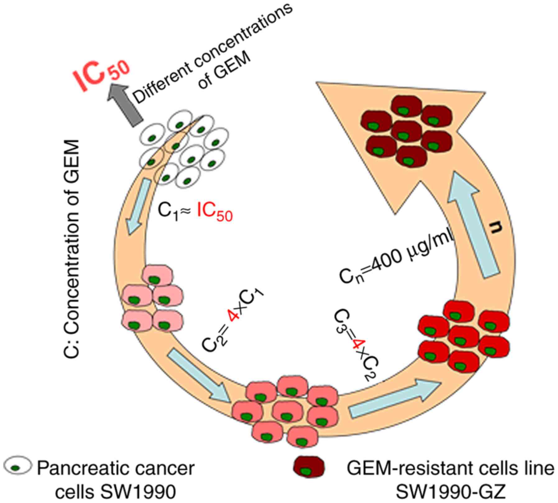
In the present study, the GEM-resistant pancreatic
cancer cell line SW1990-GZ was established by exposing parental
SW1990 cells, which cannot tolerate GEM, to increasing
concentrations of GEM. The GEM-resistant cell line SW1990-GZ was
established and the characteristics of SW1990 and SW1990-GZ cells,
including the protein expression of p53, were assessed and
compared.
Materials and methods
Materials
The human pancreatic cancer cell line SW1990 was
obtained from the Cell Bank of the Chinese Academy of Sciences
(Shanghai, China). MTT and dimethyl sulfoxide (DMSO) were purchased
from Sigma-Aldrich (Merck KGaA, Darmstadt, Germany). Also,
2,2-difluorodeoxycytidine (gemcitabine, GEM) was purchased from Eli
Lilly Company. Phosphate-buffered saline (PBS), fetal bovine serum
(FBS), and RPMI-1640 were obtained from Invitrogen (Thermo Fisher
Scientific, Inc., Waltham, MA, USA).
Establishment of the GEM-resistant
cell line SW1990-GZ
The GEM-resistant cell line SW1990-GZ was
established by exposing SW1990 cells to increasing concentrations
(0.01 µg/ml to 0.5 µg/ml) of GEM during the growth phase at 37°C
for 1 week. SW1990 cells were cultured in RPMI-1640 containing 10%
FBS and different concentrations of GEM. Cell apoptosis was
assessed using an MTT assay, and the median lethal dose of SW1990
cells was identified to be 0.07 µg/ml. SW1990 cells were the
cultured in the RPMI-1640 containing 0.1 µg/ml GEM at 37°C for 48
h, following which the culture and dead cells were replaced with
fresh drug-free medium. The remaining cells were cultured under the
aforementioned conditions until the logarithmic phase of cell
growth was reached. The cells were passaged twice and re-cultured
in medium containing GEM at 0.1 µg/ml until they stabilized. The
medium was subsequently replaced with culture medium containing 0.4
µg/ml GEM and cultured with a cycle progress as mentioned
previously, according to a four-fold increase in the drug
concentration. Finally, the cells were cultured in medium
containing 400 µg/ml GEM. The remaining viable cells were
determined to be stably resistant to high concentrations of GEM.
After 10 months, a stable gemcitabine-resistant cell line was
successfully acquired and designated as SW1990-GZ. The general
induction process is illustrated in Fig.
1.
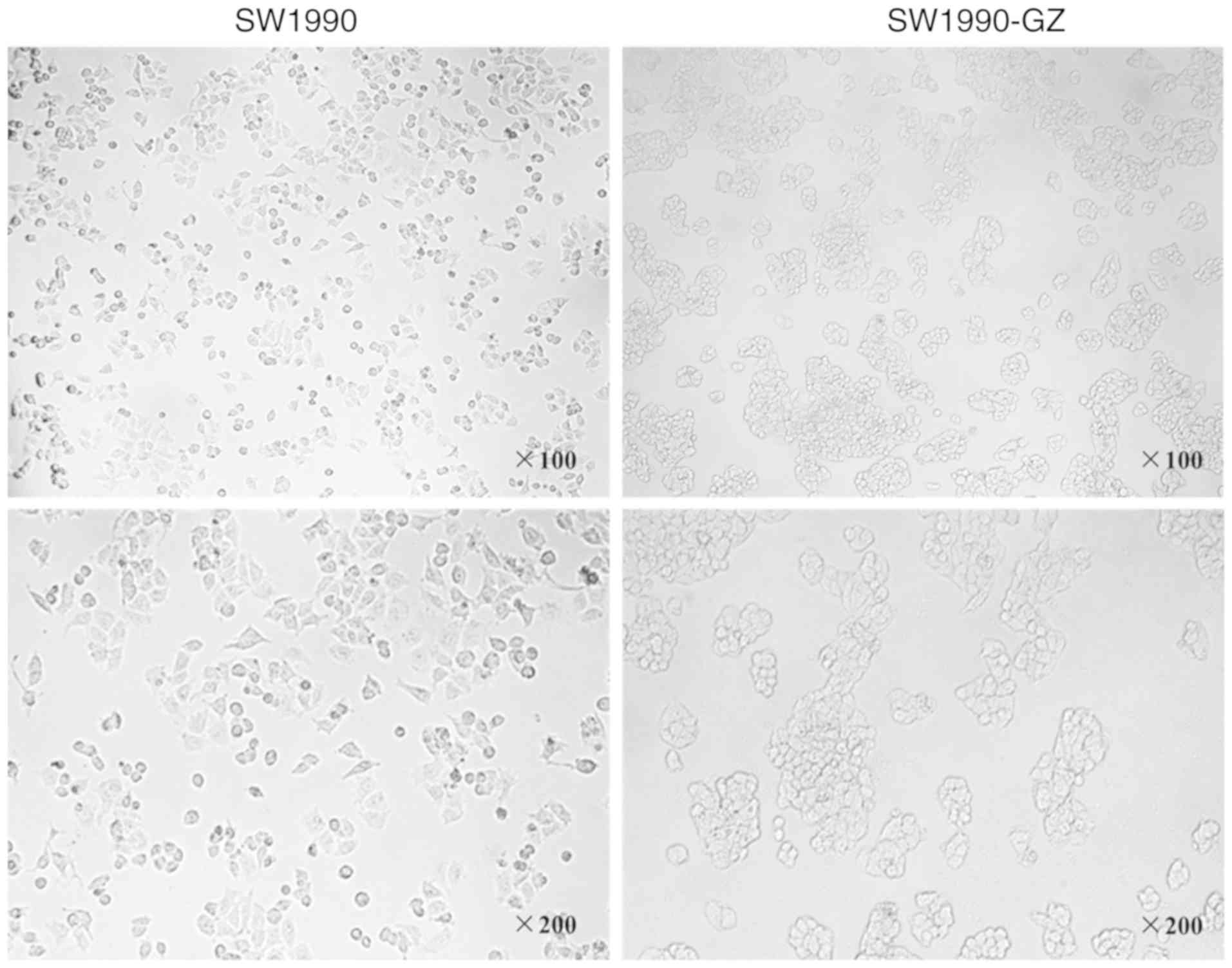
Morphologic appearance of SW1990 cells
and SW1990-GZ cells
SW1990 cells and SW1990-GZ cells were separately
cultured in 25 cm2 culture flasks in an atmosphere
containing 5% CO2, 37°C. When cells entered the
logarithmic phase, the morphologic appearance of SW1990 cells and
SW1990-GZ cells were visualized under an optical microscope (×100
magnification) and kept for further analysis.
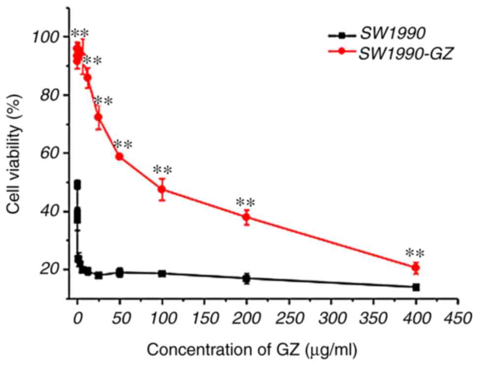
MTT assay to assess GEM sensitivity in
SW1990 and SW1990-GZ cells
To measure GEM sensitivity, SW1990 and SW1990-GZ
cells were seeded in 96-well plates (5.0×10³ cells/well)
and incubated at 37°C for 24 h. The medium was then replaced with
100 µl medium containing different concentrations of GEM (the
concentrations were 400, 200, 100, 50, 25, 12.5, 6.25, 3.125,
1.5625, and 0.7815 µg/ml) and cultured at 37°C for an additional 72
h. Subsequently, 20 µl per well of MTT solution (5 mg/ml;
Sigma-Aldrich; Merck KGaA) was added to the cells followed by
incubation at 37°C for 4 h. Then, 150 µl DMSO was substituted for
the supernatant, followed by oscillation for 10 min. Absorbance at
490 nm was detected using a microplate reader (Multiskan MK3;
Thermo Labsystems, Santa Rosa, CA, USA). Cell viability (%) =
(ODwith drug-ODwithout
drug)/(ODcontrol-ODwithout drug) × 100.
The resistance coefficient (R) of SW1990-GZ cells was calculated as
follows: R=IC50 [half maximal (50%) inhibitory
concentration]SW1990-GZ cells/IC50
SW1990 cells. Finally, dose-cell survival curves were
drawn. Each experiment was repeated at least 3 times.
Growth curves of SW1990 and SW1990-GZ
cell lines
To compare the viability of SW1990 and SW1990-GZ
cells, cells were seeded in 24-well plates (5.0×10³
cells/well) and incubated for 24 h at 37°C in an atmosphere
containing 5% CO2. Cells in three apertures were then
selected and the number of viable cells was calculated using an MTT
assay. This measurement was repeated each day until day 8 and the
growth curve was drawn.
Cellular uptake of GEM measured using
HPLC
To detect the cellular uptake of GEM, SW1990 and
SW1990-GZ cells were seeded in 24-well plates (1.0×10⁵
cells/well) and incubated for 24 h. The medium was then replaced
with 0.5 ml serum-free medium containing 200 µg/ml GEM and
incubated for 2 or 4 h. The cells were washed three times (5 min
each time) with PBS, lysed, dissolved in methanol and centrifuged
at 7,620 × g for 10 min at 4°C. Finally, the supernatant was
obtained and dried, following which the concentration of GEM in
SW1990 and SW1990-GZ cells was determined using a Waters HPLC
system (Waters Corporation, Milford, MA, USA) comprising a 1,525
binary pump, 2,487 UV/visible detector, 1,500 column heater and a
Symmetry C18 column. The UV/visible detector was set at 405 nm and
linked to Breeze 2 software (Shenzhen, China) for data analysis.
HPLC grade ammonium acetate buffer (0.05 M, pH 5.5) with methanol
at a ratio of 85:15 (v/v) was used as the mobile phase at 30°C with
a flow rate of 1.0 ml/min. Linear calibration curves for
concentrations in the range of 0.1–3.0 µg/ml were constructed by
linear regression analysis using the peak areas. The concentration
of GEM in the solution was calculated based the standard curve.
Western blot analysis
SW1990 and SW1990-GZ cells were homogenized in a
radio immunoprecipitation assay (RIPA) buffer (50 mM Tris-HCl, pH
7.4, 0.1% SDS, 1% NP-40, 0.25% sodium deoxycholate, 150 mM NaCl, 1
mM EDTA, 1 mM EGTA, and 1 mM Na3VO4). Prior
to homogenization, a protease inhibitor cocktail (Sigma-Aldrich;
Merck KGaA; cat. no. P2714) was added. The protein concentration
was measured using the Bradford assay. Proteins (50 µg) were
separated on 12% SDS-PAGE gels and were then transferred to PVDF
membranes (EMD Millipore, Billerica, MA, USA; cat. no. IPVH00010).
Polyvinylidene fluoride membranes were blocked in 5% bovine serum
albumin (Sigma-Aldrich; Merck KGaA; cat. no. A4737) for 1 h at room
temperature and probed with the primary mouse anti-human antibody
against p53 (1:1,000; cat. no. 2524; Cell Signaling Technology,
Inc., Danvers, MA, USA), and with rabbit anti-human antibody
against β-actin (1:1,000; cat. no. BS1002; Bioworld Technology,
Inc., St. Louis Park, MN, USA) as an internal control. These
samples were then followed by incubation with horseradish
peroxidase conjugated rabbit anti-mouse (1:10,000; cat. no.
TA130002; OriGene Technologies, Inc., Beijing, China) and goat
anti-rabbit (1:10,000; cat. no. TA130015; OriGene Technologies,
Inc., Beijing, China) at 37°C for 2 h. The results were visualized
using an ECL assay kit (Pierce; Thermo Fisher Scientific, Inc.).
The protein levels were analyzed using ImageJ software (v1.8.0;
National Institutes of Health, Bethesda, MD, USA) and normalized
relative to the internal control.
Statistical analysis
All the statistical analyses were performed using
SPSS, (version 12.0.1; SPSS Inc., Chicago, IL, USA). Quantitative
data are expressed as the mean ± standard deviation unless
otherwise stated. Statistical analyses of between-group effects on
the cytotoxic effects of GEM, the cellular uptake of GEM, and the
protein expression of p53 in SW1990-GZ and SW1990 cells were
performed using an unpaired Student's t-test. The growth of
SW1990-GZ and SW1990 cells were assessed using repeated measures
two-way analysis of variance (ANOVA) with group as between factor
and day as within factor followed by a least significant difference
(LSD). P<0.05 was considered to indicated a statistically
significant difference.
Results
Morphologic changes in SW1990-GZ
cells
SW1990 cells were exposed to increasing
concentrations of GEM for 10 months to establish a stable
gemcitabine-resistant cell line, SW1990-GZ. Compared with SW1990
cells, the gap junction was increased in SW1990-GZ cells, while
more granular substances were also observed (Fig. 2).
Cytotoxic effects of GEM
To examine the cytotoxicity of GEM, an MTT viability
assay was performed for SW1990 and SW1990-GZ cells following
treatment with the aforementioned concentrations of GEM. Cell
viability was decreased by treatment with GEM in a dose-dependent
manner. However, SW1990-GZ cells had a significantly lower
sensitivity to GEM compared with SW1990 cells (P<0.01). These
results suggest that SW1990-GZ cells showed obvious GEM resistance
(Fig. 3).
Growth of SW1990-GZ and SW1990
cells
As presented in Fig.
4, repeated measures ANOVA revealed that time (number of days
cultivated) and group (SW1990 vs. SW1990-GZ) affected the growth of
SW1990-GZ and SW1990 cells (time effect: F=12.00, P<0.01; group
effect: F=212.17, P<0.01; interaction effect: F=12.00,
P<0.01). The results of LSD analysis demonstrated that on days
7, 8 and 9, the growth rate of SW1990-GZ cells was low compared
with SW1990 cells.
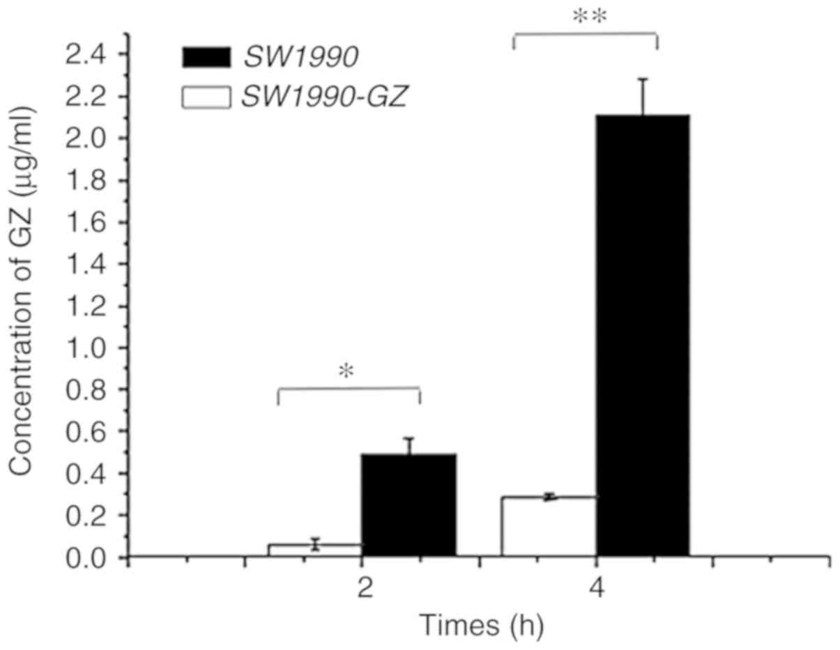
Cellular uptake of GEM in SW1990-GZ
and SW1990 cells
Following incubation in culture medium containing
200 µg/ml GEM for 2 or 4 h, the uptake efficiency of SW1990 and
SW1990-GZ cells was assessed using HPLC. The extraction efficiency
of SW1990-GZ and SW1990 cells was found to be 80.26 and 83.12%,
respectively. As illustrated in Fig.
5, following incubation with GEM for 2 or 4 h, the amount of
intracellular GEM was significantly lower in SW1990-GZ cells
compared with in SW1990 cells, suggesting that SW1990-GZ cells had
a reduced intake capacity compared with SW1990 cells (At 2 h,
t=7.047, P<0.05; at 4 h, t=14.81, P<0.01).
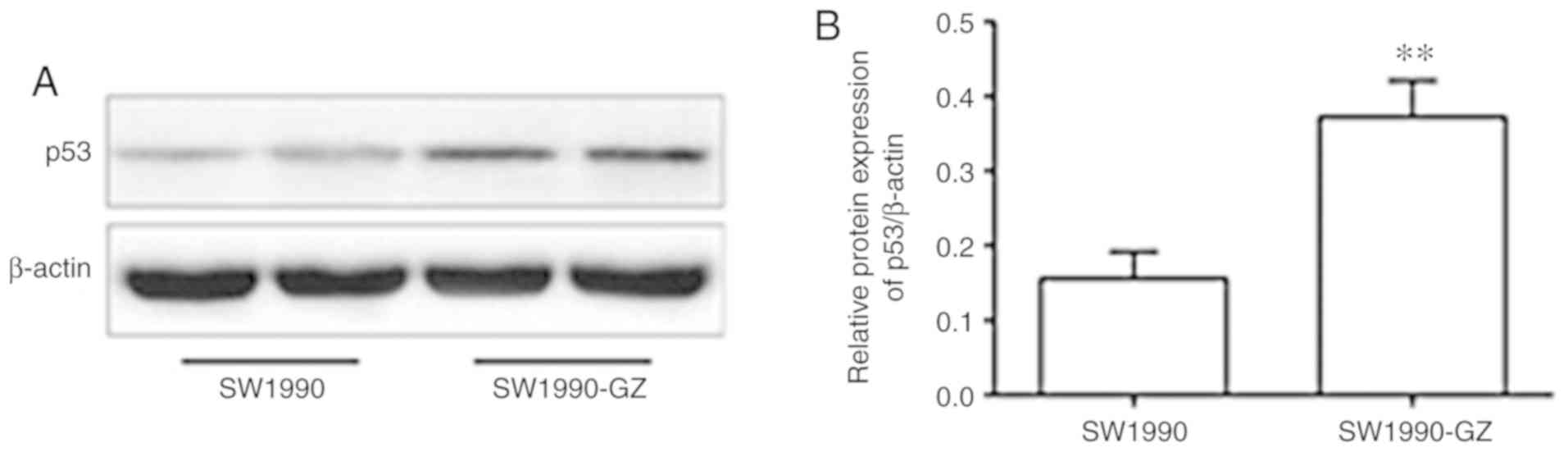
Protein expression of p53 in SW1990-GZ
and SW1990 cells
Compared with the SW1990 cells, the protein
expression of p53 was significantly increased in the GEM-resistant
SW1990-GZ cells (Fig. 6).
Discussion
There are a number of methods that may be used to
generate drug-resistant cancer cell lines, including gene
transfection and drug-induction (14–16). In
the present study, the GEM-resistant SW1990-GZ cell line was
established by exposing pancreatic SW1990 cells to gradually
increasing concentrations of GEM for 10 months. During the
induction process, the following problems should be addressed;
firstly, the IC50 of parental SW1990 cells should be
measured and a safe initial concentration of GEM should then be
selected. Secondly, prior to increasing the concentration of GEM,
the culture medium should be replaced with GEM-free medium to keep
cells in a stable growth condition. If the cells are cultured in
medium containing different concentrations of GEM, cell viability
may be damaged. Thirdly, the concentration of GEM should be
increased by an appropriate gradient. Concentration changes that
are too large or too small are not conducive to the formation of
stably resistant cells. Finally, during the induction process,
basic knowledge of cell culture techniques and technologies is
critical. The induction of GEM resistance must be performed with
great care in order to successfully establish a resistant cell
line.
In the present study, cell sensitivity to GEM and
GEM uptake capacity were assessed using MTT and HPLC, respectively.
The results indicated that the SW1990-GZ cells were stably
resistant to GEM. As such, SW1900-GZ cells may be an ideal cell
model for studies aiming to elucidate the mechanisms of
drug-resistance in pancreatic cancer. The gradual increase in GEM
concentration used in the present study is similar to that observed
in chemotherapy. As such, the results of the present study may have
clinical applications in cancer therapy.
In the present study, a growth curve was constructed
and it was determined that the growth rate of SW1990-GZ cells was
significantly lower compared with SW1990 cells. This may be
associated with the fact that GEM kills tumor cells primarily via
affecting DNA synthesis (17).
During the establishment of SW1990-GZ cells, GEM is used to treat
the growing cells intermittently, so that the cells in the
proliferative phase with active DNA synthesis are inhibited
earlier. Continuously carrying out such a screening, the pancreatic
cancer cells with poor growth and slow sensitivity to GEM are left
behind to form GEM resistant pancreatic cancer cell lines. It has
previously been demonstrated that tumor formation and growth are
maintained by a heterogeneous group of tumor cells and/or a subset
of tumor stem cells (5). One of the
important mechanisms contributing to the formation of
drug-resistant tumor cells is that stem cells are not killed, while
the majority of tumor cells are killed by chemotherapy drugs
(18,19). Another study revealed that cancer
stem cells grow slowly or stay dormant for long periods (20). In the present study, SW1990-GZ cell
growth was much slower compared with the parental SW1990 cell line,
which supports previous findings.
The results of the present study also revealed that
the GEM uptake capacity was significantly reduced in SW1990-GZ
cells compared with SW1990 cells. This suggests that GEM intake was
somehow limited in drug-resistant cells or that the drug was
rapidly removed from cells after uptake. This eventually leads to a
reduced intracellular GEM concentration, resulting in GEM-resistant
tumor cells. GEM is a pro-drug which required nucleotides to
introduce into cells and exerts a low toxic effect on the cell
(21–24). GEM intake decreases if the expression
of human equilibrative nucleoside transporter (hENT1) is
downregulated, which diminishes the cytotoxicity of GEM (25,26).
Previous studies have reported that patients with low hENT1
expression respond poorly to GEM (21,27,28).
Tanaka et al (21) analyzed
gene transporter polymorphisms in 149 cases of advanced local
pancreatic cancer, and the results demonstrated that the low
expression of hENT1 was associated with grade III and IV
neutropenia, chemoresistance to GEM (P=0.17) and a poor prognosis
(progression-free survival of 4.2 vs. 8.3 months). In the present
study, the GEM uptake ability of SW1990-GZ cells was significantly
lower than SW1990 cells (P<0.05). This may be a result of low
hENT1 expression, and so hENT1 may be an appropriate treatment
target for reducing GEM-resistance in pancreatic cancer. In
addition, several lines of evidence indicated that abnormal changes
in p53 protein levels are considered to be associated with
resistance to tumor cells (29). The
results demonstrated that the protein expression of p53 was
significantly increased in the GEM-resistant SW1990-GZ cells
compared with the SW1990 cells (Fig.
1), indicating that increased gemcitabine chemoresistance in
SW1990-GZ cells may due to the up-regulated protein expression of
p53, which needs to be confirmed in future studies.
Several studies have indicated that
gemcitabine-resistant pancreatic cancer cell strain SW1990-GZ,
induced by increasing drug dosage intermittently from SW1990, was
extremely stable (30,31). Consistently, the morphological
observation and drug resistance testing confirmed the stability of
SW1990-GZ cell in the present study. Taken together the above
results, we concluded that SW1990-GZ cell may be an ideal tool to
investigate the molecular basis of the serious GEM-resistant
phenotype of pancreatic cancer cells. However, this conclusion
should be verified by repeated experiments.
Several limitations in the present study should be
acknowledged. First, since the focus of the present study was on
how to establish drug-resistant strains, the mechanisms of drug
resistance have not been further examined. For example, due to the
fact that GEM introduces DNA damage in cells as detected by g-H2AX,
the amounts of g-H2AX between the parental SW1990 cells and
SW1990-GZ cells in response to GEM should be checked and compared
in subsequent experiments. Secondly, sequencing technology and
professional institutions to distinguish the difference between the
two cell lines should be conducted. Third, to confirm the
hypothesis that GEM resistance of SW1990-GZ cells may be due to
their lower uptake of GEM, the expression changes of the
transporter genes such as ABC transporters should be investigated.
Finally, the present study only carried out drug resistance
experiments on SW1990 cells, and not in other pancreatic cancer
cell lines.
In summary, the present study successfully
established the GEM-resistant SW1990-GZ cell line. By assessing
cytotoxicity, cell growth and drug-uptake capacity, the present
study confirmed that SW1990-GZ cells are stably resistant to GEM
and may be used to explore the possible mechanisms of GEM
resistance. Further studies may utilize SW1990-GZ cells to further
investigate the underlying mechanisms of chemoresistance and
develop novel therapeutic targets for pancreatic cancer.
Acknowledgements
Not applicable.
Funding
The present study was supported by the External
Science and Technology Cooperation Planning Projects of Anhui
Province of China (grant no. 1604b0602021).
Availability of data and materials
All data generated and analyzed during the present
study are included in this published article.
Authors' contributions
YY, FD and MG performed the experiments, contributed
to data analysis and wrote the manuscript. YY, FD, YFJ and LR
analyzed the data. YY conceptualized the study design and
contributed to experimental materials. All authors read and
approved the final manuscript.
Ethics approval and consent to
participate
All experiments performed on animals were approved
by the Animal Ethics Committee and complied with the Principles of
Laboratory Animal Use and Care of Animal Ethics Committee of Anhui
Medical University.
Patient consent for publication
Not applicable.
Competing interests
The authors declare that they have no competing
interests.
References
|
1
|
Siegel RL, Miller KD and Jemal A: Cancer
statistics, 2016. CA Cancer J Clin. 66:7–30. 2016. View Article : Google Scholar : PubMed/NCBI
|
|
2
|
Edwards BK, Noone AM, Mariotto AB, Simard
EP, Boscoe FP, Henley SJ, Jemal A, Cho H, Anderson RN, Kohler BA,
et al: Annual Report to the Nation on the status of cancer,
1975–2010, featuring prevalence of comorbidity and impact on
survival among persons with lung, colorectal, breast, or prostate
cancer. Cancer. 120:1290–1314. 2014. View Article : Google Scholar : PubMed/NCBI
|
|
3
|
Chen YW, Liu JY, Lin ST, Li JM, Huang SH,
Chen JY, Wu JY, Kuo CC, Wu CL, Lu YC, et al: Proteomic analysis of
gemcitabine-induced drug resistance in pancreatic cancer cells. Mol
Biosyst. 7:3065–3074. 2011. View Article : Google Scholar : PubMed/NCBI
|
|
4
|
Chen W, Zheng R, Baade PD, Zhang S, Zeng
H, Bray F, Jemal A, Yu XQ and He J: Cancer statistics in China,
2015. CA Cancer J Clin. 66:115–132. 2016. View Article : Google Scholar : PubMed/NCBI
|
|
5
|
D'Angelo F, Antolino L, Farcomeni A,
Sirimarco D, Kazemi Nava A, De Siena M, Petrucciani N, Nigri G,
Valabrega S, Aurello P and Ramacciato G: Neoadjuvant treatment in
pancreatic cancer: Evidence-based medicine? A systematic review and
meta-analysis. Med Oncol. 34:852017. View Article : Google Scholar : PubMed/NCBI
|
|
6
|
Rakhra S, Strauss JB, Robertson J, McGinn
CJ, Kim T, Huang J, Blake A, Helenowski I, Hayes JP, Mulcahy M and
Small W Jr: Hypofractionated conformal radiotherapy with concurrent
full-dose gemcitabine versus standard fractionation radiotherapy
with concurrent fluorouracil for unresectable pancreatic cancer: A
multi-institution experience. J Gastrointest Cancer. 47:196–201.
2016. View Article : Google Scholar : PubMed/NCBI
|
|
7
|
Żmijewska-Tomczak M, Milecki P, Olek-Hrab
K, Hojan K, Golusiński W, Rucińska A and Adamska A: Factors
influencing quality of life in patients during radiotherapy for
head and neck cancer. Arch Med Sci. 10:1153–1159. 2014. View Article : Google Scholar : PubMed/NCBI
|
|
8
|
Rajabpour A, Rajaei F and Teimoori-Toolabi
L: Molecular alterations contributing to pancreatic cancer
chemoresistance. Pancreatology. 17:310–320. 2017. View Article : Google Scholar : PubMed/NCBI
|
|
9
|
Otake A, Tsuji D, Taku K, Kawasaki Y,
Yokoi M, Nakamori H, Osada M, Matsumoto M, Inoue K, Hirai K and
Itoh K: Chemotherapy-induced neutropenia as a prognostic factor in
patients with metastatic pancreatic cancer treated with
gemcitabine. Eur J Clin Pharmacol. 73:1033–1039. 2017. View Article : Google Scholar : PubMed/NCBI
|
|
10
|
Long J, Zhang Y, Yu X, Yang J, LeBrun DG,
Chen C, Yao Q and Li M: Overcoming drug resistance in pancreatic
cancer. Expert Opin Ther Targets. 15:817–828. 2011. View Article : Google Scholar : PubMed/NCBI
|
|
11
|
Soussi T, Dehouche K and Béroud C: p53
website and analysis of p53 gene mutations in human cancer: Forging
a link between epidemiology and carcinogenesis. Hum Mutat.
15:105–113. 2000. View Article : Google Scholar : PubMed/NCBI
|
|
12
|
Coggi G, Bosari S, Roncalli M, Graziani D,
Bossi P, Viale G, Buffa R, Ferrero S, Piazza M, Blandamura S, et
al: p53 protein accumulation and p53 gene mutation in esophageal
carcinoma. A molecular and immunohistochemical study with
clinicopathologic correlations. Cancer. 79:425–432. 1997.
View Article : Google Scholar : PubMed/NCBI
|
|
13
|
Dhayat SA, Mardin WA, Seggewiß J, Ströse
AJ, Matuszcak C, Hummel R, Senninger N, Mees ST and Haier J:
MicroRNA profiling implies new markers of gemcitabine
chemoresistance in mutant p53 pancreatic ductal adenocarcinoma.
PLoS One. 10:e01437552015. View Article : Google Scholar : PubMed/NCBI
|
|
14
|
Mezencev R, Matyunina LV, Wagner GT and
McDonald JF: Acquired resistance of pancreatic cancer cells to
cisplatin is multifactorial with cell context-dependent involvement
of resistance genes. Cancer Gene Ther. 23:446–453. 2016. View Article : Google Scholar : PubMed/NCBI
|
|
15
|
Elaskalani O, Razak NB, Falasca M and
Metharom P: Epithelial-mesenchymal transition as a therapeutic
target for overcoming chemoresistance in pancreatic cancer. World J
Gastrointest Oncol. 9:37–41. 2017. View Article : Google Scholar : PubMed/NCBI
|
|
16
|
Azmi AS, Bao B and Sarkar FH: Exosomes in
cancer development, metastasis, and drug resistance: A
comprehensive review. Cancer Metastasis Rev. 32:623–642. 2013.
View Article : Google Scholar : PubMed/NCBI
|
|
17
|
Ando T, Ichikawa J, Okamoto A, Tasaka K,
Nakao A and Hamada Y: Gemcitabine inhibits viability, growth, and
metastasis of osteosarcoma cell lines. J Orthop Res. 23:964–969.
2005. View Article : Google Scholar : PubMed/NCBI
|
|
18
|
Hong SP, Wen J, Bang S, Park S and Song
SY: CD44-positive cells are responsible for gemcitabine resistance
in pancreatic cancer cells. Int J Cancer. 125:2323–2331. 2009.
View Article : Google Scholar : PubMed/NCBI
|
|
19
|
Qiu H, Fang X, Luo Q and Ouyang G: Cancer
stem cells: A potential target for cancer therapy. Cell Mol Life
Sci. 72:3411–3424. 2015. View Article : Google Scholar : PubMed/NCBI
|
|
20
|
Bao Q, Zhao Y, Renner A, Niess H, Seeliger
H, Jauch KW and Bruns CJ: Cancer stem cells in pancreatic cancer.
Cancers (Basel). 2:1629–1641. 2010. View Article : Google Scholar : PubMed/NCBI
|
|
21
|
Tanaka M, Javle M, Dong X, Eng C,
Abbruzzese JL and Li D: Gemcitabine metabolic and transporter gene
polymorphisms are associated with drug toxicity and efficacy in
patients with locally advanced pancreatic cancer. Cancer.
116:5325–5335. 2010. View Article : Google Scholar : PubMed/NCBI
|
|
22
|
Garcia-Manteiga J, Molina-Arcas M, Casado
FJ, Mazo A and Pastor-Anglada M: Nucleoside transporter profiles in
human pancreatic cancer cells: Role of hCNT1 in
2′,2′-difluorodeoxycytidine-induced cytotoxicity. Clin Cancer Res.
9:5000–5008. 2003.PubMed/NCBI
|
|
23
|
Maréchal R, Mackey JR, Lai R, Demetter P,
Peeters M, Polus M, Cass CE, Young J, Salmon I, Devière J and Van
Laethem JL: Human equilibrative nucleoside transporter 1 and human
concentrative nucleoside transporter 3 predict survival after
adjuvant gemcitabine therapy in resected pancreatic adenocarcinoma.
Clin Cancer Res. 15:2913–2919. 2009. View Article : Google Scholar : PubMed/NCBI
|
|
24
|
Nordh S, Ansari D and Andersson R: hENT1
expression is predictive of gemcitabine outcome in pancreatic
cancer: A systematic review. World J Gastroenterol. 20:8482–8490.
2014. View Article : Google Scholar : PubMed/NCBI
|
|
25
|
Hagmann W, Jesnowski R and Löhr JM:
Interdependence of gemcitabine treatment, transporter expression,
and resistance in human pancreatic carcinoma cells. Neoplasia.
12:740–747. 2010. View Article : Google Scholar : PubMed/NCBI
|
|
26
|
Hesler RA, Huang JJ, Starr MD, Treboschi
VM, Bernanke AG, Nixon AB, McCall SJ, White RR and Blobe GC:
TGF-β-induced stromal CYR61 promotes resistance to gemcitabine in
pancreatic ductal adenocarcinoma through downregulation of the
nucleoside transporters hENT1 and hCNT3. Carcinogenesis.
37:1041–1051. 2016. View Article : Google Scholar : PubMed/NCBI
|
|
27
|
Sheikh R, Walsh N, Clynes M, O'Connor R
and McDermott R: Challenges of drug resistance in the management of
pancreatic cancer. Expert Rev Anticancer Ther. 10:1647–1661. 2010.
View Article : Google Scholar : PubMed/NCBI
|
|
28
|
Maréchal R, Bachet JB, Mackey JR, Dalban
C, Demetter P, Graham K, Couvelard A, Svrcek M, Bardier-Dupas A,
Hammel P, et al: Levels of gemcitabine transport and metabolism
proteins predict survival times of patients treated with
gemcitabine for pancreatic adenocarcinoma. Gastroenterology.
143:664–674.e6. 2012. View Article : Google Scholar : PubMed/NCBI
|
|
29
|
Cascinu S, Graziano F, Del Ferro E,
Staccioli MP, Ligi M, Carnevali A, Muretto P and Catalano G:
Expression of p53 protein and resistance to preoperative
chemotherapy in locally advanced gastric carcinoma. Cancer.
83:1917–1922. 1998. View Article : Google Scholar : PubMed/NCBI
|
|
30
|
An Y, Yao J, Wei JS, Lu ZP, Cai HH, Dai
CC, Qian ZY, Xu ZK and Miao Y: Establish a gemcitabine-resistant
pancreatic cancer cell line SW1990/GZ and research the relationship
between SW1990/GZ and pancreatic cancer stem cell. Zhonghua Wai Ke
Za Zhi. 48:999–1003. 2010.(In Chinese). PubMed/NCBI
|
|
31
|
Niu BZ, Chen G, Li LJ, Wu YD and Zhao YP:
Drug resistance and activity changes of thioredoxin reductase in
pancreatic cancer cells strain SW1990 induced by gemcitabine.
Zhongguo Yi Xue Ke Xue Yuan Xue Bao. 27:606–610. 2005.(In Chinese).
PubMed/NCBI
|