Introduction
Lung cancer is the main cause of cancer-related
mortality among males in developed and less developed countries
(1) and is one of the malignant
tumors that pose a great threat to human health. Especially with
the increase of smoking, obesity and physical inactivity, the
incidence rate of some low molecular lung cancer is increasing, and
the incidence rate and mortality rate of lung cancer are on the
rise (2). The obscure symptoms in
its early stage are often misdiagnosed as other respiratory
diseases, resulting in a more serious condition when diagnosed.
Undifferentiated lung carcinoma, a common type of lung cancer with
poor differentiation of tumor cells, can be divided into small cell
undifferentiated carcinoma and large cell undifferentiated
carcinoma according to the histological morphology of tumor cells.
Small cell undifferentiated carcinoma (small cell cancer) is the
most malignant cancer, accounting for ~13–15% of the total lung
cancer (3), which mainly occurs in
the large bronchus near the segmental bronchus, with its cancer
cells having the characteristics of fast growth, rapid development,
strong invasiveness, and easy migration to the brain, liver,
adrenal gland, and bone. It is sensitive to radiotherapy and
chemotherapy, but with a poor prognosis (4). Whereas large cell undifferentiated
carcinoma (large cell cancer) has larger cancer cells, lower
incidence, higher malignant degree and poorer prognosis (5).
It has been found that thyroid transcription
factor-1 (TTF-1), as a member of NKx2 family of nucleoprotein
transcription factors in the same region, plays a crucial role in
lung development, cell growth and differentiation, and is one of
the suppressor genes of lung adenocarcinoma. As a highly sensitive
and specific molecular marker of lung adenocarcinoma, TTF-1 can
help to differentiate lung adenocarcinoma (6,7). Hara
et al (8) considered that
TTF-1 was a favorable prognostic factor for lung adenocarcinoma.
Matzke-Ogi et al (9) also
considered that TTF-1 was overexpressed in 95% of primary lung
adenocarcinoma, but TTF-1 was also a favorable prognostic factor
for non-phosphorous non-small cell lung cancer patients. Although
more attention has been paid to the study of TTF-1 in lung cancer
in recent years, there are few studies on the expression and
prognosis of TTF-1 in undifferentiated lung cancer. CD44, located
in the short arm of chromosome 11, is mainly involved in
heterotypic adhesion (10), that is,
the adhesion of tumor cells to host cells and host mechanisms, and
promotes the invasion and metastasis of tumor cells (11). CD44v6, a member of CD44 family having
a close relationship with tumor cell invasion and metastasis
(12), has been found to play an
important role in the occurrence, metastasis and prognosis of
various malignant tumors in recent years (13–15).
Matzke-Ogi et al (9) found
that elevated levels of CD44v6 in patients with metastatic
pancreatic tumors were associated with shorter survival time.
However, we still do not know the expression and prognosis of
CD44v6 and TTF-1 in undifferentiated lung cancer.
The present study explored the correlation between
expression and prognosis of TTF-1 and CD44v6 in undifferentiated
lung cancer to provide reference and direction for clinical
practice.
Patients and methods
General data
One hundred and sixteen patients with large cell
cancer admitted to Penglai Traditional Chinese Medicine Hospital
(Yantai, China), from June 2011 to February 2013 were collected as
group A, 120 cases with small cell cancer as group B, and 80 normal
individuals as group C. There were 66 males and 50 females in group
A aged from 44 to 75 (61.5±10.4) years, 71 males and 49 females in
group B aged from 43 to 78 years (62.1±10.6), 46 males and 34
females in group C aged from 45 to 78 years (61.8±10.6). This study
was approved by the Medical Ethics Committee of Penglai Traditional
Chinese Medicine Hospital and the patients were informed. Signed
informed consents were obtained from the patients or their
guardians. Inclusion criteria: i) The patients were pathologically
diagnosed with undifferentiated lung cancer; ii) no radiotherapy or
chemotherapy was performed before the surgery; iii) the clinical
data were complete and the patients could be followed up by
telephone. Exclusion criteria: i) Patients with suspicious
pathology and mixed pathology; ii) patients with serious heart and
lung function diseases and other diseases; iii) pregnant or
lactating women.
Reagents and instruments
CD44v6 protein ELISA test kit (Shanghai J&l
Biotechnology Co., Ltd., JL19068); TTF-1 ELISA test kit (Shanghai
LMAI Biology Co., Ltd., LM-TTF1-Hu).
Detection method
Five milliliters of sterile venous blood was
collected at 7 a.m. the day after admission, and serum was
immediately separated by centrifugation at 3,000 × g for 15 min at
4°C, and anticoagulated with ethylenediaminetetraacetic acid
(EDTA), and stored at −80°C, and cancer tissues and adjacent
tissues were collected from group A and group B. Concentration of
the standard: TTF-1 dilution concentration: 8, 4, 2, 1 and 0.5
ng/ml; CD44v6 dilution concentration: 80, 40, 20, 10 and 5 ng/ml.
Blank, standard and testing sample wells were set. Fifty
microliters of standard with different concentrations was added to
the standard wells, 50 µl of testing sample to the testing sample
wells, and 100 µl of enzyme labeled antibody to all the wells. Then
the reaction wells were sealed with a closure plate membrane,
incubated for 60 min at 37°C in a water bath or constant
temperature box. Next, the liquid was discarded and dried with
absorbent paper, and the washing buffer (350 µl) was added to each
well for 1 min, then discarded and dried with absorbent paper. The
plate was washed five times. Afterwards, 50 µl of each substrate A
and B was added to each well, and incubated at 37°C in the dark for
15 min. Fifty microliters of stop solution was added to each well,
then the optical density (OD) value was measured at 450 nm
wavelength within 15 min. A standard curve was drawn and a linear
regression equation was obtained, and the OD value of the sample
was substituted into the equation to calculate the concentration of
the sample.
Outcome measures
Main outcome measures: The expression of TTF-1 and
CD44v6 in the serum of groups A, B and C and the tissues of group A
and group B were compared. The patients were followed up by
telephone at 1, 3, 6, 12, 24, 36, 48 and 60 months after admission
to record their 5-year survival and to divide them into the
survival group and the deceased group. Then the expression of TTF-1
and CD44v6 in the two groups was compared and the mortality risk
factors were analyzed by multivariate logistic regression.
Secondary outcome measures: Receiver operating
characteristic (ROC) curve was used to analyze the diagnostic value
of TTF-1 and CD44v6 and the best cut-off value in undifferentiated
cancer mortality. The patients were divided into the high and low
expression groups according to the values in order to observe the
5-year mortality and the Kaplan-Meier (K-M) survival curve was
drawn.
Statistical analysis
SPSS 20.0 (Shanghai Cabit Information Technology
Co., Ltd.) medical statistical analysis software was used to carry
out statistical analysis on the collected data. GraphPad Prism 7
(Shenzhen Soft Head Technology Co., Ltd.) was used to draw figures.
The enumeration data expressed as a rate (%) were analyzed using
the Chi-squared test (denoted by χ2). The
Kolmogorov-Smirnov (K-S) test was used to analyze the data
distribution, and the measurement data were expressed as the mean ±
standard deviation (mean ± SD). The independent samples t-test was
used for comparison of the normal distribution data between two
groups (denoted by t), and one-way analysis of variance (ANOVA) was
used for the comparison between multiple groups (denoted by F).
LSD-t test was the post hoc test. Rank sum test was used for ranked
data. Pearsons test was used to analyze the correlation between
TTF-1 and CD44v6 expression in serum of group A and group B and
cancer tissues. ROC was used for evaluation of the ability of TTF-1
and CD44v6 to diagnose undifferentiated cancer and mortality; K-M
was used for 5-year survival analysis with log rank test, and
Logistic regression analysis for multivariate analysis. P<0.05
was regarded as statistically significant.
Results
Basic information and clinical data of
patients
Basic information and clinical data were compared in
the three groups. There were 116 patients in group A, 66 males and
50 females, with an average age of 61.5±10.4 years, an average
disease course of 3.52±1.64 years, an average body mass index (BMI)
of 21.15±3.18 kg/m2, 43 with smoking history, 25 with
history of alcohol abuse, 84 urban residents and 32 rural
residents. Whereas, there were 120 patients in group B, 71 males
and 49 females, with an average age of 62.1±10.6 years, an average
disease course of 3.21±1.34 years, an average BMI of 22.08±3.47
kg/m2, 48 with smoking history, 24 with history of
alcohol abuse, 87 urban residents and 33 rural residents. Group C
consisted of 80 individuals, 46 males and 34 females with an
average age of 61.8±10.6 years, an average BMI of 21.93±3.25
kg/m2, 27 with smoking history, 11 with history of
alcohol abuse, 60 urban residents and 20 rural residents. There was
no statistical difference in clinical data between the two groups
(P>0.05) (Table I).
 | Table I.Clinical data of patients. |
Table I.
Clinical data of patients.
| Factors | Group A (n=116) | Group B (n=120) | Group C (n=80) |
t/χ2/F/Z | P-value |
|---|
| Sex |
| Male | 66 (56.90) | 71 (59.17) | 46 (57.50) | 1.647 | 0.329 |
|
Female | 50 (43.10) | 49 (40.83) | 34 (42.50) |
|
|
| Age (years) | 61.5±10.4 | 62.1±10.6 | 62.8±10.6 | 0.363 | 0.696 |
| Course of disease
(years) | 3.52±1.64 | 3.21±1.34 |
| 1.545 | 0.124 |
| BMI
(kg/m2) | 21.15±3.18 | 22.08±3.47 | 21.93±3.25 | 2.585 | 0.077 |
| Smoking history |
| Yes | 43 (37.07) | 48 (40.00) | 27 (33.75) | 0.677 | 0.572 |
| No | 73 (62.93) | 72 (60.00) | 53 (66.25) |
|
|
| History of alcohol
abuse |
| Yes | 25 (21.55) | 24 (20.00) | 11 (13.75) | 0.113 | 0.897 |
| No | 91 (78.45) | 96 (80.00) | 69 (86.25) |
|
|
| Residence |
|
Urban | 84 (66.67) | 87 (71.43) | 60 (75.00) | 0.202 | 0.828 |
|
Rural | 32 (33.33) | 33 (28.57) | 20 (25.00) |
|
|
| TNM stage |
| I | 19 (16.38) | 21 (17.50) |
|
|
|
| II | 42 (25.86) | 44 (25.84) |
| 0.215 | 0.829 |
|
III | 50 (34.48) | 49 (33.33) |
|
|
|
| IV | 5 (4.32) | 6 (5.00) |
|
|
|
| Distal
metastasis |
|
Yes | 48 | 51 |
| 0.030 | 0.862 |
| No | 68 | 69 |
|
|
|
Expression of TTF-1 and CD44v6 in the
serum of groups A, B and C and the tissues of groups A and B
The results of enzyme linked immunosorbent assay
(ELISA) showed that the expression of CD44v6 in the serum of group
A and group B was higher than that in group C, and the expression
in group B was higher than that in group A (P<0.05). TTF-1
expression in group A and group B was higher than that in group C,
and the expression in group A was higher than that in group B
(P<0.05) (Table II). The
expression of TTF-1 and CD44v6 in group A and group B was
significantly higher than those in adjacent tissues (P<0.05)
(Tables III and IV). The expression of TTF-1 in group A was
significantly higher than that in group B (P<0.05), and the
expression of CD44v6 was significantly lower than that in group B
(P<0.05), as shown in Table
V.
| Table II.TTF-1 and CD44v6 expression in groups
A, B and C. |
Table II.
TTF-1 and CD44v6 expression in groups
A, B and C.
| Groups | Group A
(n=116) | Group B
(n=120) | Group C (n=80) | t value | P-value |
|---|
| TTF-1 (ng/ml) | 157.34±45.91 |
110.79±21.35a |
93.18±11.23a,b | 312.93 | <0.001 |
| CD44v6 (ng/ml) | 154.28±40.17 |
196.44±59.10a |
112.31±25.27a,b | 96.59 | <0.001 |
| Table III.Expression of TTF-1 and CD44v6 in
group A and adjacent tissues. |
Table III.
Expression of TTF-1 and CD44v6 in
group A and adjacent tissues.
| Group A | Cancer tissue
(n=116) | Paracancerous
tissue (n=116) | t value | P-value |
|---|
| TTF-1 (ng/ml) | 159.07±45.37 | 96.24±10.32 | 13.495 | <0.001 |
| CD44v6 (ng/ml) | 153.54±39.84 | 114.25±23.45 | 9.166 | <0.001 |
| Table IV.Expression of TTF-1 and CD44v6 in
group B and adjacent tissues. |
Table IV.
Expression of TTF-1 and CD44v6 in
group B and adjacent tissues.
| Group B | Cancer tissue
(n=120) | Paracancerous
tissue (n=120) | t value | P-value |
|---|
| TTF-1 (ng/ml) | 112.95±21.24 | 93.18±11.23 | 9.883 | <0.001 |
| CD44v6 (ng/ml) | 196.43±56.10 | 112.31±25.27 | 14.254 | <0.001 |
| Table V.Comparison of TTF-1 and CD44v6
expression in groups A and B cancer tissues. |
Table V.
Comparison of TTF-1 and CD44v6
expression in groups A and B cancer tissues.
| Cancer tissues | Group A
(n=116) | Group B
(n=120) | t value | P-value |
|---|
| TTF-1 (ng/ml) | 159.07±45.37 | 112.95±21.24 | 8.189 | <0.001 |
| CD44v6 (ng/ml) | 153.54±39.84 | 196.43±56.10 | 5.808 | <0.001 |
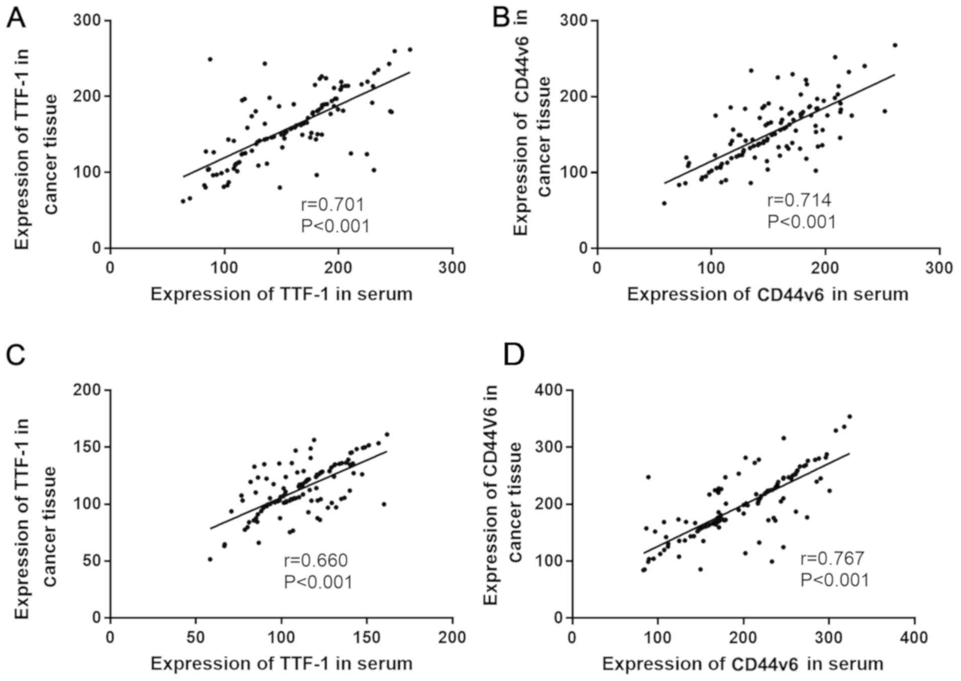
Correlation between the expression of
TTF-1 and CD44v6 in serum of groups A and B and cancer tissues
Pearsons correlation analysis showed that TTF-1
expression was positively correlated with TTF-1 expression in
cancer tissues (r=0.701, P<0.001), and CD44v6 expression in
serum was positively correlated with CD44v6 expression in cancer
tissues (r=0.714, P<0.001). The expression of TTF-1 in serum of
group B was positively correlated with the expression of TTF-1 in
cancer tissues (r=0.660, P<0.001). The expression of CD44v6 in
serum was positively correlated with the expression of CD44v6 in
cancer tissues (r=0.767, P<0.001) (Fig. 1).
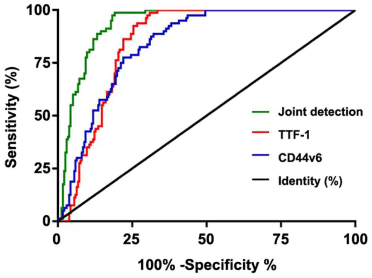
Diagnostic value of TTF-1 and CD44v6
in undifferentiated cancer
ROC curve of the expression of TTF-1 and CD44v6 in
the serum of three groups of patients was plotted in order to
analyze their diagnostic value in undifferentiated cancer. The
results showed that the area under curve (AUC) and 95% confidence
interval (CI) of TTF-1 were 0.852 and 0.812–0.893, respectively;
those of CD44v6 were 0.840 and 0.797–0.883; and those of joint
detection were 0.934 and 0.908–0.960, respectively (Table VI and Fig. 2).
| Table VI.ROC curve data. |
Table VI.
ROC curve data.
| Indexes | AUC | 95% CI | Specificity | Sensitivity | Youden index | Cut-off |
|---|
| TTF-1 | 0.852 | 0.812–0.893 | 68.64% | 98.75% | 67.39% | <112.981 |
| CD44v6 | 0.840 | 0.797–0.883 | 67.37% | 88.75% | 56.12% | <144.333 |
| Joint
detection | 0.934 | 0.908–0.960 | 81.70% | 96.25% | 79.55% | <0.228 |
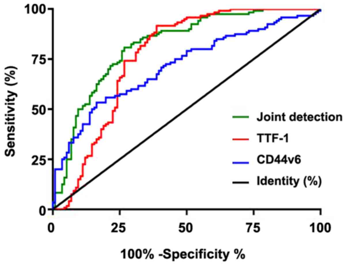
Diagnostic value of TTF-1 and CD44v6
in large and small cell cancers
ROC curve analysis of TTF-1 and CD44v6 expression in
group A and group B was plotted to analyze their diagnostic value
in large and small cell cancers. The results showed that AUC and
95% CI of TTF-1 were 0.766 and 0.702–0.831, respectively; those of
CD44v6 were 0.715 and 0.649–0.780, and those of joint detection
curve were 0.820 and 0.766–0.875, respectively (Table VII and Fig. 3).
| Table VII.ROC curve data. |
Table VII.
ROC curve data.
| Indexes | AUC | 95% CI | Specificity | Sensitivity | Youden index | Cut-off |
|---|
| TTF-1 | 0.766 | 0.702–0.831 | 60.34% | 91.67% | 52.01% | <147.148 |
| CD44v6 | 0.715 | 0.649–0.780 | 83.62% | 52.50% | 36.12% | <197.000 |
| Joint
detection | 0.820 | 0.766–0.875 | 73.28% | 80.00% | 53.28% | <0.502 |
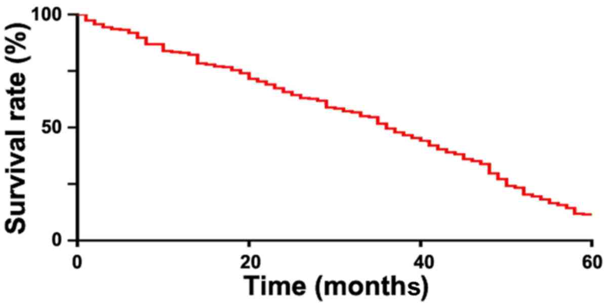
Survival of patients
The 5-year survival of patients in group A and group
B were recorded. All the patients (n=236) were followed up
successfully, 209 died and 27 survived at 5 years, with a survival
rate of 11.44% (Fig. 4).
Expression of TTF-1 and CD44v6 in
deceased and survival groups
The patients were divided into the survival group
(n=27) and the deceased group (n=209) according to the 5-year
survival of patients in group A and group B. ELISA showed that the
expression of TTF-1 and CD44v6 in the serum of the deceased group
were higher than that in the survival group (P<0.05), as shown
in Table VIII.
| Table VIII.Expression of TTF-1 and CD44v6 in
mortality and survival groups. |
Table VIII.
Expression of TTF-1 and CD44v6 in
mortality and survival groups.
| Groups | Deceased group
(n=209) | Survival group
(n=27) | t value | P-value |
|---|
| TTF-1 (ng/ml) | 182.96±51.27 | 121.04±27.67 | 6.153 | <0.001 |
| CD44v6 (ng/ml) | 197.04±60.22 | 144.34±51.88 | 4.342 | <0.001 |
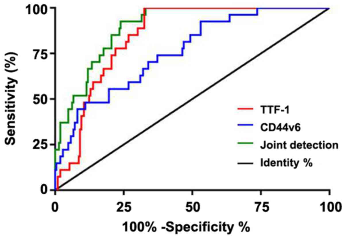
Diagnostic value of TTF-1 and CD44v6
in undifferentiated lung cancer mortality
ROC curve of the expression of TTF-1 and CD44v6 in
surviving and non-survivng patients was plotted to analyze their
diagnostic value in undifferentiated lung cancer. The results
showed that the AUC and 95% CI of TTF-1 were 0.866 and 0.820–0.912,
respectively, those of CD44v6 were 0.746 and 0.684–0.808, and those
of the joint detection were 0.897 and 0.857–0.936, respectively
(Table IX and Fig. 5).
| Table IX.Diagnostic value of TTF-1 and CD44v6
in undifferentiated lung cancer mortality. |
Table IX.
Diagnostic value of TTF-1 and CD44v6
in undifferentiated lung cancer mortality.
| Indexes | AUC | 95% CI | Specificity | Sensitivity | Youden index | Cut-off |
|---|
| TTF-1 | 0.866 | 0.820–0.912 | 66.99% | 100.00% | 66.99% | <163.312 |
| CD44v6 | 0.746 | 0.684–0.808 | 46.41% | 92.56% | 38.97% | <199.417 |
| Joint
detection | 0.897 | 0.857–0.936 | 75.60% | 92.59% | 68.19% | <0.914 |
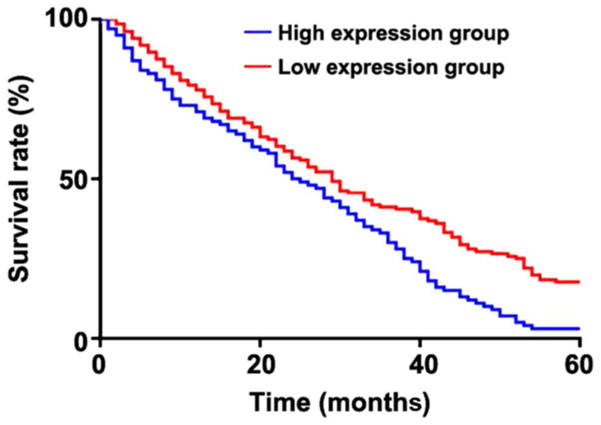
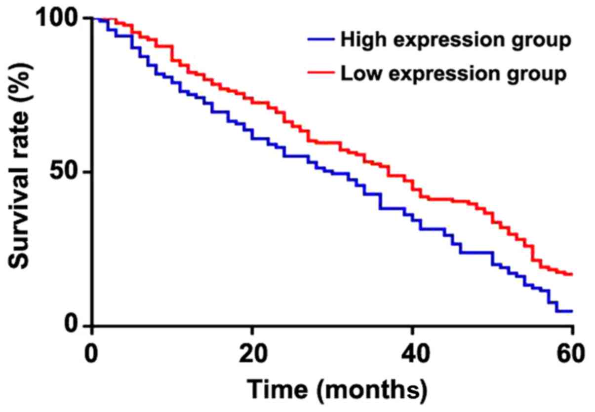
Correlation between the expression of
TTF-1 and CD44v6 and the 5-year survival of patients
The patients were divided into high and low
expression groups according to the cut-off points of expression of
TTF-1 and CD44v6 in undifferentiated lung cancer mortality. The K-M
survival curve found that the survival in TTF-1 high expression
group was significantly lower than that in the low expression group
(P=0.0005), and the survival in the CD44v6 high expression group
was significantly lower than that in the low expression group
(P=0.0041) (Figs. 6 and 7).
Univariate analysis of survival
The clinical data of patients in the deceased and
survival groups were collected for univariate analysis. It was
found that there was no difference in sex, BMI, age, history of
alcohol abuse and residence between the two groups (P>0.05).
However, there was statistical difference in the course of disease,
smoking history or TNM stage (P<0.05) (Table X).
| Table X.Univariate analysis. |
Table X.
Univariate analysis.
| Factor | Deceased group
(n=209) | Survival group
(n=27) |
t/χ2/Z | P-value |
|---|
| Sex |
|
Male | 119 (66.51) | 18 (66.67) | 0.929 | 0.335 |
|
Female | 90 (33.49) | 9 (33.33) |
|
|
| Age (years) | 60.7±10.2 | 61.2±10.5 | 0.239 | 0.811 |
| Course of disease
(year) | 3.79±2.03 | 2.67±1.82 | 2.728 | 0.007 |
| BMI
(kg/m2) | 23.55±1.34 | 23.06±1.90 | 1.695 | 0.091 |
| Smoking
history |
|
Yes | 87 (41.63) | 4 (14.81) | 7.256 | 0.007 |
| No | 122 (58.37) | 23 (85.19) |
|
|
| History of alcohol
abuse |
|
Yes | 41 (19.62) | 8 (29.63) | 1.457 | 0.227 |
| No | 168 (80.38) | 19 (70.37) |
|
|
| Residence |
|
Urban | 151 (72.25) | 20 (74.07) | 0.040 | 0.842 |
|
Rural | 58 (27.75) | 7 (25.93) |
|
|
| TNM stage |
| I | 33 (15.79) | 7 (25.93) |
|
|
| II | 73 (34.93) | 13 (48.14) | 2.368 | 0.018 |
|
III | 92 (44.02) | 7 (25.93) |
|
|
| IV | 11 (5.26) | 0 (0) |
|
|
| TTF-1 |
|
<161.900 | 113 (54.07) | 23 (85.19) | 9.482 | 0.002 |
|
≥161.900 | 96 (45.93) | 4 (14.81) |
|
|
| CD44v6 |
|
<185.600 | 116 (55.50) | 22 (81.48) | 6.646 | 0.010 |
|
≥185.600 | 93 (44.50) | 5 (18.52) |
|
|
| Distal
metastasis |
|
Yes | 92 (44.02) | 7 (25.93) | 3.867 | 0.049 |
| No | 117 (55.98) | 20 (74.07) |
|
|
Multivariate analysis of survival
The indicators with differences in univariate
analysis were included into an assignment (Table XI). Then a multivariate logistic
regression analysis (forward: LR) was carried out and showed that
smoking history and distal metastasis were not independent risk
factors of undifferentiated lung cancer mortality during the course
of disease (OR, 0.349; 95% CI, 0.160–0.761), TNM stage (OR, 3.183;
95% CI, 1.514–6.695), TTF-1 (OR, 0.110; 95% CI, 0.050–0.242) and
CD44v6 (OR, 0.262; 95% CI, 0.124–0.552) (Table XII).
| Table XI.Assignment table. |
Table XI.
Assignment table.
| Factors | Assignment |
|---|
| Course of
disease | ≥2 years=1, <2
years=0 |
| Smoking
history | Yes=1, No=0 |
| TNM stage | III, IV=1; I,
II=0 |
| Distal
metastasis | Yes=1, No=0 |
| TTF-1 | <163.312=1,
≥163.312=0 |
| CD44v6 | <199.417=1,
≥199.417=0 |
| Mortality
status | Deceased=1,
Survival=0 |
| Table XII.Multivariate analysis of
survival. |
Table XII.
Multivariate analysis of
survival.
|
|
|
|
|
|
| 95% CI of EXP
(B) |
|---|
|
|
|
|
|
|
|
|
|---|
| Factors | B | SE | Wald | Sig. | Exp (B) | Lower limit | Upper limit |
|---|
| Course of
disease | −1.053 | 0.398 | 7.012 | 0.008 | 0.349 | 0.160 | 0.761 |
| TNM stage | 1.158 | 0.379 | 9.319 | 0.002 | 3.183 | 1.514 | 6.695 |
| TTF-1 | −2.208 | 0.403 | 29.956 | <0.01 | 0.110 | 0.050 | 0.242 |
| CD44v6 | −1.340 | 0.381 | 12.38 | <0.01 | 0.262 | 0.124 | 0.552 |
Discussion
Lung cancer is the leading cause of cancer mortality
for human worldwide. The lack of specific and sensitive tools for
early diagnosis and the inadequacy of targeted treatment have
resulted in unsatisfactory treatment results (16). The cause is still unclear, but study
has shown that environmental pollution and long-term smoking are
closely related to the occurrence of lung cancer (17). Undifferentiated lung cancer,
characterized by poor differentiation of tumor cells, high degree
of malignancy, strong invasion and poor prognosis, is divided into
large cell cancer and small cell cancer according to the
histological morphology of tumor cells. TTF-1 plays an important
role in the differentiation of lung epithelial cells in the early
stage (18), and is also crucial to
the formation and structure of lung tissue. Ma et al
(19) has found that TTF-1
expression in lung squamous cell carcinoma and lung adenocarcinoma
is increased, but the specific mechanism of the increase is still
unclear. Moreover, there are few studies on the correlation between
expression and prognosis of TTF-1 in undifferentiated lung cancer.
CD44 is an important cell surface adhesion molecule and is closely
related to the invasion and metastasis of tumor cells. Studies have
shown that the abnormal expression of CD44v6, a member of the CD44
family, has a close relationship with the occurrence, development,
metastasis and prognosis of various tumor cells (9,20–22).
Some scholars found that the activity of CD44/CD44v6 depends on the
connection with integral membrane and cytosolic signaling
molecules, protease and transcriptional regulation (23), thus promoting tumor metastasis.
However, it is still unclear whether CD44v6 can be used as a
prognostic indicator for undifferentiated lung cancer.
In this study, we collected large differentiated
cancer patients, small differentiated cancer patients and normal
individuals. The expression of TTF-1 and CD44v6 in the serum of the
three groups and patient tissues were detected by ELISA. It was
found that the expression of TTF-1 and CD44v6 in the serum of
undifferentiated cancer patients were higher than that in normal
individuals, and the expression of TTF-1 and CD44v6 in cancer
tissues was also higher than that in adjacent tissues. This is
similar to the results by Perner et al (24) which showed TTF-1 was highly expressed
in various lung cancer types, and also to the study by Tran et
al (25) which suggested CD44v6
was highly expressed in squamous cell carcinoma. The expression of
TTF-1 in large cell cancer patients was higher than that in small
cell cancer patients, but the expression of CD44v6 was the
opposite, with statistical differences, which indicates that TTF-1
and CD44v6 may be potential diagnostic indicators of
undifferentiated cancer. In this study, it was found that the AUC
of TTF-1 and CD44v6 was 0.852 and 0.840, respectively through the
ROC curve for diagnosis of undifferentiated cancer. The joint curve
of TTF-1 and CD44v6 was plotted, and the AUC was 0.934. When the
cut-off point was less than 0.228, the optimal specificity and
sensitivity were 81.70 and 96.25%, which were significantly better
than the single detection. This study also found that the AUC of
TTF-1 and CD44v6 was 0.766 and 0.715 through ROC curve for
diagnosis of large and small cell cancers. The AUC of the joint
curve was 0.820. When the cut-off point was less than 0.502, the
optimal specificity and sensitivity were 81.70 and 96.25%, and the
diagnosis of large and small cell cancers was also higher than that
of single detection. Therefore, the expression of TTF-1 and CD44v6
can be diagnostic indicators of large and small cell cancers.
A follow-up survey was conducted on the survival of
patients in group A and group B. In total 236 patients were
followed up, 209 died and 27 survived at 5 years, with a survival
rate of 11.44%. The patients were divided into groups according to
their mortality conditions, and the diagnostic value of TTF-1 and
CD44v6 in undifferentiated lung cancer mortality was predicted by
ROC curve. AUC of TTF-1 was 0.848, and when the cutoff point was
less than 163.312 the optimal specificity and sensitivity were
66.99 and 100.00%; the AUC of CD44v6 was 0.762, when the cutoff
point was less than 199.417 the optimal specificity and sensitivity
were 46.41 and 92.56%, respectively. Thus, the sensitivity and
specificity of the two indicators were quite different when they
were detected separately. Therefore, joint detection was carried
out with an AUC of 0.897, and when the cutoff point was less than
0.914, the optimal specificity and sensitivity were 75.60 and
92.59%, respectively, indicating that joint detection can make up
the defects between the two factors. The patients were divided into
high and low expression groups according to the cut-off points of
TTF-1 and CD44v6, and the K-M survival curve was plotted. It was
found that the patients with high expression of TTF-1 and CD44v6
had significantly lower 5-year survival than the patients with low
expression, similar to the result of Situ et al (26) that the survival rate of patients with
high expression of CD44v6 in non-small cell cancer patients is
lower than that of patients with low expression. Shinohara et
al (27) also reported that the
5-year survival rate of CD44v6 overexpression in cancer tissues was
lower by immunohistochemistry and that the prognosis of CD44v6
overexpression in serum by ELISA was worse, which further confirmed
our view. This study also found that low expression of TTF-1 and
CD44v6 were protective factors for patients' mortality through
multivariate logistic regression analysis, which suggested that the
expression of TTF-1 and CD44v6 can be used as predictors of 5-year
mortality of patients with undifferentiated lung cancer, and the
course of disease and TNM stage of patients were independent risk
factors of undifferentiated lung cancer mortality.
However, there are some limitations in this study.
Correlation between TTF-1 and CD44v6 was not analyzed and the
specific mechanism of TTF-1 and CD44v6 on the growth, proliferation
and invasion of tumor cells was not studied. Therefore, the
relationship between TTF-1 and CD44v6 and the occurrence mechanism
need to be explored in later studies to verify the results of the
present study.
In conclusion, the course of disease, TNM stage,
TTF-1 and CD44v6 are independent mortality factors of
undifferentiated lung cancer patients. TTF-1 and CD44v6 have
certain diagnostic value in undifferentiated lung cancer and can be
used as mortality predictors of undifferentiated lung cancer.
Acknowledgements
Not applicable.
Funding
No funding was received.
Availability of data and materials
The datasets used and/or analyzed during the present
study are available from the corresponding author on reasonable
request.
Authors' contributions
YW wrote the manuscript and was also involved in the
design and conception of the study. XY was responsible for ELISA.
ML analyzed and interpreted the patients' data. BW helped with
statistical analysis. All authors read and approved the final
manuscript.
Ethics approval and consent to
participate
The study was approved by the Medical Ethics
Committee of Penglai Traditional Chinese Medicine Hospital (Yantai,
China). Patients, who participated in this research, had complete
clinical data. Signed informed consents were obtained from the
patients or their guardians.
Patient consent for publication
Not applicable.
Competing interests
The authors declare that they have no competing
interests.
References
|
1
|
Torre LA, Bray F, Siegel RL, Ferlay J,
Lortet-Tieulent J and Jemal A: Global cancer statistics, 2012. CA
Cancer J Clin. 65:87–108. 2015. View Article : Google Scholar : PubMed/NCBI
|
|
2
|
Aristarco V, Serrano D, Gandini S,
Johansson H, Macis D, Guerrieri-Gonzaga A, Lazzeroni M, Feroce I,
Pruneri G, Pagani G, et al: A randomized, placebo-controlled, phase
II, presurgical biomarker trial of celecoxib versus exemestane in
postmenopausal breast cancer patients. Cancer Prev Res (Phila).
9:349–356. 2016. View Article : Google Scholar : PubMed/NCBI
|
|
3
|
Alvarado-Luna G and Morales-Espinosa D:
Treatment for small cell lung cancer, where are we now? - a review.
Transl Lung Cancer Res. 5:26–38. 2016.PubMed/NCBI
|
|
4
|
Rudin CM, Ismaila N, Hann CL, Malhotra N,
Movsas B, Norris K, Pietanza MC, Ramalingam SS, Turrisi AT III and
Giaccone G: Treatment of small-cell lung cancer: American Society
of Clinical Oncology Endorsement of the American College of Chest
Physicians Guideline. J Clin Oncol. 33:4106–4111. 2015. View Article : Google Scholar : PubMed/NCBI
|
|
5
|
Hanagiri T, Oka S, Takenaka S, Baba T,
Yasuda M, Ono K, So T, Uramoto H, Takenoyama M and Yasumoto K:
Results of surgical resection for patients with large cell
carcinoma of the lung. Int J Surg. 8:391–394. 2010. View Article : Google Scholar : PubMed/NCBI
|
|
6
|
Sumi T, Hirai S, Yamaguchi M, Tanaka Y,
Tada M, Yamada G, Hasegawa T, Miyagi Y, Niki T, Watanabe A, et al:
Survivin knockdown induces senescence in TTF 1-expressing,
KRAS-mutant lung adenocarcinomas. Int J Oncol. 53:33–46.
2018.PubMed/NCBI
|
|
7
|
Puglisi F, Aprile G, Bruckbauer M, Barbone
F, Damante G, Guerra S, Beltrami CA and Di Loreto C: Combined
analysis of MIB-1 and thyroid transcription factor-1 predicts
survival in non-small cell lung carcinomas. Cancer Lett.
162:97–103. 2001. View Article : Google Scholar : PubMed/NCBI
|
|
8
|
Hara K, Saito T, Hayashi T, Mitani K,
Takamochi K, Oh S, Suzuki K and Yao T: Inverse correlation between
galectin-4 and TTF-1 in lung adenocarcinoma. Virchows Arch.
471:375–382. 2017. View Article : Google Scholar : PubMed/NCBI
|
|
9
|
Matzke-Ogi A, Jannasch K, Shatirishvili M,
Fuchs B, Chiblak S, Morton J, Tawk B, Lindner T, Sansom O, Alves F,
et al: Inhibition of tumor growth and metastasis in pancreatic
cancer models by interference with CD44v6 signaling.
Gastroenterology. 150:513–525.e10. 2016. View Article : Google Scholar : PubMed/NCBI
|
|
10
|
Ssadh HA, Spencer PS, Alabdulmenaim W,
Alghamdi R, Madar IH, Miranda-Sayago JM and Fernández N:
Measurements of heterotypic associations between cluster of
differentiation CD74 and CD44 in human breast cancer-derived cells.
Oncotarget. 8:92143–92156. 2017. View Article : Google Scholar : PubMed/NCBI
|
|
11
|
Okayama H, Kumamoto K, Saitou K, Hayase S,
Kofunato Y, Sato Y, Miyamoto K, Nakamura I, Ohki S, Sekikawa K, et
al: CD44v6, MMP-7 and nuclear Cdx2 are significant biomarkers for
prediction of lymph node metastasis in primary gastric cancer.
Oncol Rep. 22:745–755. 2009.PubMed/NCBI
|
|
12
|
Ekici S, Cerwinka WH, Duncan R, Gomez P,
Civantos F, Soloway MS and Lokeshwar VB: Comparison of the
prognostic potential of hyaluronic acid, hyaluronidase (HYAL-1),
CD44v6 and microvessel density for prostate cancer. Int J Cancer.
112:121–129. 2004. View Article : Google Scholar : PubMed/NCBI
|
|
13
|
Chen JQ, Zhan WH, He YL, Peng JS, Wang JP,
Cai SR and Ma JP: Expression of heparanase gene, CD44v6, MMP-7 and
nm23 protein and their relationship with the invasion and
metastasis of gastric carcinomas. World J Gastroenterol.
10:776–782. 2004. View Article : Google Scholar : PubMed/NCBI
|
|
14
|
Liu YJ, Yan PS, Li J and Jia JF:
Expression and significance of CD44s, CD44v6, and nm23 mRNA in
human cancer. World J Gastroenterol. 11:6601–6606. 2005. View Article : Google Scholar : PubMed/NCBI
|
|
15
|
Wang Z, von Au A, Schnölzer M, Hackert T
and Zöller M: CD44v6-competent tumor exosomes promote motility,
invasion and cancer-initiating cell marker expression in pancreatic
and colorectal cancer cells. Oncotarget. 7:55409–55436.
2016.PubMed/NCBI
|
|
16
|
Ferlay J, Soerjomataram I, Dikshit R, Eser
S, Mathers C, Rebelo M, Parkin DM, Forman D and Bray F: Cancer
incidence and mortality worldwide: Sources, methods and major
patterns in GLOBOCAN 2012. Int J Cancer. 136:E359–E386. 2015.
View Article : Google Scholar : PubMed/NCBI
|
|
17
|
Hori M, Tanaka H, Wakai K, Sasazuki S and
Katanoda K: Secondhand smoke exposure and risk of lung cancer in
Japan: A systematic review and meta-analysis of epidemiologic
studies. Jpn J Clin Oncol. 46:942–951. 2016. View Article : Google Scholar : PubMed/NCBI
|
|
18
|
Tanaka Y, Yamaguchi M, Hirai S, Sumi T,
Tada M, Saito A, Chiba H, Kojima T, Watanabe A, Takahashi H, et al:
Characterization of distal airway stem-like cells expressing
N-terminally truncated p63 and thyroid transcription factor-1 in
the human lung. Exp Cell Res. 372:141–149. 2018. View Article : Google Scholar : PubMed/NCBI
|
|
19
|
Ma Y, Fan M, Dai L, Kang X, Liu Y, Sun Y,
Yan W, Liang Z, Xiong H and Chen K: The expression of TTF-1 and
Napsin A in early-stage lung adenocarcinoma correlates with the
results of surgical treatment. Tumour Biol. 36:8085–8092. 2015.
View Article : Google Scholar : PubMed/NCBI
|
|
20
|
Yu Q and Stamenkovic I: Localization of
matrix metalloproteinase 9 to the cell surface provides a mechanism
for CD44-mediated tumor invasion. Genes Dev. 13:35–48. 1999.
View Article : Google Scholar : PubMed/NCBI
|
|
21
|
Amirghofran Z, Jalali SA, Hosseini SV,
Vasei M, Sabayan B and Ghaderi A: Evaluation of CD44 and CD44v6 in
colorectal carcinoma patients: Soluble forms in relation to tumor
tissue expression and metastasis. J Gastrointest Cancer. 39:73–78.
2008. View Article : Google Scholar : PubMed/NCBI
|
|
22
|
Marzese DM, Liu M, Huynh JL, Hirose H,
Donovan NC, Huynh KT, Kiyohara E, Chong K, Cheng D, Tanaka R, et
al: Brain metastasis is predetermined in early stages of cutaneous
melanoma by CD44v6 expression through epigenetic regulation of the
spliceosome. Pigment Cell Melanoma Res. 28:82–93. 2015. View Article : Google Scholar : PubMed/NCBI
|
|
23
|
Wang Z, Zhao K, Hackert T and Zöller M:
CD44/CD44v6 a reliable companion in cancer-initiating cell
maintenance and tumor progression. Front Cell Dev Biol. 6:972018.
View Article : Google Scholar : PubMed/NCBI
|
|
24
|
Perner S, Wagner PL, Soltermann A,
LaFargue C, Tischler V, Weir BA, Weder W, Meyerson M, Giordano TJ,
Moch H, et al: TTF1 expression in non-small cell lung carcinoma:
Association with TTF1 gene amplification and improved survival. J
Pathol. 217:65–72. 2009. View Article : Google Scholar : PubMed/NCBI
|
|
25
|
Tran TA, Kallakury BV, Sheehan CE and Ross
JS: Expression of CD44 standard form and variant isoforms in
non-small cell lung carcinomas. Hum Pathol. 28:809–814. 1997.
View Article : Google Scholar : PubMed/NCBI
|
|
26
|
Situ D, Long H, Lin P, Zhu Z, Wang J,
Zhang X, Xie Z and Rong T: Expression and prognostic relevance of
CD44v6 in stage I non-small cell lung carcinoma. J Cancer Res Clin
Oncol. 136:1213–1219. 2010. View Article : Google Scholar : PubMed/NCBI
|
|
27
|
Shinohara S, Hanagiri T, Taira A, Takenaka
M, Oka S, Chikaishi Y, Uramoto H, So T, Yamada S and Tanaka F:
Immunohistochemical expression and serum levels of CD44 as
prognostic indicators in patients with non-small cell lung cancer.
Oncology. 90:327–338. 2016. View Article : Google Scholar : PubMed/NCBI
|