Introduction
According to the cancer statistics database from the
American Cancer Society (ACS) liver cancer is one of the six most
frequent malignancies and ranks the fifth leading cause of
cancer-associated mortalities in the US (1). Globally, liver cancer is the 3rd
leading cause of cancer-related death with ~700,000 deaths
occurring annually (2). Despite the
combined use of chemotherapy and surgery, the prognosis and
survival rates of liver cancer patients remain poor (3).
The early stage diagnosis of liver cancer and the
prediction of liver cancer patient prognosis are considered
imperative for the prevention of this disease (4). Certain tumor markers including
α-fetoprotein, carcinoembryonic antigen, transforming growth
factor-β and microRNA-500/29/112 have been previously used for
early diagnosis and prognosis of liver cancer (5,6).
However, the use of these tumor markers remains limited. Therefore,
several research groups have focused on the development of novel
biomarkers for the early detection and prognosis of liver
cancer.
NAD(P)(H)-dependent oxidoreductase genes of the
aldo-keto reductase type 1C (AKR1C) family, including
AKR1C1, AKR1C2, AKR1C3 and AKR1C4 are located on
chromosome 10p15-p14 (7). Normally,
AKR1C1, AKR1C2, AKR1C3 and AKR1C4 enzymes are implicated in steroid
metabolism and their expression levels are localized in the normal
tissues of the lung, liver, prostate, testis and mammary glands.
The upregulation or downregulation of AKR1C1, AKR1C2, AKR1C3 and
AKR1C4 are associated with several human tumors including ovarian,
breast, colon, prostate, gastric and leukemia (8–12).
However, the exact functions of the different AKR1C1, AKR1C2,
AKR1C3 and AKR1C4 members in combination with tumorigenesis and
liver cancer prognosis have not been fully identified.
In the present study, the expression levels of the
AKRIC family isoforms were examined in liver cancer, in order to
determine the expression pattern of distinct AKR1C family members
in liver cancer tissues compared with that noted in normal tissues.
Moreover, the corresponding prognostic value of AKR1C1-3 was
examined in liver cancer. The present analysis can improve the
early diagnosis of liver cancer and support the individual care for
liver cancer treatment.
Materials and methods
Validation AKR1C1, AKR1C2, AKR1C3 and
AKR1C4 genes expression by Oncomine analysis
Oncomine gene expression array datasets (https://www.oncomine.org/) is an online cancer
microarray database (13) that
contains 19 cancer types, 715 datasets and 86,733 samples
corresponding to ~48 million gene expression measurements.
Differential expression analyses comparing most cancer tissue types
with the corresponding normal tissues were available for
exploration. The data could be queried and visualized for a
selected gene across all analyses (13). Regarding the differential analysis of
AKR1C1, AKR1C2, AKR1C3 and AKR1C4 between liver
cancer tissues and normal liver samples, the thresholds were set as
follows: Analysis type: Cancer vs. normal; cancer type: Liver
cancer; sample type: Clinical specimen; data type: mRNA. The
individual mRNA levels of AKR1C1, AKR1C2, AKR1C3 and
AKR1C4 in different types of cancers were determined from
the Oncomine database in comparison with the levels of the
corresponding normal tissues. In the present study, the top 10%
gene rank with a P<0.001 and fold-change >2 were used as
thresholds.
Evaluation of AKR1C1, AKR1C2, AKR1C3
and AKR1C4 mRNA levels by Cancer Cell lines Encyclopedia (CCLE)
analysis
The CCLE project is a collaboration between the
Broad Institute, the Novartis Institutes for Biomedical Research
and the Genomics Novartis Foundation that aims to conduct a
detailed genetic and pharmacological characterization of a large
panel of human cancer models in order to develop integrated
computational analyses that link distinct pharmacological
vulnerabilities to genomic patterns (14). Furthermore, the CCLE project aims to
translate cell line integrative genomics into cancer patient
stratification and consists of a compilation of gene expression,
chromosomal copy number and parallel sequencing data (14). All raw and processed data are
available at an integrated portal on www.broadinstitute.org/ccle. The CCLE provides public
access to genomic data, analysis and visualization for 1,062 cell
lines representing 37 distinct cancer types. The investigation of
the mRNA levels of AKR1C1, AKR1C2, AKR1C3 and AKR1C4
in a series of cancer cell lines was conducted by CCLE analysis.
The gene expression data were derived from human cancer cell lines,
in order to identify whether and/or to what extent the AKR1C1,
AKR1C2, AKR1C3 and AKR1C4 genes were expressed in liver
cancer cell lines compared with their expression in other types of
cancer cell lines.
Comparison of the AKR1C family gene
expression levels in liver cancer and paired normal tissues by the
Gene Expression Profiling Interactive Analysis (GEPIA)
database
GEPIA is a newly developed interactive web database
including 9,736 tumor and 8,587 normal samples from the TCGA and
GTEx projects, which can be used for RNA sequencing and RNA
expression analyses. GEPIA provides customizable functions, such as
tumor and/or normal differential expression analysis, profiling
according to cancer types or pathological stages, patient survival
analysis, detection of gene expression similarities, correlation
analysis and dimensionality reduction analysis. GEPIA fills in the
gap between cancer genomics big data and the delivery of integrated
information to end users, thereby facilitating the prognostic value
of the current data resources. GEPIA is available at http://gepia.cancer-pku.cn/. In the present study,
this online tool was used to analyze the levels of the AKR1C
family members between liver cancer and normal control specimens.
Statistical analysis was performed using the Student's t-test to
and the cut-off P<0.01.
Prognostic value assessment of
AKR1C1-3 using the Kaplan-Meier plotter survival analysis
The prognostic significance of the mRNA expression
levels of the AKR1C1-3 in several cancer types was evaluated
using the Kaplan-Meier plotter (http://www.kmplot.com), an online database that can be
used to assess the effect of genes in cancer prognosis. The
Kaplan-Meier plotter can assess the effects of 54,675 genes on
survival using 10,461 cancer samples. The analysis was applied to
evaluate the prognostic value of the gene expression levels of the
AKR1C1-3 in liver cancer. In this database, the data were
extracted regarding lung (15),
ovarian (16), liver (17), gastric (18) and breast cancers (19). The patient samples were divided into
two cohorts according to the median expression of each gene (high
vs. low expression). The overall survival (OS) of liver cancer
patients was analyzed using a Kaplan-Meier survival plot. Briefly,
the three genes (AKR1C1, AKR1C2 and AKR1C3) were
uploaded into the database to obtain the Kaplan-Meier survival
plots, in which the number-at-risk was shown below the main plot.
Subsequently, the plot data was exported as text and the
Kaplan-Meier survival curves for the expression of each gene of
interest.
Co-expression and protein-protein
interaction (PPI) network construction
The top 100 co-expression genes were extracted of
AKR1C3 across all tumor samples from the GEPIA database
(http://gepia.cancerpku.cn/index.html)
and then the Search Tool for the Retrieval of Interacting Genes
(STRING) database (http://string-db.org/) was designed in order to
analyze the PPI information (20).
To evaluate the potential PPI relationship, the previously
identified DEGs were mapped to the STRING database. The PPI pairs
were extracted with a combined score of 0.4. Subsequently, the PPI
network was visualized by the Cytoscape software (version 3.6.1;
www.cytoscape.org/). The nodes with a higher
degree of connectivity appeared more essential in maintaining the
stability of the entire network.
Statistical analysis
AKR1C1-4 genes were queried in Oncomine
database and the results were filtered by selecting ‘liver cancer’,
‘mRNA and cancer vs. normal analysis’, ‘2-fold change’,
‘P<0.001’ and ‘top 10% gene rank’. In CCLE database, the line
within a box indicatesthe mean. The following settings for GEPIA
were used as: ‘Expression on Box Plots’, ‘Gene=FNDC1’, ‘|log2FC|
Cutoff=1’, ‘P-value Cutoff=0.01’, ‘Datasets=STAD’, ‘Log
Scale=log2(TPM + 1)’, ‘Jitter Size=0.4’ and ‘Match TCGA
normal and GTEx data’. The prognostic value of the AKR1C1-3
genes in liver cancer was analyzed using the Kaplan-Meier Plotter.
The settings were used for the analysis: ‘Overall survival’, ‘auto
select best cutoff’, ‘censored at threshold’, (patients surviving
over the selected threshold are censored instead of excluded),
‘tumor stage all’, ‘tumor stage Tall’, ‘tumor stage N all’, ‘tumor
stage M all’, ‘Lauren classification all’ and ‘differentiation
all’. The AKR1C genes probe set was 48–315996, 17–101636,
226–101485 and 226–101485 respectively. Patients were split
according to median expression or expression at best cutoff for the
probe. The results of the Kaplan-Meier analysis are presented with
hazard ratio (HR) and log-rank P-values from a log-rank test.
P<0.05 was considered to indicate a statistically significant
difference. The data were extracted from the Oncomine database,
CCLE, GEPIA website and Kaplan-Meier Plotter between December 2017
and March 2019.
Results
The diverse expression patterns of
AKR1C1, AKR1C2, AKR1C3 and AKR1C4 genes as determined by analysis
of the Oncomine database
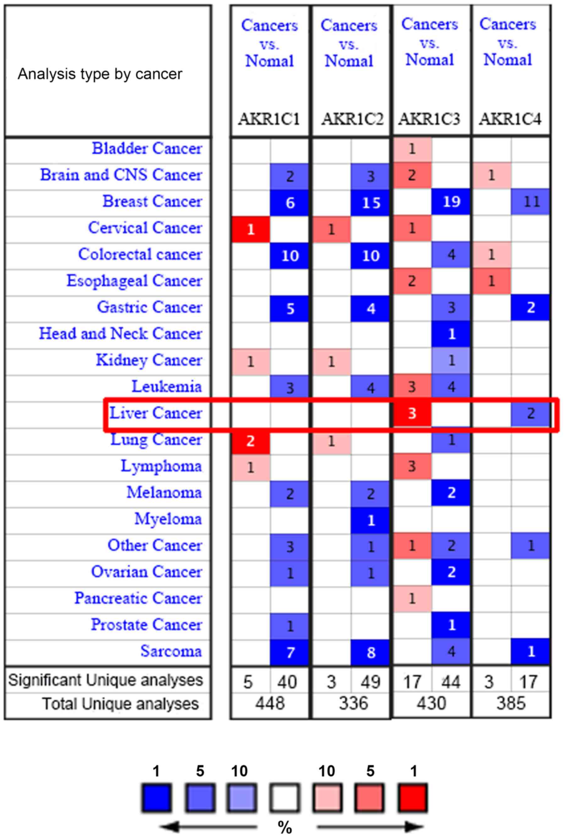
Using Oncomine analysis, the mRNA levels of the
AKR1C family of genes in various human cancer types
including hematogenous malignancies and solid tumors were
investigated. The number in each cell represents the number of
analyses that meet the threshold within those analysis and cancer
types. There were 3 and 2 analyses that meet the thresholds for
AKR1C3 and AKR1C4 respectively (Fig.
1). No analysis met the thresholds for AKR1C1 and AKR1C2.
To further examine the expression status of
AKR1C1, AKR1C2, AKR1C3 and AKR1C4 genes in liver
cancer, the data corresponding to the four genes with regard to the
liver cancer tissue number, normal tissue number, fold-change,
t-test T, P-value and rank were summarized (Table I). Among the AKR1C family of
genes, AKR1C3 was overexpressed at the highest levels in
liver cancer (n=386) compared with those noted in normal tissues
(n=327) in the studies of Chen et al (21), Wurmbach et al (22) and Roessler et al (23) with fold-changes between 1.774 and
3.438. The differential expression analysis of AKR1C1 and
AKR1C2 exhibited fold-changes of 1.435 and 1.543 for liver
cancer (n=35) and normal tissues (n=10), respectively, as
determined in the Wurmbach et al (22) study. However, AKR1C4 was
downregulated in liver cancer (n=225) compared with its
corresponding expression in normal tissues (n=220) with a fold
change of −2.594 as shown in the Roessler et al (23) study.
 | Table I.The mRNA expression of AKR1C
family genes in normal and liver cancer tissue. |
Table I.
The mRNA expression of AKR1C
family genes in normal and liver cancer tissue.
| Gene | Liver cancer,
n | Normal, n | Fold-change | t-test, T | P-value | Gene
ranka, % | (Refs.) |
|---|
| AKR1C1 | 35 | 10 | 1.435 | 2.533 |
8.00×10⁻³ | 21 | (21) |
| AKR1C2 | 35 | 10 | 1.543 | 3.626 |
4.53×10⁻⁴ | 11 | (21) |
| AKR1C3 | 225 | 220 | 3.438 | 22.18 |
3.86×10−71 | 1 | (22) |
|
| 22 | 21 | 2.965 | 6.185 |
1.44×10⁻⁷ | 4 | (22) |
|
| 35 | 10 | 2.470 | 6.309 |
1.62×10⁻⁶ | 3 | (21) |
|
| 104 | 76 | 1.774 | 6.140 |
2.81×10⁻⁹ | 7 | (22) |
| AKR1C4 | 225 | 220 | −2.594 | −9.710 |
7.89×10⁻²⁰ | 9 | (21) |
The expression levels of AKR1C3 were
differentially overexpressed between liver cancer and normal
tissues, whereas the expression levels of AKR1C4 were
downregulated in liver cancer tissues. The expression levels of
AKR1C1 and AKR1C2 in liver cancer tissues were not
increased significantly.
The expression levels of AKR1C1,
AKR1C2, AKR1C3 and AKR1C4 are upregulated in liver cancer cell
lines as determined by CCLE analysis
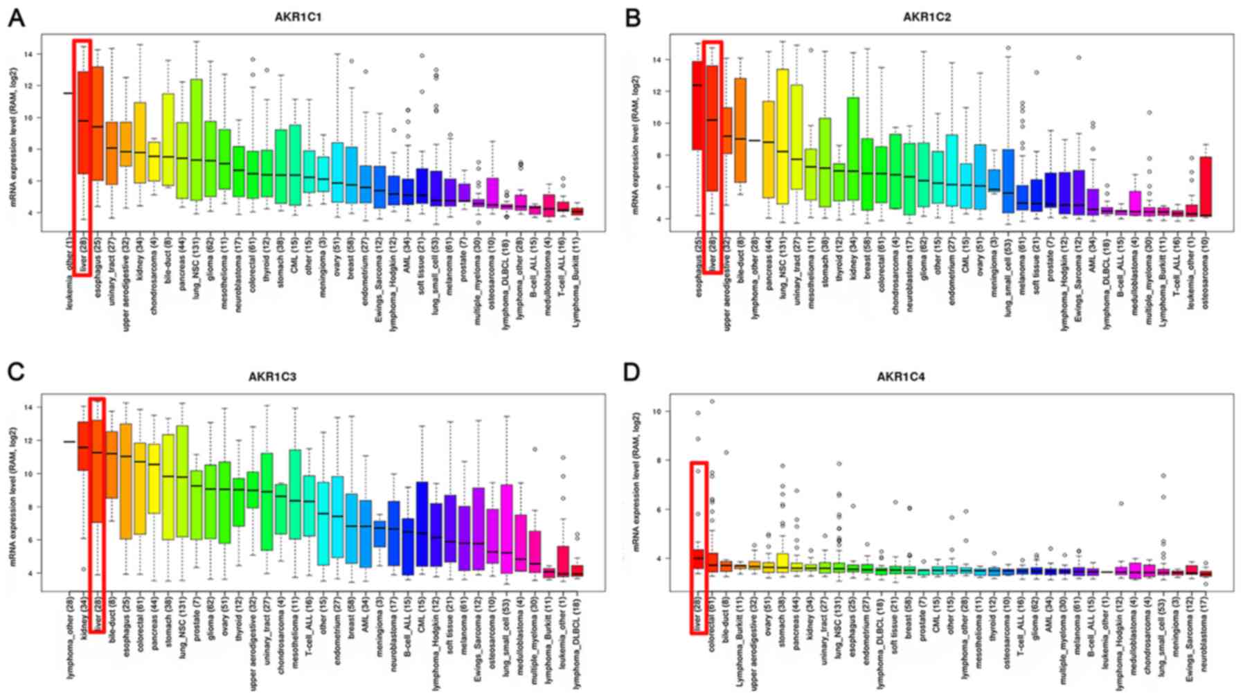
The expression levels of AKR1C1, AKR1C2,
AKR1C3 and AKR1C4 in liver cancer cell lines were ranked
as the top 3 out of 37 distinct cancer types in the CCLE databases
(Fig. 2). The total of their
expression levels was higher than that of the other cancer types
(Fig. 2). These results were
consistent with those for AKR1C1-3 indicating that these
genes were upregulated in liver cancer as determined by the
Oncomine database. In contrast to AKR1C1-3, the expression
levels of AKR1C4 were significantly downregulated in liver
cancer samples derived from the Oncomine database but were highly
expressed in the CCLE database (Table
I; Fig. 1). It was implied that
AKR1C1-3, especially AKR1C3 may play roles in the
development of liver cancer. The role of AKR1C4 in liver
cancer should be further examined.
AKR1C1, AKR1C2, AKR1C3 and AKR1C4 are
differentially expressed in liver cancer tissues as determined by
GEPIA analysis
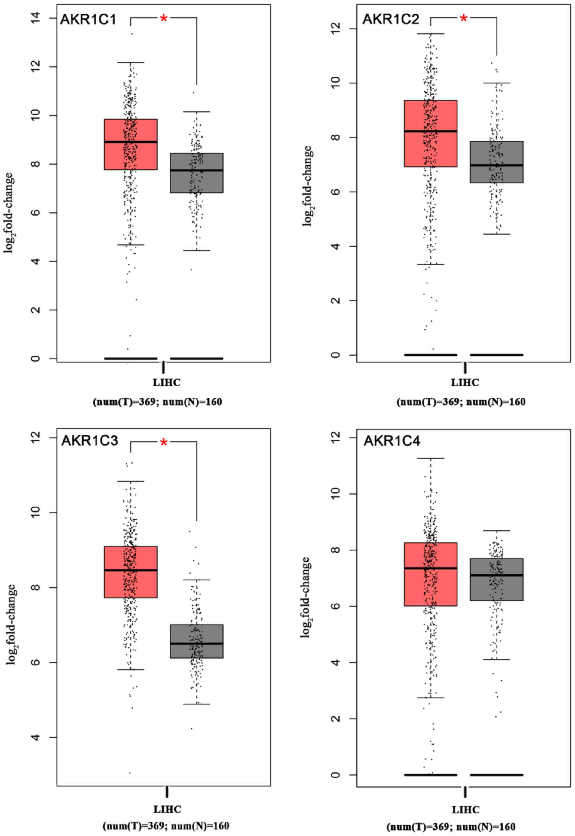
To examine the expression levels of AKR1C
members in liver cancer, their mRNA levels were compared between
liver cancer and normal liver samples using GEPIA. The GEPIA
database included 369 liver cancer and 160 normal liver tissues,
which were used for the expression analysis (24). The data indicated that the
AKR1C1-4 gene members were overexpressed in liver cancer
samples compared with normal liver samples. The boxplot (P<0.05;
Fig. 3) revealed consistent data
with those obtained by the CCLE database analysis.
Association of AKR1C1, AKR1C2 and
AKR1C3 with survival time in liver cancer patients
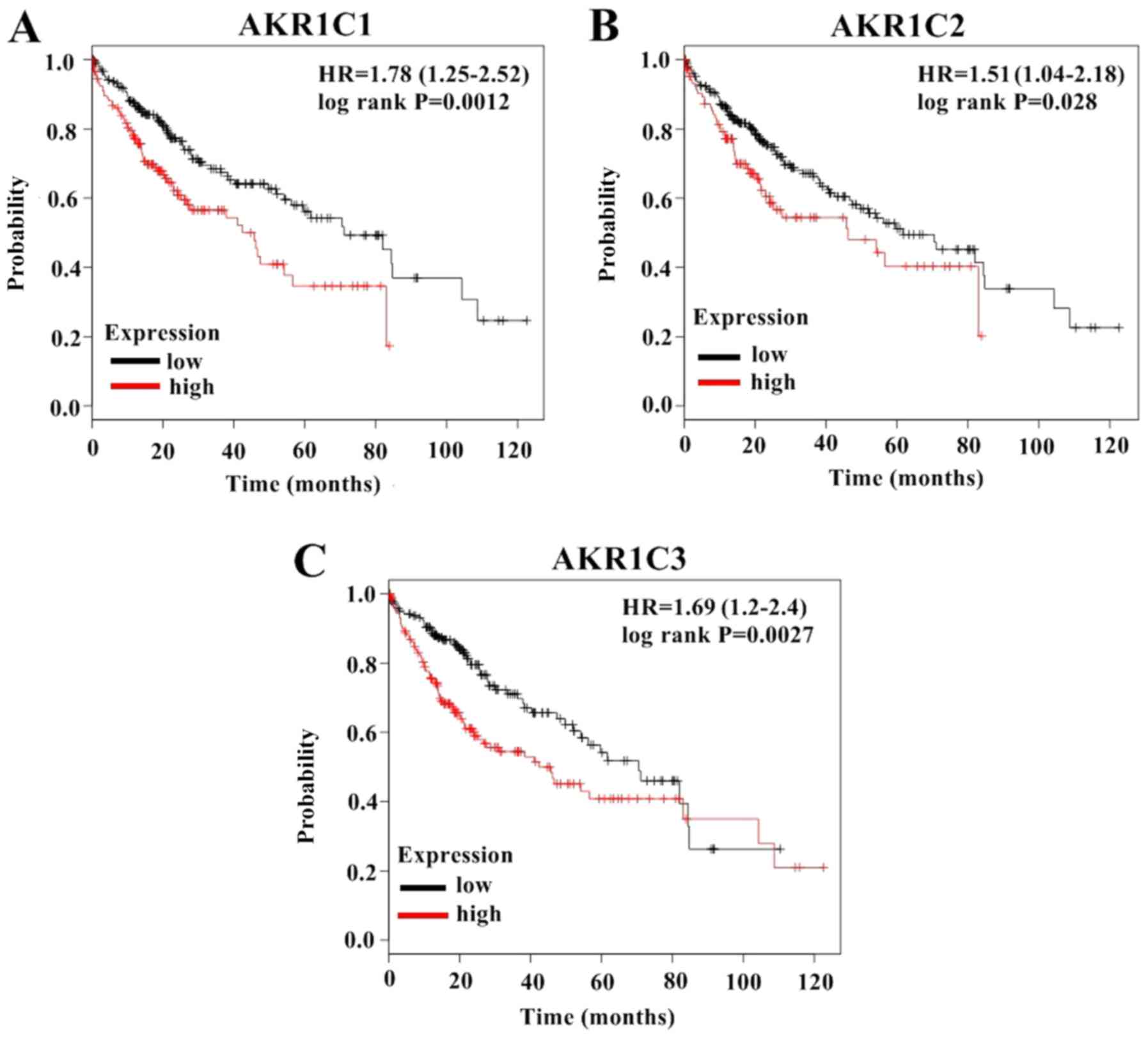
It was found that the expression levels of AKR1C1-3
were elevated in liver cancer tissues and cells as demonstrated by
the Oncomine, CCLE and GEPIA databases. However, the expression
levels of the AKR1C4 gene as determined by the Oncomine, CCLE and
GEPIA databases were not consistent. Therefore, the association
between the liver cancer patient survival time and the mRNA
expression levels of the AKRIC1-3 genes was identified using
a Kaplan-Meier plotter. The survival curves were plotted for all
liver cancer patients (Fig. 4).
Liver cancer patients with high expression of AKR1C1 (HR=1.78;
P=0.0012), AKR1C2 (HR=1.51; P=0.028) and AKR1C3 (HR=1.69; P=0.027)
exhibited a significant association with lower OS. High mRNA
expression levels of AKR1C1, AKR1C2 and AKR1C3 were significantly
associated with low OS in all liver cancer patients.
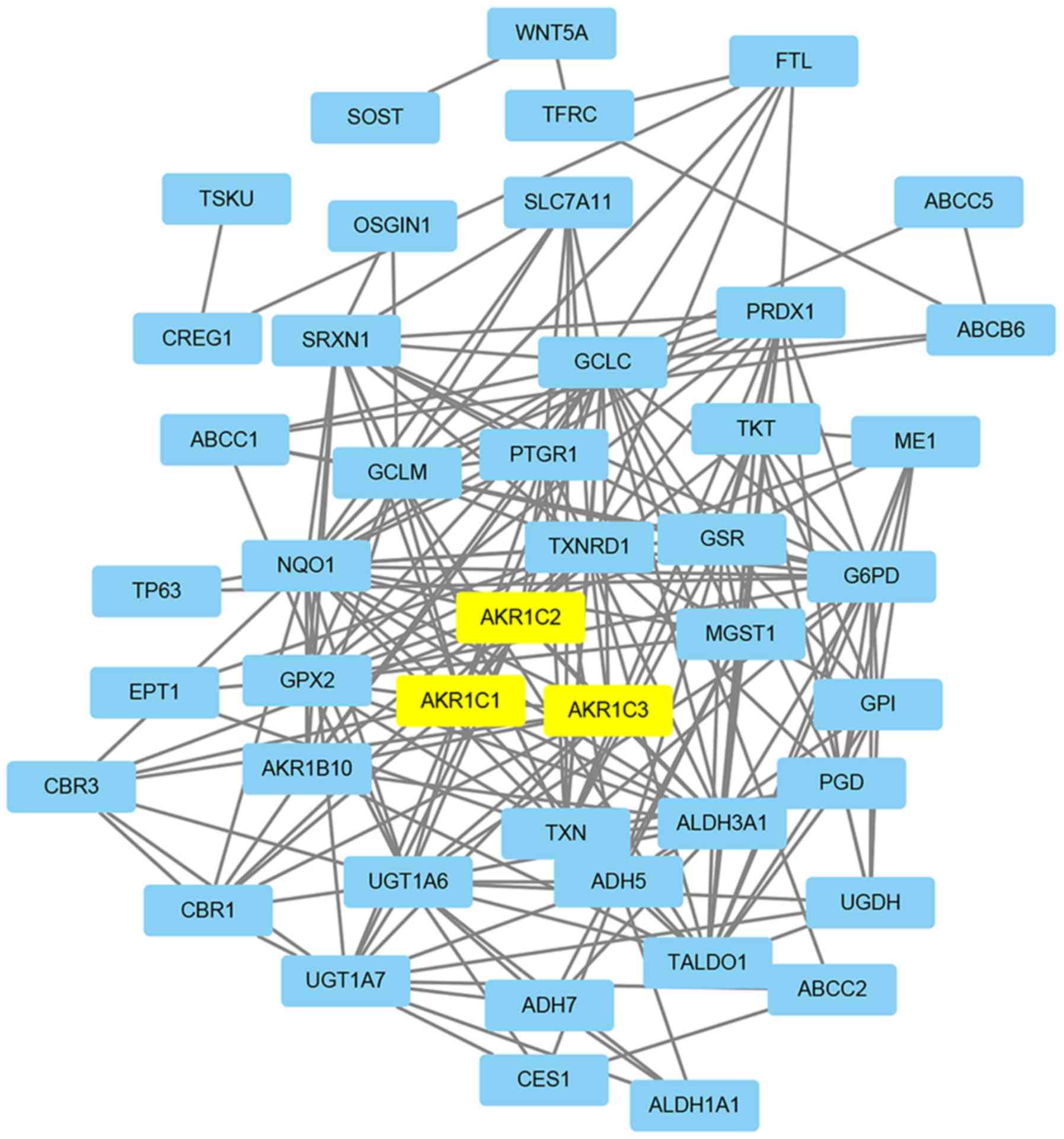
The co-expression and PPI network
construction
Co-expression gene analysis was conducted by the PPI
networks of AKR1C3, which were differentially expressed in liver
cancer (Fig. 5). It was shown that
AKR1C1-3 was involved in the same pathway displaying 44
total unique interactors.
Discussion
The aldo-keto reductase type 1C comprises the
isoforms AKR1C1-AKR1C4 that serve important roles in the metabolism
of steroid hormones, conjugated steroids, neurosteroids and bile
acids (10,12,25–27).
These enzymes are also involved in the modulation of carcinogen
metabolism (10,12,25–27).
AKR1C1 is an important contributor in the proliferation and
migration of tumor cells including small-cell lung cancer and
breast cancer (12,26). The AKR1C2 gene may contribute
to the incidence, progression and invasion of breast cancer
(12) and hepatocellular carcinoma
(28). AKR1C3 overexpression
is associated with the progression and aggressiveness of
non-small-cell lung cancer, esophageal cancer and prostate cancer
(10,29,30). The
catalytic-dependent and-independent function of the AKR1C isoforms
demonstrated critical roles in the proliferation and migration of
cells and in tumor drug resistance (31). To the best of our knowledge, the
association between the expression levels of the AKR1C isoforms and
the incidence of liver cancer has not been examined to date.
Liver cancer is one of the leading causes of
cancer-related deaths worldwide. The 5-year OS rate of liver cancer
patients is <5% (32). Early
diagnosis provides the only cure for these patients. In addition to
surgical resection, ablation and liver transplantation are used
frequently as therapeutic modalities for liver cancer patients.
Molecular-targeted therapies are considered potential emerging
treatment of advanced liver cancer.
The present study initially demonstrated that high
expression levels of AKR1C1-3, notably AKR1C3,
predicted low survival. Using the Oncomine, CCLE and GEPIA
databases, the expression levels of the AKR1C family members
were analyzed in liver cancer samples and compared with the
corresponding expression levels of the normal samples. The results
demonstrated that the expression levels of AKR1C3 were
elevated in liver cancer tissues compared with those of the normal
tissues. The expression levels of AKR1C1 and AKR1C2 in liver cancer
tissues were not increased significantly in the Oncomine database
while expression was high in CCLE and GEPIA databases. However, the
expression levels of the AKR1C4 gene that were noted in the
Oncomine, CCLE and GEPIA databases were not consistent. The role of
AKR1C4 in liver cancer requires further examination. The
elevated expression of AKR1C3 in the Oncomine, CCLE and
GEPIA databases suggested that AKR1C3 may serve as a
potential diagnostic, therapeutic biomarker for liver cancer
patients.
Survival analysis of the levels of AKR1C1-3
demonstrated similar results indicating that overexpression of
AKR1C1-3 was associated with lower survival in patients with
liver cancer. These results suggested the critical role of AKR1C1-3
regarding their contribution in liver cancer initiation and/or
progression.
To the best of our knowledge, the regulation of the
expression levels of AKR1C in liver cancer has not been
explored. A limited number of reports have shown that the
overexpression of AKR1C2 and AKR1C3 are associated
with liver cancer progression (28,33).
Although the AKR1C1-3 enzymes are involved in the same pathway, the
exact mechanism of their contribution to the occurrence,
development and prognosis of liver cancer is still unknown.
AKR1C1-3 can act as promising biomarkers for liver
cancer diagnosis and prognosis. The high expression of AKR1C1-3,
notably AKR1C3 may be associated with the incidence,
development and prognosis of liver cancer. Nevertheless, the
current study presented results derived solely from bioinformatics
analyses and further experimental evidence is required to validate
these findings.
Acknowledgements
Not applicable.
Funding
No funding was received.
Availability of data and materials
The datasets used and/or analyzed during the present
study are available from the corresponding author on reasonable
request.
Authors' contributions
SZ conceived and designed the study. SW, ZZ and WL
made substantial contributions to the design of the current study,
acquisition of data, interpretation of data and revising the
manuscript. All authors read and approved the final manuscript.
Ethics statement and consent to
participate
Not applicable.
Patient consent for publication
Not applicable.
Competing interests
The authors declare that they have no competing
interests.
References
|
1
|
Siegel RL, Miller KD and Jemal A: Cancer
statistics, 2018. CA Cancer J Clin. 68:7–30. 2018. View Article : Google Scholar : PubMed/NCBI
|
|
2
|
Heimbach JK, Kulik LM, Finn RS, Sirlin CB,
Abecassis MM, Roberts LR, Zhu AX, Murad MH and Marrero JA: Aasld
guidelines for the treatment of hepatocellular carcinoma.
Hepatology. 67:358–380. 2018. View Article : Google Scholar : PubMed/NCBI
|
|
3
|
Liu S, Miao R, Zhai M, Pang Q, Deng Y, Liu
S, Qu K, Liu C and Zhang J: Effects and related mechanisms of
serotonin on malignant biological behavior of hepatocellular
carcinoma via regulation of Yap. Oncotarget. 8:47412–47424.
2017.PubMed/NCBI
|
|
4
|
Black AP and Mehta AS: The search for
biomarkers of hepatocellular carcinoma and the impact on patient
outcome. Curr Opin Pharmacol. 41:74–78. 2018. View Article : Google Scholar : PubMed/NCBI
|
|
5
|
Behne T and Copur MS: Biomarkers for
hepatocellular carcinoma. Int J Hepatol. 2012:8590762012.
View Article : Google Scholar : PubMed/NCBI
|
|
6
|
Zhao YJ, Ju Q and Li GC: Tumor markers for
hepatocellular carcinoma. Mol Clin Oncol. 1:593–598. 2013.
View Article : Google Scholar : PubMed/NCBI
|
|
7
|
Barski OA, Mindnich R and Penning TM:
Alternative splicing in the Aldo-Keto reductase superfamily:
Implications for protein nomenclature. Chem Biol Interact.
202:153–158. 2013. View Article : Google Scholar : PubMed/NCBI
|
|
8
|
Rizner TL and Penning TM: Role of
Aldo-Keto reductase family 1 (AKR1) enzymes in human steroid
metabolism. Steroids. 79:49–63. 2014. View Article : Google Scholar : PubMed/NCBI
|
|
9
|
Frycz BA, Murawa D, Borejsza-Wysocki M,
Wichtowski M, Spychała A, Marciniak R, Murawa P, Drews M and
Jagodziński PP: Transcript level of AKR1C3 is down-regulated in
gastric cancer. Biochem Cell Biol. 94:138–146. 2016. View Article : Google Scholar : PubMed/NCBI
|
|
10
|
Sun SQ, Gu X, Gao XS, Li Y, Yu H, Xiong W,
Yu H, Wang W, Li Y, Teng Y and Zhou D: Overexpression of AKR1C3
significantly enhances human prostate cancer cells resistance to
radiation. Oncotarget. 7:48050–48058. 2016.PubMed/NCBI
|
|
11
|
Lewis MJ, Wiebe JP and Heathcote JG:
Expression of progesterone metabolizing enzyme genes (AKR1C1,
AKR1C2, AKR1C3, SRD5A1, SRD5A2) is altered in human breast
carcinoma. BMC Cancer. 4:272004. View Article : Google Scholar : PubMed/NCBI
|
|
12
|
Wenners A, Hartmann F, Jochens A, Roemer
AM, Alkatout I, Klapper W, van Mackelenbergh M, Mundhenke C, Jonat
W and Bauer M: Stromal markers AKR1C1 and AKR1C2 are prognostic
factors in primary human breast cancer. Int J Clin Oncol.
21:548–556. 2016. View Article : Google Scholar : PubMed/NCBI
|
|
13
|
Rhodes DR, Yu J, Shanker K, Deshpande N,
Varambally R, Ghosh D, Barrette T, Pandey A and Chinnaiyan AM:
ONCOMINE: A cancer microarray database and integrated Data-Mining
platform. Neoplasia. 6:1–6. 2004. View Article : Google Scholar : PubMed/NCBI
|
|
14
|
Barretina J, Caponigro G, Stransky N,
Venkatesan K, Margolin AA, Kim S, Wilson CJ, Lehár J, Kryukov GV,
Sonkin D, et al: The cancer cell line encyclopedia enables
predictive modelling of anticancer drug sensitivity. Nature.
483:603–607. 2012. View Article : Google Scholar : PubMed/NCBI
|
|
15
|
Győrffy B, Surowiak P, Budczies J and
Lánczky A: Online survival analysis software to assess the
prognostic value of biomarkers using transcriptomic data in
Non-small-cell lung cancer. PLoS One. 8:e822412013. View Article : Google Scholar : PubMed/NCBI
|
|
16
|
Győrffy B, Lánczky A and Szállási Z:
Implementing an online tool for genome-wide validation of
survival-associated biomarkers in ovarian-cancer using microarray
data from 1287 patients. Endocr Relat Cancer. 19:197–208. 2012.
View Article : Google Scholar : PubMed/NCBI
|
|
17
|
Menyhart O, Nagy A and Gyorffy B:
Determining consistent prognostic biomarkers of overall survival
and vascular invasion in hepatocellular carcinoma. R Soc Open Sci.
5:1810062018. View Article : Google Scholar : PubMed/NCBI
|
|
18
|
Szász AM, Lánczky A, Nagy Á, Förster S,
Hark K, Green JE, Boussioutas A, Busuttil R, Szabó A and Győrffy B:
Cross-validation of survival associated biomarkers in gastric
cancer using transcriptomic data of 1,065 patients. Oncotarget.
7:49322–49333. 2016. View Article : Google Scholar : PubMed/NCBI
|
|
19
|
Györffy B, Lanczky A, Eklund AC, Denkert
C, Budczies J, Li Q and Szallasi Z: An online survival analysis
tool to rapidly assess the effect of 22,277 genes on breast cancer
prognosis using microarray data of 1,809 patients. Breast Cancer
Res Treat. 123:725–731. 2010. View Article : Google Scholar : PubMed/NCBI
|
|
20
|
Szklarczyk D, Franceschini A, Wyder S,
Forslund K, Heller D, Huerta-Cepas J, Simonovic M, Roth A, Santos
A, Tsafou KP, et al: STRING v10: Protein-protein interaction
networks, integrated over the tree of life. Nucleic Acids Res.
43((Database Issue)): D447–D452. 2015. View Article : Google Scholar : PubMed/NCBI
|
|
21
|
Chen X, Cheung ST, So S, Fan ST, Barry C,
Higgins J, Lai KM, Ji J, Dudoit S, Ng IO, et al: Gene expression
patterns in human liver cancers. Mol Biol Cell. 13:1929–1939. 2002.
View Article : Google Scholar : PubMed/NCBI
|
|
22
|
Wurmbach E, Chen YB, Khitrov G, Zhang W,
Roayaie S, Schwartz M, Fiel I, Thung S, Mazzaferro V, Bruix J, et
al: Genome-wide molecular profiles of HCV-induced dysplasia and
hepatocellular carcinoma. Hepatology. 45:938–947. 2007. View Article : Google Scholar : PubMed/NCBI
|
|
23
|
Roessler S, Jia HL, Budhu A, Forgues M, Ye
QH, Lee JS, Thorgeirsson SS, Sun Z, Tang ZY, Qin LX and Wang XW: A
unique metastasis gene signature enables prediction of tumor
relapse in early-stage hepatocellular carcinoma patients. Cancer
Res. 70:10202–10212. 2010. View Article : Google Scholar : PubMed/NCBI
|
|
24
|
Tang Z, Li C, Kang B, Gao G, Li C and
Zhang Z: GEPIA: A web server for cancer and normal gene expression
profiling and interactive analyses. Nucleic Acids Res. 45:W98–W102.
2017. View Article : Google Scholar : PubMed/NCBI
|
|
25
|
Hevir N, Vouk K, Sinkovec J, Ribic-Pucelj
M and Rizner TL: Aldo-Keto reductases AKR1C1, AKR1C2 and AKR1C3 may
enhance progesterone metabolism in ovarian endometriosis. Chem Biol
Interact. 191:217–226. 2011. View Article : Google Scholar : PubMed/NCBI
|
|
26
|
Tian H, Li X, Jiang W, Lv C, Sun W, Huang
C and Chen R: High expression of AKR1C1 is associated with
proliferation and migration of small-cell lung cancer cells. Lung
Cancer (Auckl). 7:53–61. 2016.PubMed/NCBI
|
|
27
|
Tiryakioglu NO and Tunali NE: Association
of AKR1C3 Polymorphisms with bladder cancer. Urol J. 13:2615–2621.
2016.PubMed/NCBI
|
|
28
|
Lu D, Zhang X and Cao X: The effect of
AKR1C2 gene on hepatocarcinogenesis and its abnormal expression in
hepatocellular carcinoma from Qidong, China, a liver cancer high
risk area. Progress Biochemistry Biophysics. 30:906–918. 2003.
|
|
29
|
Zhong T, Xu F, Xu J, Liu L and Chen Y:
Aldo-Keto reductase 1C3 (AKR1C3) is associated with the doxorubicin
resistance in human breast cancer via PTEN loss. Biomed
Pharmacother. 69:317–325. 2015. View Article : Google Scholar : PubMed/NCBI
|
|
30
|
Xiong W, Zhao J, Yu H, Li X, Sun S, Li Y,
Xia Q, Zhang C, He Q, Gao X, et al: Elevated expression of AKR1C3
increases resistance of cancer cells to ionizing radiation via
modulation of oxidative stress. PLoS One. 9:e1119112014. View Article : Google Scholar : PubMed/NCBI
|
|
31
|
Zeng CM, Chang LL, Ying MD, Cao J, He QJ,
Zhu H and Yang B: Aldo-Keto Reductase AKR1C1-AKR1C4: Functions,
regulation, and intervention for Anti-cancer therapy. Front
Pharmacol. 8:1192017. View Article : Google Scholar : PubMed/NCBI
|
|
32
|
Dhanasekaran R, Limaye A and Cabrera R:
Hepatocellular carcinoma: Current trends in worldwide epidemiology,
risk factors, diagnosis, and therapeutics. Hepat Med. 4:19–37.
2012.PubMed/NCBI
|
|
33
|
Abbattista MR, Jamieson SM, Gu Y, Nickel
JE, Pullen SM, Patterson AV, Wilson WR and Guise CP: Pre-clinical
activity of PR-104 as monotherapy and in combination with sorafenib
in hepatocellular carcinoma. Cancer Biol Ther. 16:610–622. 2015.
View Article : Google Scholar : PubMed/NCBI
|