Introduction
Ovarian cancer is the most lethal gynecologic
cancer, usually bearing a poor outcome (1). In 2019, a total of 22,530 new ovarian
cancer cases and 13,980 ovarian cancer-associated mortalities were
predicted to occur in the United States (1). Although aggressive surgery and
combination chemotherapy have improved survival, the 5-year overall
survival (OS) rate remains only 30% (2). Thus, identification of prognostic
markers is of great need to allow improved risk prediction.
A metabolic hallmark of the majority of cancer cells
is the shift from oxidative phosphorylation to aerobic glycolysis
even with abundant oxygen, which is known as the Warburg effect
(3). Since inhibiting the entry of
pyruvate into the mitochondria leads to the redirection of
glycolytic intermediates into the pentose phosphate pathway, which
provides precursors for the synthesis of nucleotides and lipids,
the Warburg effect contributes to the highly anabolic phenotype of
cancer (4). Therefore, elevated
expression of key enzymes that catalyze the pentose phosphate
pathway has been observed in several types of cancer, including
hepatocellular cancer (5) and
colorectal carcinoma (6). The
pentose phosphate pathway is responsible for the production of
ribose-5-phosphate and NADPH (7).
Ribose-5-phosphate is necessary for the synthesis of nucleic acids
to meet the needs of rapidly proliferating tumor cells (8). The pentose phosphate pathway comprises
two branches, oxidative and non-oxidative. The non-oxidative branch
of the pentose phosphate pathway provides >85% of ribose for
nucleic acids synthesis of certain tumor cells, including
pancreatic adenocarcinoma (9). The
transketolase family genes encode thiamine-dependent enzymes, which
are key enzymes in the non-oxidative part of the pentose phosphate
pathway (10). The transketolase
family comprises three human genes, transketolase (TKT) and
TKT-like genes 1 and 2 (TKTL1 and TKTL2) (11).
Previous studies have revealed that the
transketolase family genes serve important roles in the initiation,
progression and cross-talk with other signaling pathways in cancer.
Upregulated transketolase family genes have been identified in
multiple types of cancer, including hepatocellular carcinoma
(5), esophageal cancer (12), lung cancer (13), head and neck cancer (14), and colorectal carcinoma (6) and endometrial carcinoma (15). Overexpression of these genes is
associated with poor outcome of patients with esophageal (16), lung (17), gastric (18), breast (19), colon and urothelial (20) cancer. Aberrant expression of
transketolase family genes has also been observed in ovarian cancer
(21). However, studies on the
prognostic value of the transketolase family genes in ovarian
cancer are limited (22). In
addition, none of these studies have comprehensively assessed the
prognostic effects of individual transketolase family components in
ovarian cancer at the mRNA level. The aim of the present study was
to determine the expression levels and prognostic significance of
transketolase family genes in patients with ovarian carcinoma.
Materials and methods
Datasets
Oncomine is an online collection of datasets with
data mining platform (23). The
expression of three transketolase family genes (TKT, TKTL1 and
TKTL2) in normal ovarian tissues and ovarian cancer were analyzed
by Oncomine. Bonome, Lu, Hendrix and the Cancer Genome Atlas cancer
(TCGA) ovarian datasets were analyzed in the present study.
Survival analysis
Kaplan-Meier plotter (24) is a widely used online database that
integrates gene expression and clinical data of various types of
cancer such as breast (24), lung
(25) and ovarian (26,27)
cancer. In the present study, the progression-free survival (PFS)
of 1,435 patients and OS of 1,656 patients with ovarian cancer were
assessed using the Kaplan-Meier plotter database (www.kmplot.com). The information about gene expression
and survival of patients with ovarian cancer was collected from the
Gene Expression Omnibus (GEO), Cancer Biomedical Informatics Grid
(http://cabig.cancer.gov/) and TCGA cancer
datasets (28). Clinical data,
including stage, histology, grade, tumor protein p53 (TP53)
mutation status and treatment, were also obtained. The association
between transketolase gene mRNA levels and clinicopathological
parameters was determined in patients with ovarian cancer with the
corresponding clinical information. Transketolase family genes TKT,
TKTL1 and TKTL2 were entered into the database to produce
Kaplan-Meier survival plots. Patients were classified into ‘low’
and ‘high’-expression groups according to the quantile expression
of the proposed biomarker. Hazard ratio (HR) with 95% confidence
intervals (CI) and log-rank P-value were calculated.
Validation dataset
The GSE9891 dataset, which was part of the
Kaplan-Meier plotter, was downloaded separately from GEO as a
validation dataset (29). Expression
data and clinic information of patients with ovarian cancer from
GSE9891 were used to validate the prognostic values of
transketolase family genes in ovarian cancer.
Statistical analysis
Unpaired Student's-test was performed to compare the
expression level of transketolase family genes between ovarian
cancer and normal ovarian tissues from Oncomine. Results were
presented in box plots. Kaplan-Meier curve was used to assess the
association between expression of transketolase family genes and
the survival rate in ovarian cancer. The log-rank test was used to
compare the survival differences between the low and
high-expression groups from the Kaplan-Meier plotter. P<0.05 was
considered to indicate a statistically significant difference.
Results
Transcription level of TKT in ovarian
cancer compared with that in normal tissues
The transketolase family involves three human genes:
TKT, TKTL1 and TKTL2. The differential expression of these three
genes between normal ovarian tissue and ovarian cancer was analyzed
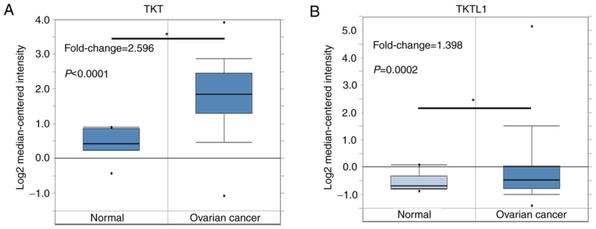
using the Oncomine database. In the Bonome dataset, which comprised
185 ovarian cancer tissues and 10 normal ovarian tissues from
patients with other gynecological diseases, the mRNA level of TKT
in ovarian cancer tissue was higher compared with that in normal
tissue (Fig. 1A). In the TCGA
dataset, which comprised 586 serous ovarian cancer samples and 8
adjacent normal ovarian tissues from patients with ovarian cancer,
no difference in TKT expression was observed between serous ovarian
cancer and normal ovarian tissue (Table
I). Since other types of ovarian cancer were rare, TKT
expression was systematically analyzed by combining the Lu and
Hendrix datasets, in which ovarian cancer cases were classified by
histological types. The results of the analysis demonstrated that
TKT was upregulated in endometrioid, clear cell and mucinous
ovarian cancer compared with its expression in normal ovarian
tissue (Table I).
 | Table I.Comparison of the expression profile
of transketolase family genes in normal ovarian tissue vs. various
types of ovarian cancer. |
Table I.
Comparison of the expression profile
of transketolase family genes in normal ovarian tissue vs. various
types of ovarian cancer.
|
|
| Tissues, n |
|
|---|
|
|
|
|
|
|---|
| Transketolase
family gene | Cancer type | Normal | Cancer | P-value |
|---|
| TKT | Serous | 8 | 586 | 0.409 |
|
| Endometrioid | 9 | 46 |
<0.001a (upregulated) |
|
| Clear cell | 9 | 15 | 0.007a (upregulated) |
|
| Mucinous | 9 | 22 | 0.001a (upregulated) |
| TKTL1 | Serous | 8 | 586 |
<0.001a (upregulated) |
|
| Endometrioid | 9 | 46 | 0.164 |
|
| Clear cell | 9 | 15 | 0.463 |
|
| Mucinous | 9 | 22 | 0.324 |
| TKTL2 | Serous | 5 | 20 | 0.311 |
|
| Endometrioid | 5 | 9 | 0.113 |
|
| Clear cell | 5 | 7 | 0.066 |
|
| Mucinous | 5 | 9 | 0.147 |
Transcription level of TKTL1 in
ovarian cancer compared with that in normal tissues
Compared with that in normal ovarian tissue, TKTL1
expression was increased by 1.398-fold in all ovarian cancer
samples in the Bonome dataset (Fig.
1B). According to the TCGA dataset, TKTL1 expression was
increased by 2.384-fold in serous ovarian cancer compared with that
in normal ovarian tissue (P<0.001; Table I). However, no difference in TKTL1
expression was observed between endometrioid, clear cell or
mucinous ovarian cancer and normal ovarian tissue (Table I).
Transcription level of TKTL2 in
ovarian cancer compared with that in normal tissues
No dataset compared TKTL2 expression between all
ovarian cancer and normal ovarian tissues. Only the Lu dataset that
separated ovarian cancer cases into different histological types
was accessible in Oncomine; no significant differences in TKTL2
expression levels were observed between serous, endometrioid, clear
cell or mucinous ovarian cancer and normal ovarian tissue (Table I).
Prognostic value of TKT in ovarian
cancer
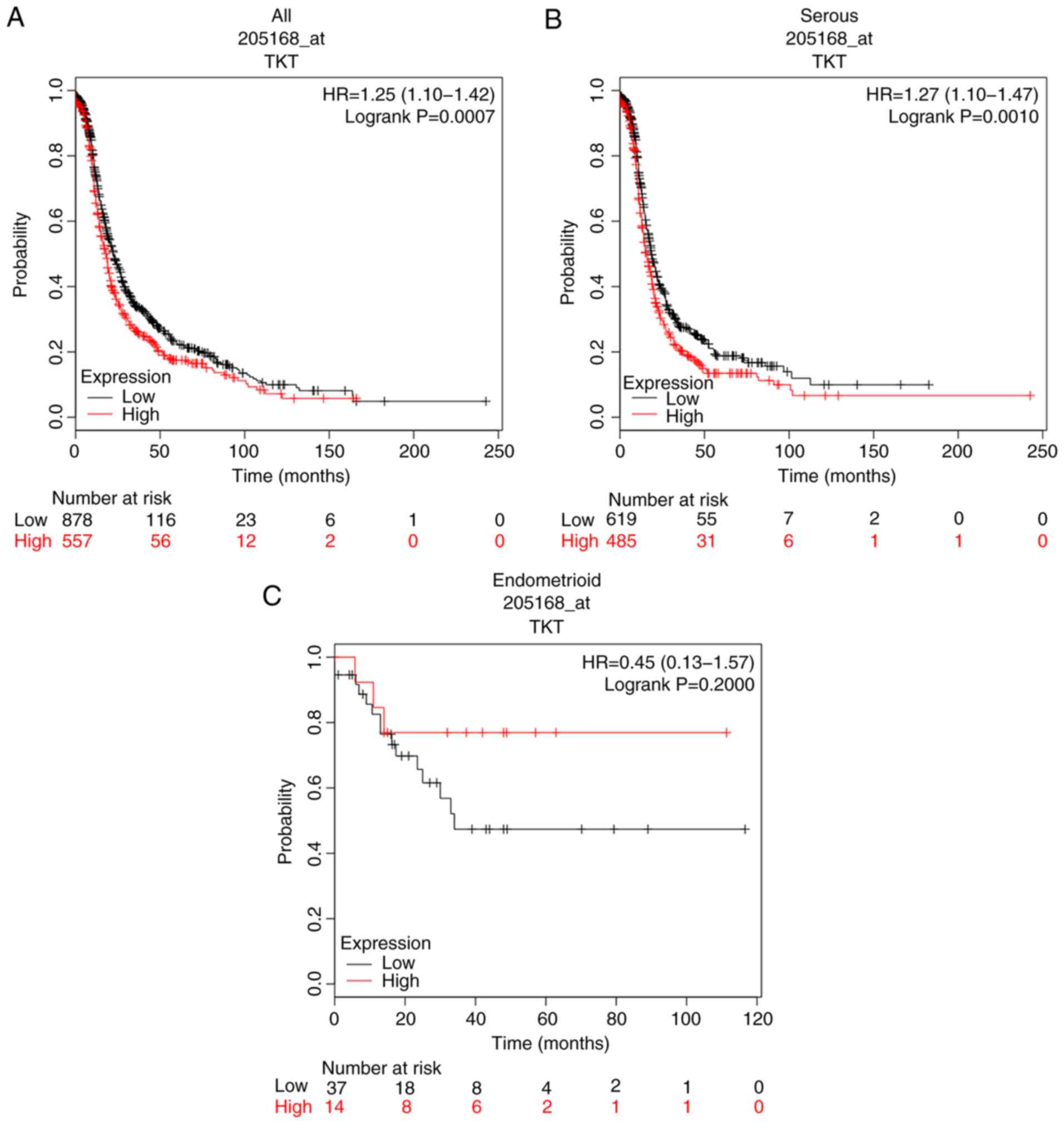
The prognostic value of transketolase family genes
was assessed. Survival information for the three transketolase
genes was obtained from the Kaplan-Meier plotter. The prognostic
value of TKT mRNA expression in ovarian cancer was evaluated; the
Affymetrix identifier (ID) was 205168_at TKT. PFS curves were
plotted for all patients with ovarian cancer (N=1,435; Fig. 2A), patients with serous cancer
(N=1,104; Fig. 2B) and endometrioid
cancer (N=51; Fig. 2C). Increased
expression levels of TKT mRNA were identified to be associated with
poorer PFS in all patients with ovarian cancer (HR, 1.25; 95% CI,
1.10–1.42; P=0.0007). High TKT expression was also associated with
poor PFS in serous ovarian cancer (HR, 1.27; 95% CI, 1.10–1.47;
P=0.0010). However, TKT mRNA expression levels were not associated
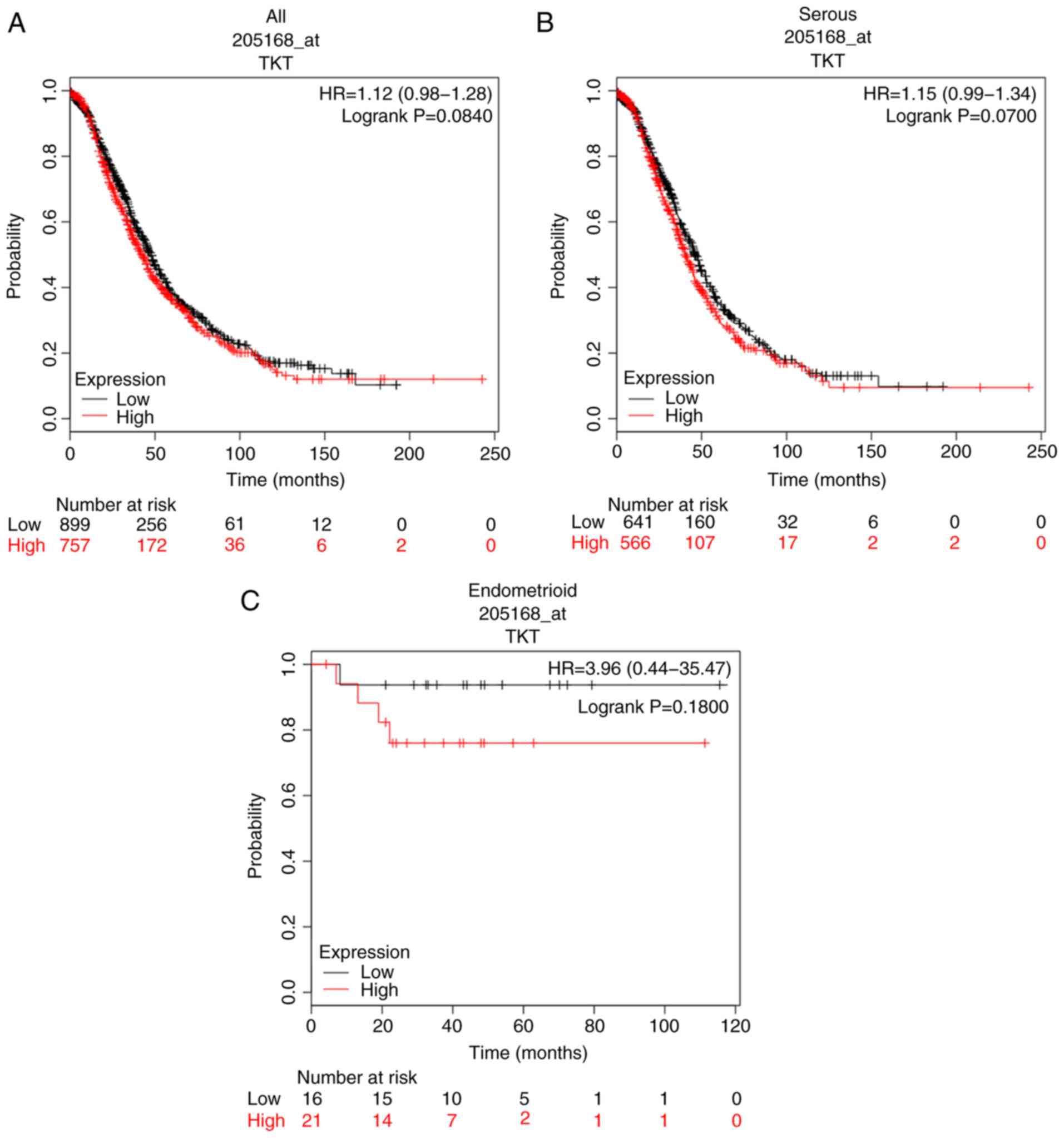
with PFS in patients with endometrioid cancer. The OS between the
high and low-TKT expression groups in all ovarian cancer and serous
ovarian cancer cases was not significantly different (Fig. 3A and B). TKT mRNA expression was not
associated with OS in patients with endometrioid cancer (Fig. 3C).
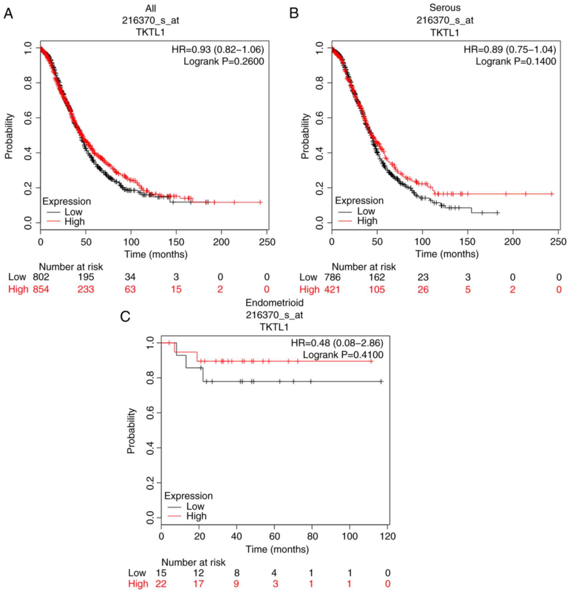
Prognostic value of TKTL1 in ovarian
cancer
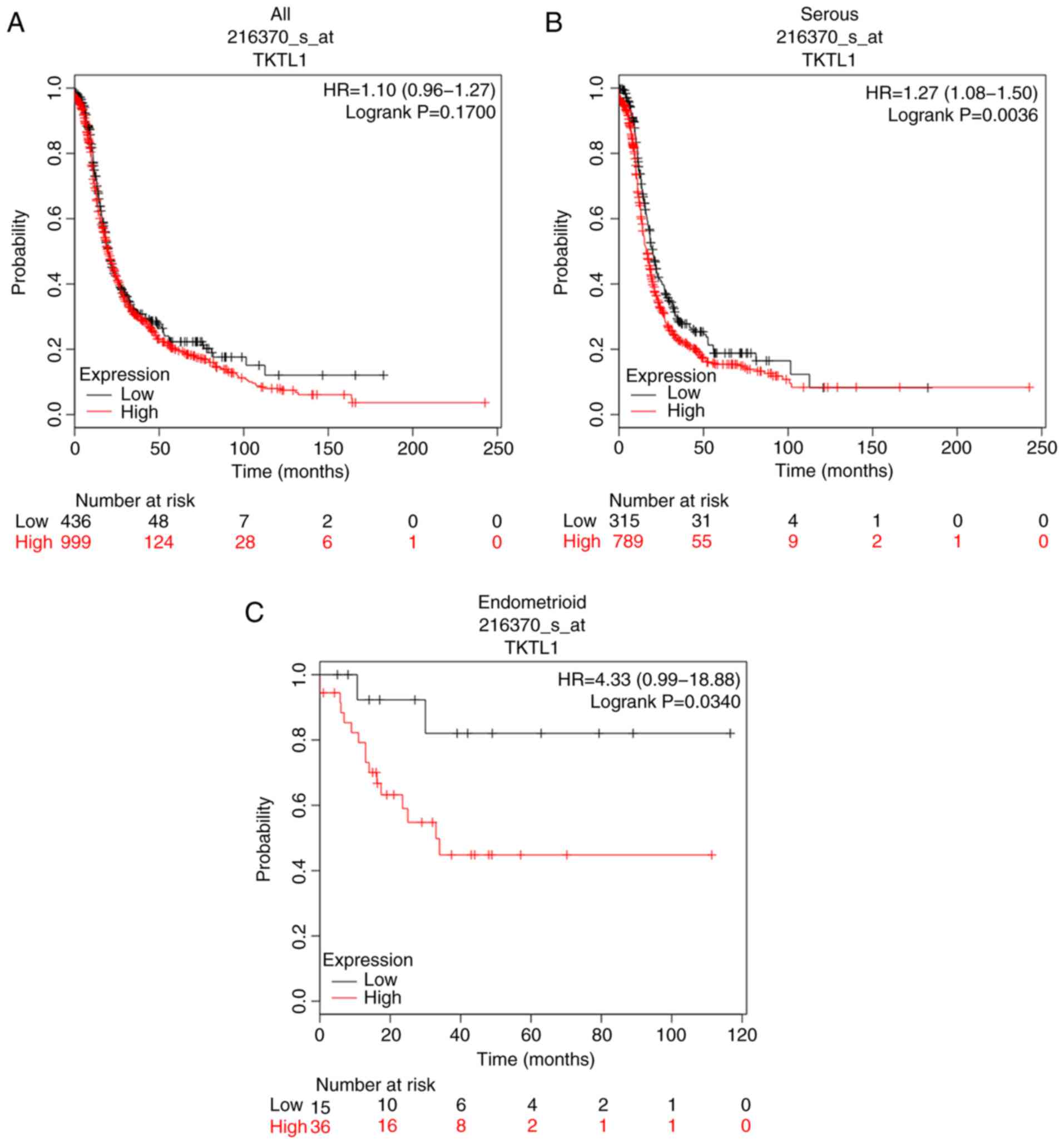
The prognostic value of TKTL1 mRNA expression in
ovarian cancer was evaluated; the Affymetrix ID was 216370_s_at
TKTL1. No differences were observed in PFS between the high and
low-TKTL1 expression groups in all patients with ovarian cancer
(Fig. 4A). However, stratified
analyses demonstrated that high expression of TKTL1 was associated
with poor PFS in patients with serous ovarian cancer (HR, 1.27; 95%
CI, 1.08–1.50; P=0.0036; Fig. 4B),
as well as in patients with endometrioid ovarian cancer (HR, 4.33;
95% CI, 0.99–18.88; P=0.0340; Fig.
4C). TKTL1 mRNA expression levels were not associated with OS
in all patients with ovarian cancer (Fig. 5A), nor in patients with serous
ovarian cancer (Fig. 5B) or
endometrioid ovarian cancer (Fig.
5C).
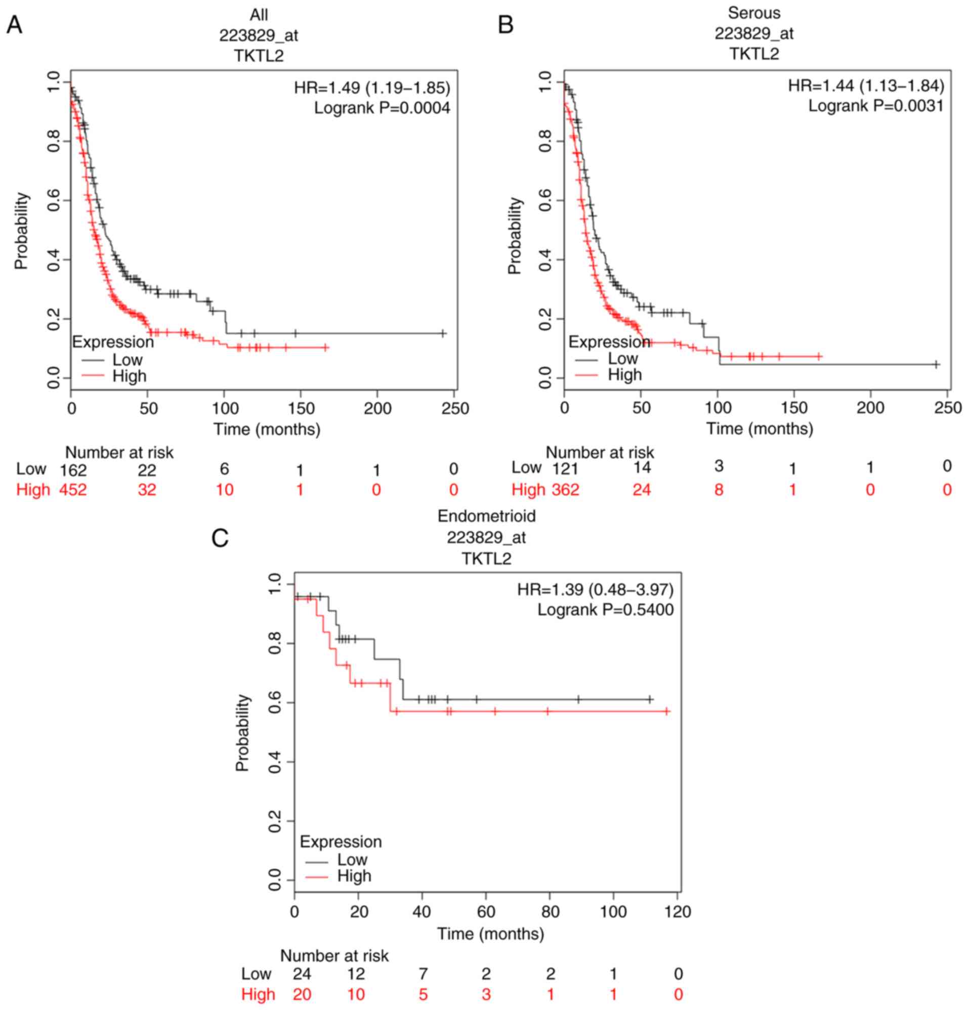
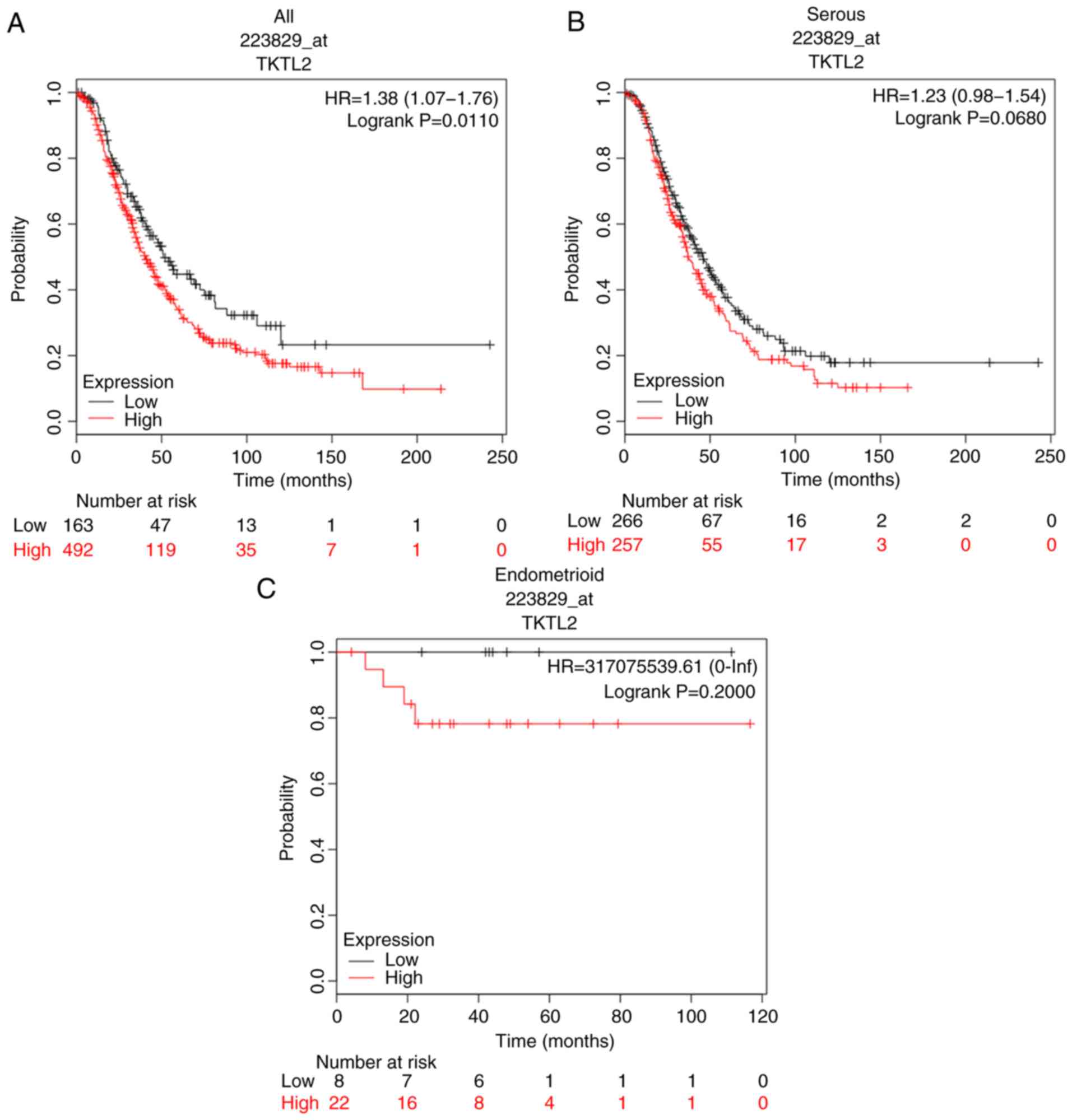
Prognostic value of TKTL2 in ovarian
cancer
The prognostic value of TKTL2 mRNA expression in
ovarian cancer was evaluated; the Affymetrix ID was 223829_at
TKTL2. High TKTL2 mRNA expression indicated unfavorable PFS for all
patients with ovarian cancer (HR, 1.49; 95% CI, 1.19–1.85;
P=0.0004; Fig. 6A) and patients with
serous ovarian cancer (HR, 1.44; 95% CI, 1.13–1.84; P=0.0031;
Fig. 6B). However, TKTL2 mRNA
expression levels were not associated with PFS in patients with
endometrioid ovarian cancer (Fig.
6C). High expression of TKTL2 was associated with OS in all
patients with ovarian cancer (HR, 1.38; 95% CI, 1.07–1.76;
P=0.0110; Fig. 7A). TKTL2 exhibited
no association with OS in patients with serous ovarian cancer
(Fig. 7B) or endometrioid ovarian
cancer (Fig. 7C).
Prognostic value of transketolase
family genes in different subtypes of ovarian cancer
The association between individual transketolase
gene mRNA expression levels and clinicopathological features of
patients with ovarian cancer was analyzed. Only samples with the
relevance clinical information were included. The association with
pathological clinical stage (Table
II), grade (Table III),
chemotherapy treatment (Table IV)
and TP53 mutation status (Table V)
of patients with ovarian cancer was determined. As presented in
Table II, the three transketolase
genes were associated with poor PFS in patients with stage III/IV
ovarian cancer, but not stage I/II ovarian cancer. The individual
transketolase genes were associated with poor PFS in patients with
grade I/II and grade III ovarian cancer (Table III). TKT and TKTL2 were associated
with unfavorable PFS in patients with ovarian cancer treated with
platinum chemotherapy (Table IV).
In addition, high levels of TKTL1 and TKTL2 were associated with
poor PFS in patients with ovarian cancer treated with taxol
chemotherapy (Table IV). The
association between transketolase family gene expression levels and
PFS of ovarian cancer patients was also evaluated based on the TP53
mutation status; the individual transketolase genes were only
significantly associated with poor PFS in patients with
TP53-mutated ovarian cancer, but not in those with TP53 wild-type
ovarian cancer.
| Table II.Association of transketolase family
genes with progression-free survival in patients with different
clinical stages of ovarian cancer. |
Table II.
Association of transketolase family
genes with progression-free survival in patients with different
clinical stages of ovarian cancer.
| Transketolase
family gene | Clinical stage | Patients, n | HR (95% CI) | P-value |
|---|
| TKT | I+II | 163 | 0.75
(0.42–1.34) | 0.3300 |
|
| III+IV | 1,081 | 1.24
(1.08–1.43) | 0.0025a |
| TKTL1 | I+II | 163 | 1.40
(0.76–2.59) | 0.2800 |
|
| III+IV | 1,081 | 1.32
(1.12–1.56) | 0.0008a |
| TKTL2 | I+II | 115 | 2.06
(0.84–5.08) | 0.1100 |
|
| III+IV | 494 | 1.36
(1.08–1.70) | 0.0079a |
| Table III.Association of transketolase family
genes with progression-free survival in patients with different
pathological grades of ovarian cancer. |
Table III.
Association of transketolase family
genes with progression-free survival in patients with different
pathological grades of ovarian cancer.
| Transketolase
family gene | Pathological
grade | Patients, n | HR (95% CI) | P-value |
|---|
| TKT | I+II | 293 | 1.46
(1.09–1.96) | 0.0120a |
|
| III | 837 | 1.21
(1.03–1.43) | 0.0220a |
| TKTL1 | I+II | 293 | 1.41
(1.04–1.91) | 0.0250a |
|
| III | 837 | 1.37
(1.12–1.67) | 0.0020a |
| TKTL2 | I+II | 189 | 1.53
(1.03–2.28) | 0.0350a |
|
| III | 315 | 1.51
(1.18–1.95) | 0.0011a |
| Table IV.Association of transketolase family
genes with progression-free survival in patients with ovarian
cancer who received chemotherapy. |
Table IV.
Association of transketolase family
genes with progression-free survival in patients with ovarian
cancer who received chemotherapy.
| Transketolase
family gene | Chemotherapy | Patients, n | HR (95% CI) | P-value |
|---|
| TKT | Platinum | 1,259 | 1.19
(1.05–1.36) | 0.0078a |
|
| Taxol | 715 | 1.16
(0.95–1.40) | 0.1400 |
|
| Taxol +
platinum | 698 | 1.14
(0.94–1.39) | 0.1900 |
| TKTL1 | Platinum | 1,259 | 0.91
(0.80–1.04) | 0.1500 |
|
| Taxol | 715 | 1.23
(1.03–1.48) | 0.0250a |
|
| Taxol +
platinum | 698 | 1.24
(1.03–1.50) | 0.0240a |
| TKTL2 | Platinum | 502 | 1.31
(1.08–1.59) | 0.0063a |
|
| Taxol | 381 | 1.44
(1.13–1.84) | 0.0031a |
|
| Taxol +
platinum | 380 | 1.44
(1.13–1.83) | 0.0034a |
| Table V.Association of transketolase family
genes with progression-free survival in patients with ovarian
cancer with different TP53 mutation status. |
Table V.
Association of transketolase family
genes with progression-free survival in patients with ovarian
cancer with different TP53 mutation status.
| Transketolase
family gene | TP53 mutation | Patients, n | HR (95% CI) | P-value |
|---|
| TKT | No | 84 | 0.59
(0.32–1.07) | 0.0780 |
|
| Yes | 483 | 1.53
(1.22–1.92) | 0.0002a |
| TKTL1 | No | 84 | 1.59
(0.91–2.79) | 0.1000 |
|
| Yes | 483 | 1.37
(1.07–1.75) | 0.0130a |
| TKTL2 | No | 19 | 2.03
(0.64–6.38) | 0.2200 |
|
| Yes | 124 | 1.88
(1.24–2.86) | 0.0027a |
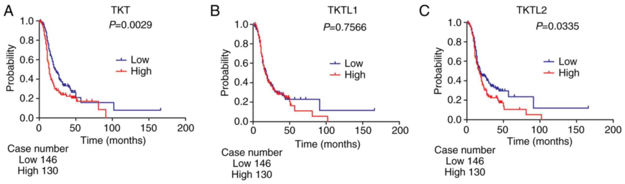
Validation of prognostic values of
transketolase family genes in ovarian cancer
For the verification of the prognostic roles of
transketolase family genes, the GSE9891 dataset was analyzed by
log-rank test (Fig. 8). Similar
results to the Kaplan-Meier plotter analysis were observed: In all
patients with ovarian cancer, high expression levels of TKT and
TKTL2 were associated with poor PFS, whereas TKTL1 was not
associated with PFS.
Discussion
Transketolases, which are key enzymes of the
non-oxidative branch of the pentose phosphate pathway, are
associated with the tumorigenesis and development of a variety of
tumors (10). The aim of the current
study was to investigate the expression levels and prognostic
values of transketolase genes in ovarian cancer.
TKT is not only involved in cancer development and
progression, but also contributes to patient survival. For example,
TKT has been reported to be upregulated in hepatocellular carcinoma
(5). In addition, high TKT
expression levels indicate poor survival in hepatocellular
carcinoma and esophageal cancer (5,16). A
number of previous studies have indicated carcinogenic roles for
TKT in breast cancer and gynecological tumors. In breast cancer,
Tseng et al (30)
demonstrated that TKT expression levels were upregulated in breast
cancer compared with those in normal tissue, and were higher in
metastatic lymphoid tissue compared with those in the primary site.
Further analysis of the association between TKT expression and
clinical parameters identified that TKT expression was associated
with breast cancer stage and grade, patient age, and tumor size and
type; in addition, patients with high TKT expression exhibited a
worse outcome compared with that observed in the low-TKT expression
group (30). A study by Yang et
al (31) revealed that TKT was
highly expressed in 81.1% of cervical cancer samples and was
significantly upregulated in cervical cancer compared with its
expression in normal cervical tissue. Chen et al (32) compared serum peptide profiles among
healthy control subjects and patients with cervical cancer prior
and subsequent to surgery. The results demonstrated that TKT
expression levels were higher in patients with cervical cancer
prior to surgery compared with those in healthy subjects and
patients following surgery. In addition, TKT is expressed in
ascites fluid from patients with ovarian cancer, but not from
individuals with benign lesions (21). Yi et al (33) identified TKT in exosomes derived from
two late-stage ovarian cancer cell lines and suggested that TKT may
serve a dominant role in exosome-mediated carcinogenicity and
cancer progression. Depletion of TKT by TKT siRNA inhibited
proliferation in ovarian cancer cells in vitro (22). Oxythiamine, a transketolase
inhibitor, also decreased tumor cell proliferation in ovarian
cancer cell lines and primary serous ovarian cancer cells (22). Only one study is available concerning
the prognostic values of TKT in ovarian cancer, which reported that
patients with high nuclear TKT expression in peritoneal metastases
exhibited shorter OS time compared with that of patients with low
TKT protein levels (22). However,
no correlation was identified between PFS and TKT expression in
either primary cancer or peritoneal metastases (22). Thus, whether or not TKT mRNA has a
prognostic role in patients with ovarian cancer remains to be
determined. In the present study, a significant upregulation of TKT
expression was observed in ovarian cancer compared with that in
normal ovarian tissue. In addition, high TKT mRNA expression was
associated with poor PFS in all patients with ovarian cancer, as
well as in patients with serous-type ovarian cancer. Although the
OS between the high and low-TKT expression groups in all ovarian
cancer and serous ovarian cancer samples was not significantly
different, the P-value was <0.1.
The mechanism underlying the carcinogenic roles of
TKT in tumors has been investigated in a number of studies. A
recent study has reported that TKT interacts with SH2
domain-containing 5 and activates STAT3 phosphorylation, thus
promoting the proliferation of liver cancer cells (34). This result suggested that STAT3 may
be involved in the regulation of liver cancer cell proliferation by
TKT. Lin et al (35)
demonstrated that the knockdown of TKT prevents vascular
endothelial growth factor secretion and inhibits cell proliferation
in lung cancer. To the best of our knowledge, no conclusive results
are available on the mechanism of TKT in ovarian cancer, which
needs further investigation.
Previous studies have demonstrated that TKTL1 serves
crucial roles in cancer prognosis. High expression levels of TKTL1
have been reported to be a significant indicator of poor outcome in
colorectal (36) and gastric
(18) cancer, esophageal squamous
cell carcinoma (37), and locally
advanced rectal (38) and non-small
cell lung (17) cancer. In ovarian
cancer, TKTL1 expression was observed in 81% of granulosa cell
tumors of the ovary, but not in normal ovarian tissue (39). Krockenberger et al (40) also demonstrated that the expression
of TKTL1 is significantly higher in tumor tissues of serous
papillary ovarian adenocarcinomas compared with that in borderline
tumors or normal tissues. In addition, increased expression of
TKTL1 was associated with higher Federation International of
Gynecology and Obstetrics stage, initial ascites and higher
histopathological grade in serous papillary ovarian carcinoma
(40). These results indicated that
TKTL1 may be an oncogene in ovarian carcinogenesis and development.
However, to the best of our knowledge, no studies on the prognostic
significance of TKTL1 in ovarian cancer are currently available. In
the present study, upregulation of TKTL1 expression was
demonstrated in all ovarian cancer cases, especially in serous
ovarian cancer. In addition, high TKTL1 mRNA expression levels were
significantly associated with poor PFS in patients with serous and
endometrioid ovarian cancer. However, when the histological types
of ovarian cancer were not distinguished, TKTL1 exhibited no
effects on PFS in all patients with ovarian cancer due to the
different cut-off values in different histological types. The
cut-off values used for serous and endometrioid ovarian cancer in
the present study were 10 and 24, respectively, which meant that
patients with serous and endometrioid ovarian cancer were split
differently to separate the high and low-expression groups.
According to this divisional standard, patients with serous ovarian
cancer with transcription level >10 comprised the high
expression group, whereas patients with serous ovarian cancer with
transcription level <10 comprised the low expression group. In
endometrioid ovarian cancer, patients whose expression value is
>24 should be classified in the high expression group, whereas
patients whose expression value is <24 should be classified in
the low expression group. However, if this study used the same
cut-off, such as 15, to separate the high and low groups, ignoring
the histological types of ovarian cancer, serous ovarian cancer
patients with actually high expression would be mistakenly defined
as low group, and endometrioid ovarian cancer patients with
actually low expression would be mistakenly classified as high
expression, thereby affecting the results of analysis. Therefore,
the histology of ovarian cancer should be considered when assessing
the prognostic roles of TKTL1 in ovarian cancer.
Studies on the function of TKTL2 in malignancies are
limited. Chen et al (41)
compared TKTL2 expression between uterine cervix cancer cells and
normal human endocervical epithelial cells, and no significant
differences between the two types of cell lines were observed. A
similar result was observed in nasopharyngeal carcinoma (42). To the best of our knowledge, no
studies on TKTL2 in ovarian cancer are currently available. In the
present study, TKTL2 expression levels were compared between
serous, endometrioid, clear cell or mucinous ovarian cancer tissues
and normal ovarian tissues; however, no differences were observed.
By contrast, high TKTL2 mRNA expression was significantly
associated with poor PFS and OS in all patients with ovarian
cancer. The negative result of the relative expression of TKTL2 in
ovarian cancer may be due to the limited sample size, as the Lu
dataset, which was used to compare the differential expression of
TKTL2 between tumor and normal tissue, only included 5 normal
ovarian tissue samples. Differential expression of TKTL2 between
ovarian cancer and normal ovarian tissue requires further
study.
The prognostic value of the transketolase family
genes in ovarian cancer with different clinical stages was further
investigated. The three transketolase genes were associated with
poor PFS in patients with stage III/IV ovarian cancer, but
exhibited no effects on PFS among patients with stage I/II ovarian
cancer. These results suggested that the transketolase family genes
may be a negative prognostic indicator for late-stage ovarian
cancer.
Platinum and taxol are the two most frequently used
chemotherapeutic drugs for treating ovarian cancer (43). However, a number of patients
ultimately progress to recurrent, chemotherapy-resistant tumors,
and the majority of these patients succumb to the disease within
one year (44). There is a great
need to identify prognostic markers to predict the outcome of
patients with ovarian cancer treated with platinum and paclitaxel.
The results of the present study demonstrated that the
transketolase family genes were associated with chemotherapy
sensitivity in ovarian cancer. Transketolase family genes served
different roles in chemotherapy sensitivity. The results revealed
that enhanced TKT expression was associated with unfavorable PFS in
patients with ovarian cancer treated with platinum, whereas high
TKTL1 expression was associated with poor PFS in patients treated
with taxol. TKTL2 was associated with poor outcomes in patients
treated with platinum plus taxol. These results were consistent
with those from previous studies. Taoka et al (45) compared differential protein
expression profiles between platinum-naïve and platinum-resistant
bladder cancer cell lines to identify the candidate molecules
associated with platinum resistance, and revealed that the
expression of TKT was increased by 1.5-fold in platinum-resistant
bladder cancer compared with that in platinum-naïve cells. Yang
et al (31) also demonstrated
that silencing of TKT significantly reduced cell viability and
increased apoptosis in the presence of platinum via glutathione
depletion and reactive oxygen species generation in cervical
cancer. In addition, overexpression of TKT increased resistance to
platinum chemotherapy by increasing cell viability and decreasing
apoptosis in cervical cancer in vitro (31). A study on the role of TKTL1 in taxol
sensitivity revealed that the inhibition of TKTL1 significantly
decreased the proliferation rate and the IC50 of taxol
in ovarian cancer (46).
Mechanistically, following TKTL1 knockdown, NADPH levels were
reduced and NADP+ levels were increased compared with
the levels in the control group, which led to taxol sensitivity in
ovarian cancer (46).
The TP53 gene, located on chromosome 17p13, encodes
the tumor-suppressor protein p53 (47). TP53 serves an important role in the
regulation of the cell cycle, DNA repair and cell death through
apoptosis (48). Mutation or loss of
function of the TP53 gene is a frequent and important genetic
alteration in the development of ovarian cancer (49). Harami-Papp et al (50) compared gene expression between
TP53-mutated and wild-type tissue samples from patients with breast
cancer and revealed that transketolase was upregulated in
TP53-mutated breast cancer compared with its expression in TP53
wild-type breast cancer. In the present study, the prognostic roles
of individual transketolase genes in patients with ovarian cancer
with different TP53 mutation status were examined; the three
transketolase family genes were associated with poor PFS in
patients with TP53-mutated, but not with TP53 wild-type, ovarian
cancer. Therefore, the transketolase family may have an effect on
the prognosis of TP53-mutated ovarian cancer.
A lack of experimental data was the main limitation
of the present study. To strengthen the conclusions, cell and
animal models with transketolase gene knockdown and overexpression
may be constructed, which would verify the tumorigenic functions of
transketolase genes in ovarian cancer. Future studies may also
collect clinical information and perform immunohistochemistry on
clinical cervical cancer samples to subsequently validate the
prognostic roles of transketolase genes.
In summary, the present study identified elevated
TKT and TKTL1 expression in ovarian cancer compared with that in
normal ovarian tissue. High mRNA expression of the above three
transketolase genes was demonstrated to be associated with poor PFS
in patients with serous ovarian cancer, especially those at an
advanced clinical stage. TKTL2 was also significantly associated
with poor OS in all patients with ovarian cancer. In addition,
transketolase family genes served a role in predicting PFS in
patients with ovarian cancer treated with platinum and/or taxol, as
well as in patients with TP53-mutated ovarian cancer. These results
suggest that transketolase genes have a prognostic value in ovarian
cancer. This may facilitate better prognostic stratification and
may be useful for the detailed understanding of the complexity and
heterogeneity implicated in the molecular biology of ovarian
cancer.
Acknowledgements
Not applicable.
Funding
The present study was supported by the Key
Laboratory of Wenzhou City-Gynecological Oncology (grant no.
ZD201603). The sponsor had no involvement in the collection,
analysis and interpretation of data or in the writing of the
manuscript.
Availability of data and materials
The datasets generated and/or analyzed during the
present study are available in Oncomine (https://www.oncomine.org/resource/login.html) and
the Kaplan-Meier plotter database (http://www.kmplot.com/analysis/index.php?p=service&cancer=ovar).
Authors' contributions
MZ and XZ conceived and designed the study. MY and
JZ collected the data. MZ and MY analyzed the data. All authors
participated in manuscript writing. All authors contributed to data
analysis, drafting and revising the manuscript, gave final approval
of the version to be published and agreed to be accountable for all
aspects of the work.
Ethics approval and consent to
participate
All data in the present study were obtained from
publicly available datasets. No additional patients were enrolled
in the study.
Patient consent for publication
Not applicable.
Competing interests
The authors declare that they have no competing
interests.
References
|
1
|
Siegel RL, Miller KD and Jemal A: Cancer
statistics, 2019. CA Cancer J Clin. 69:7–34. 2019. View Article : Google Scholar : PubMed/NCBI
|
|
2
|
Coleman MP, Forman D, Bryant H, Butler J,
Rachet B, Maringe C, Nur U, Tracey E, Coory M, Hatcher J, et al:
Cancer survival in Australia, Canada, Denmark, Norway, Sweden and
the UK, 1995–2007 (the International Cancer Benchmarking
Partnership): An analysis of population-based cancer registry data.
Lancet. 377:127–138. 2011. View Article : Google Scholar : PubMed/NCBI
|
|
3
|
Warburg O: On the origin of cancer cells.
Science. 123:309–314. 1956. View Article : Google Scholar : PubMed/NCBI
|
|
4
|
Ward PS and Thompson CB: Metabolic
reprogramming: A cancer hallmark even warburg did not anticipate.
Cancer Cell. 21:297–308. 2012. View Article : Google Scholar : PubMed/NCBI
|
|
5
|
Zheng Y, Huang Q, Ding Z, Liu T, Xue C,
Sang X and Gu J: Genome-wide DNA methylation analysis identifies
candidate epigenetic markers and drivers of hepatocellular
carcinoma. Brief Bioinform. 19:101–108. 2018.PubMed/NCBI
|
|
6
|
Diaz-Moralli S, Tarrado-Castellarnau M,
Alenda C, Castells A and Cascante M: Transketolase-like 1
expression is modulated during colorectal cancer progression and
metastasis formation. PLoS One. 6:e253232011. View Article : Google Scholar : PubMed/NCBI
|
|
7
|
Kowalik MA, Columbano A and Perra A:
Emerging role of the pentose phosphate pathway in hepatocellular
carcinoma. Front Oncol. 7:872017. View Article : Google Scholar : PubMed/NCBI
|
|
8
|
Jiang P, Du W and Wu M: Regulation of the
pentose phosphate pathway in cancer. Protein Cell. 5:592–602. 2014.
View Article : Google Scholar : PubMed/NCBI
|
|
9
|
Boros LG, Puigjaner J, Cascante M, Lee WN,
Brandes JL, Bassilian S, Yusuf FI, Williams RD, Muscarella P,
Melvin WS and Schirmer WJ: Oxythiamine and dehydroepiandrosterone
inhibit the nonoxidative synthesis of ribose and tumor cell
proliferation. Cancer Res. 57:4242–4248. 1997.PubMed/NCBI
|
|
10
|
Coy JF, Dressler D, Wilde J and Schubert
P: Mutations in the transketolase-like gene TKTL1: Clinical
implications for neurodegenerative diseases, diabetes and cancer.
Clin Lab. 51:257–273. 2005.PubMed/NCBI
|
|
11
|
Zhao J and Zhong CJ: A review on research
progress of transketolase. Neurosci Bull. 25:94–99. 2009.
View Article : Google Scholar : PubMed/NCBI
|
|
12
|
Li J, Zhu SC, Li SG, Zhao Y, Xu JR and
Song CY: TKTL1 promotes cell proliferation and metastasis in
esophageal squamous cell carcinoma. Biomed Pharmacother. 74:71–76.
2015. View Article : Google Scholar : PubMed/NCBI
|
|
13
|
Fritz P, Coy JF, Murdter TE, Ott G,
Alscher MD and Friedel G: TKTL-1 expression in lung cancer. Pathol
Res Pract. 208:203–209. 2012. View Article : Google Scholar : PubMed/NCBI
|
|
14
|
Sun W, Liu Y, Glazer CA, Shao C, Bhan S,
Demokan S, Zhao M, Rudek MA, Ha PK and Califano JA: TKTL1 is
activated by promoter hypomethylation and contributes to head and
neck squamous cell carcinoma carcinogenesis through increased
aerobic glycolysis and HIF1alpha stabilization. Clin Cancer Res.
16:857–866. 2010. View Article : Google Scholar : PubMed/NCBI
|
|
15
|
Krockenberger M, Engel JB, Schmidt M,
Kohrenhagen N, Hausler SF, Dombrowski Y, Kapp M, Dietl J and Honig
A: Expression of transketolase-like 1 protein (TKTL1) in human
endometrial cancer. Anticancer Res. 30:1653–1659. 2010.PubMed/NCBI
|
|
16
|
Chao YK, Peng TL, Chuang WY, Yeh CJ, Li
YL, Lu YC and Cheng AJ: Transketolase serves a poor prognosticator
in esophageal cancer by promoting cell Invasion via
epithelial-mesenchymal transition. J Cancer. 7:1804–1811. 2016.
View Article : Google Scholar : PubMed/NCBI
|
|
17
|
Kayser G, Sienel W, Kubitz B, Mattern D,
Stickeler E, Passlick B, Werner M and Zur Hausen A: Poor outcome in
primary non-small cell lung cancers is predicted by transketolase
TKTL1 expression. Pathology. 43:719–724. 2011. View Article : Google Scholar : PubMed/NCBI
|
|
18
|
Song Y, Liu D and He G: TKTL1 and p63 are
biomarkers for the poor prognosis of gastric cancer patients.
Cancer Biomark. 15:591–597. 2015. View Article : Google Scholar : PubMed/NCBI
|
|
19
|
Benito A, Polat IH, Noe V, Ciudad CJ,
Marin S and Cascante M: Glucose-6-phosphate dehydrogenase and
transketolase modulate breast cancer cell metabolic reprogramming
and correlate with poor patient outcome. Oncotarget.
8:106693–106706. 2017. View Article : Google Scholar : PubMed/NCBI
|
|
20
|
Langbein S, Zerilli M, Zur Hausen A,
Staiger W, Rensch-Boschert K, Lukan N, Popa J, Ternullo MP,
Steidler A, Weiss C, et al: Expression of transketolase TKTL1
predicts colon and urothelial cancer patient survival: Warburg
effect reinterpreted. Br J Cancer. 94:578–585. 2006. View Article : Google Scholar : PubMed/NCBI
|
|
21
|
Bery A, Leung F, Smith CR, Diamandis EP
and Kulasingam V: Deciphering the ovarian cancer ascites fluid
peptidome. Clin Proteomics. 11:132014. View Article : Google Scholar : PubMed/NCBI
|
|
22
|
Ricciardelli C, Lokman NA, Cheruvu S, Tan
IA, Ween MP, Pyragius CE, Ruszkiewicz A, Hoffmann P and Oehler MK:
Transketolase is upregulated in metastatic peritoneal implants and
promotes ovarian cancer cell proliferation. Clin Exp Metastasis.
32:441–455. 2015. View Article : Google Scholar : PubMed/NCBI
|
|
23
|
Rhodes DR, Kalyana-Sundaram S, Mahavisno
V, Varambally R, Yu J, Briggs BB, Barrette TR, Anstet MJ,
Kincead-Beal C, Kulkarni P, et al: Oncomine 3.0: Genes, pathways,
and networks in a collection of 18,000 cancer gene expression
profiles. Neoplasia. 9:166–180. 2007. View Article : Google Scholar : PubMed/NCBI
|
|
24
|
Gyorffy B, Lanczky A, Eklund AC, Denkert
C, Budczies J, Li Q and Szallasi Z: An online survival analysis
tool to rapidly assess the effect of 22,277 genes on breast cancer
prognosis using microarray data of 1,809 patients. Breast Cancer
Res Treat. 123:725–731. 2010. View Article : Google Scholar : PubMed/NCBI
|
|
25
|
Gyorffy B, Surowiak P, Budczies J and
Lanczky A: Online survival analysis software to assess the
prognostic value of biomarkers using transcriptomic data in
non-small-cell lung cancer. PLoS One. 8:e822412013. View Article : Google Scholar : PubMed/NCBI
|
|
26
|
Gyorffy B, Lanczky A and Szallasi Z:
Implementing an online tool for genome-wide validation of
survival-associated biomarkers in ovarian-cancer using microarray
data from 1287 patients. Endocr Relat Cancer. 19:197–208. 2012.
View Article : Google Scholar : PubMed/NCBI
|
|
27
|
Zhao M, Li S, Zhou L, Shen Q, Zhu H and
Zhu X: Prognostic values of excision repair cross-complementing
genes mRNA expression in ovarian cancer patients. Life Sci.
194:34–39. 2018. View Article : Google Scholar : PubMed/NCBI
|
|
28
|
Tian X, Han Y, Yu L, Luo B, Hu Z, Li X,
Yang Z, Wang X, Huang W, Wang H, et al: Decreased expression of
ALDH5A1 predicts prognosis in patients with ovarian cancer. Cancer
Biol Ther. 18:245–251. 2017. View Article : Google Scholar : PubMed/NCBI
|
|
29
|
Tothill RW, Tinker AV, George J, Brown R,
Fox SB, Lade S, Johnson DS, Trivett MK, Etemadmoghadam D, Locandro
B, et al: Novel molecular subtypes of serous and endometrioid
ovarian cancer linked to clinical outcome. Clin Cancer Res.
14:5198–5208. 2008. View Article : Google Scholar : PubMed/NCBI
|
|
30
|
Tseng CW, Kuo WH, Chan SH, Chan HL, Chang
KJ and Wang LH: Transketolase regulates the metabolic switch to
control breast cancer cell metastasis via the α-Ketoglutarate
signaling pathway. Cancer Res. 78:2799–2812. 2018. View Article : Google Scholar : PubMed/NCBI
|
|
31
|
Yang H, Wu XL, Wu KH, Zhang R, Ju LL, Ji
Y, Zhang YW, Xue SL, Zhang YX, Yang YF and Yu MM: MicroRNA-497
regulates cisplatin chemosensitivity of cervical cancer by
targeting transketolase. Am J Cancer Res. 6:2690–2699.
2016.PubMed/NCBI
|
|
32
|
Chen Y, Xiong X, Wang Y, Zhao J, Shi H,
Zhang H, Wang Y, Wei Y, Xue W and Zhang J: Proteomic screening for
serum biomarkers for cervical cancer and their clinical
significance. Med Sci Monit. 25:288–297. 2019. View Article : Google Scholar : PubMed/NCBI
|
|
33
|
Yi H, Zheng X, Song J, Shen R, Su Y and
Lin D: Exosomes mediated pentose phosphate pathway in ovarian
cancer metastasis: A proteomics analysis. Int J Clin Exp Pathol.
8:15719–15728. 2015.PubMed/NCBI
|
|
34
|
Zheng Y, Ming P, Zhu C, Si Y, Xu S, Chen
A, Wang J and Zhang B: Hepatitis B virus X protein-induced SH2
domain-containing 5 (SH2D5) expression promotes hepatoma cell
growth via an SH2D5-transketolase interaction. J Biol Chem.
294:4815–4827. 2019. View Article : Google Scholar : PubMed/NCBI
|
|
35
|
Lin CC, Chen LC, Tseng VS, Yan JJ, Lai WW,
Su WP, Lin CH, Huang CY and Su WC: Malignant pleural effusion cells
show aberrant glucose metabolism gene expression. Eur Respir J.
37:1453–1465. 2011. View Article : Google Scholar : PubMed/NCBI
|
|
36
|
Ahopelto K, Bockelman C, Hagstrom J,
Koskensalo S and Haglund C: Transketolase-like protein 1 expression
predicts poor prognosis in colorectal cancer. Cancer Biol Ther.
17:163–168. 2016. View Article : Google Scholar : PubMed/NCBI
|
|
37
|
Shi Z, Tang Y, Li K and Fan Q: TKTL1
expression and its downregulation is implicated in cell
proliferation inhibition and cell cycle arrest in esophageal
squamous cell carcinoma. Tumour Biol. 36:8519–8529. 2015.
View Article : Google Scholar : PubMed/NCBI
|
|
38
|
Schwaab J, Horisberger K, Ströbel P, Bohn
B, Gencer D, Kähler G, Kienle P, Post S, Wenz F, Hofmann WK, et al:
Expression of Transketolase like gene 1 (TKTL1) predicts
disease-free survival in patients with locally advanced rectal
cancer receiving neoadjuvant chemoradiotherapy. BMC Cancer.
11:3632011. View Article : Google Scholar : PubMed/NCBI
|
|
39
|
Schmidt M, Kammerer U, Segerer S, Cramer
A, Kohrenhagen N, Dietl J and Voelker HU: Glucose metabolism and
angiogenesis in granulosa cell tumors of the ovary: Activation of
Akt, expression of M2PK, TKTL1 and VEGF. Eur J Obstet Gynecol
Reprod Biol. 139:72–78. 2008. View Article : Google Scholar : PubMed/NCBI
|
|
40
|
Krockenberger M, Honig A, Rieger L, Coy
JF, Sutterlin M, Kapp M, Horn E, Dietl J and Kammerer U:
Transketolase-like 1 expression correlates with subtypes of ovarian
cancer and the presence of distant metastases. Int J Gynecol
Cancer. 17:101–106. 2007. View Article : Google Scholar : PubMed/NCBI
|
|
41
|
Chen H, Yue JX, Yang SH, Ding H, Zhao RW
and Zhang S: Overexpression of transketolase-like gene 1 is
associated with cell proliferation in uterine cervix cancer. J Exp
Clin Cancer Res. 28:432009. View Article : Google Scholar : PubMed/NCBI
|
|
42
|
Zhang S, Yang JH and Cai PC: Effects of
transketolase-like gene TKTL1 on occurrence and metastasis of human
nasopharyngeal carcinoma. Zhonghua Yi Xue Za Zhi. 88:3131–3134.
2008.(In Chinese). PubMed/NCBI
|
|
43
|
Ge L, Li N, Yuan GW, Sun YC and Wu LY:
Nedaplatin and paclitaxel compared with carboplatin and paclitaxel
for patients with platinum-sensitive recurrent ovarian cancer. Am J
Cancer Res. 8:1074–1082. 2018.PubMed/NCBI
|
|
44
|
Griffiths RW, Zee YK, Evans S, Mitchell
CL, Kumaran GC, Welch RS, Jayson GC, Clamp AR and Hasan J: Outcomes
after multiple lines of chemotherapy for platinum-resistant
epithelial cancers of the ovary, peritoneum, and fallopian tube.
Int J Gynecol Cancer. 21:58–65. 2011. View Article : Google Scholar : PubMed/NCBI
|
|
45
|
Taoka Y, Matsumoto K, Ohashi K, Minamida
S, Hagiwara M, Nagi S, Saito T, Kodera Y and Iwamura M: Protein
expression profile related to cisplatin resistance in bladder
cancer cell lines detected by two-dimensional gel electrophoresis.
Biomed Res. 36:253–261. 2015. View Article : Google Scholar : PubMed/NCBI
|
|
46
|
Zheng X and Li H: TKTL1 modulates the
response of paclitaxel-resistant human ovarian cancer cells to
paclitaxel. Biochem Biophys Res Commun. 503:572–579. 2018.
View Article : Google Scholar : PubMed/NCBI
|
|
47
|
Na K, Sung JY and Kim HS: TP53 Mutation
status of tubo-ovarian and peritoneal high-grade serous carcinoma
with a wild-type p53 immunostaining pattern. Anticancer Res.
37:6697–6703. 2017.PubMed/NCBI
|
|
48
|
Kalamanathan S, Bates V, Lord R and Green
JA: The mutational profile of sporadic epithelial ovarian
carcinoma. Anticancer Res. 31:2661–2668. 2011.PubMed/NCBI
|
|
49
|
Munksgaard PS and Blaakaer J: The
association between endometriosis and ovarian cancer: A review of
histological, genetic and molecular alterations. Gynecol Oncol.
124:164–169. 2012. View Article : Google Scholar : PubMed/NCBI
|
|
50
|
Harami-Papp H, Pongor LS, Munkacsy G,
Horvath G, Nagy AM, Ambrus A, Hauser P, Szabo A, Tretter L and
Gyorffy B: TP53 mutation hits energy metabolism and increases
glycolysis in breast cancer. Oncotarget. 7:67183–67195. 2016.
View Article : Google Scholar : PubMed/NCBI
|