Introduction
Ovarian carcinoma is one of the most lethal
malignancies of the female reproductive system worldwide (1,2). The
incidence of ovarian carcinoma has gradually increased in Asian
countries, particularly in Japan (from 3.6 to 6.5%) in recent years
(3). Despite recent advances in
surgical techniques and medical management (4–6), the
long-term prognosis of patients with ovarian carcinoma remains
unsatisfactory. Furthermore, due to the lack of early diagnostic
methods and characteristic symptoms, most patients with ovarian
carcinoma are diagnosed at advanced clinical stages [stages III and
IV according to the International Federation of Gynecology and
Obstetrics (FIGO)] and have therefore a poor prognosis. The overall
5-year survival rate of patients with ovarian carcinoma is ~20%
(7). It is therefore crucial to
determine novel biomarkers suitable for tumor early detection,
prediction of tumor behavior and targeted therapy.
AGBL2 is a cytoplasmic carboxypeptidase that
catalyzes microtubule (MT) detyrosination (8). MTs are polarized polymers comprising
α/β tubulin heterodimers (9).
Multiple posttranslational modifications on tubulins, including
detyrosination, can regulate the stability of MTs (10). MT detyrosination, which targets the
C-terminal tyrosine residual of α-tubulin and increases MT
stability (11), is frequently
observed in human malignances, such as neuroblastomas, breast
cancer and prostate cancer (12–14). In
cancer cells, detyrosinated tubulins assemble to form an invasive
front and microtentacles (15,16).
Furthermore, detyrosinated tubulins are more resistant to
microtubule-targeting agents (17).
Previous studies demonstrated that AGBL2 promotes tumorigenesis and
cancer progression in breast cancer, prostate cancer and
hepatocellular carcinoma (8,18) and could be considered as a potential
biomarker for lymph node metastasis and chemotherapy resistance in
breast cancer (19). In addition,
AGBL2 promotes hepatocellular carcinoma cancer cell growth via
IRGM-regulated autophagy. After knockdown of IRGM, AGBL2-induced
up-regulation of the crucial members in autophagy (i.e. LC3A/B,
ATG5, ATG12 and ATG3) was mainly abolished (18).
AGBL2 expression and its clinical and prognostic
relevance in ovarian carcinomas have not yet been elucidated. In
the present study, expression status of AGBL2 protein was detected
by immunohistochemistry (IHC) in a series of ovarian cancer
specimens. Furthermore, the clinicopathological and prognostic
significance of AGBL2 protein expression in ovarian carcinoma
tissues was evaluated. To the best of our knowledge, the present
study was the first to provide novel insights into the role of
AGBL2 in the development and progression of ovarian carcinoma.
Materials and methods
Patients and tissue specimens
Archived paraffin-embedded cancer specimens were
obtained from 268 patients who underwent initial surgical resection
between June 2002 and June 2010. All samples, including 30 normal
ovarian tissues, 35 cystadenoma tissues, 38 borderline ovarian
tumors tissues and 165 invasive ovarian carcinoma tissues, were
collected from the archives of the Department of Pathology from the
Cancer Center and the First Affiliated Hospital (Sun Yat-Sen
University). No patient had received preoperative chemoradiation
therapy and all patients had completed clinical follow-up (up to
December 2016), of which data were available for review in this
study. Patients with incomplete clinical follow-up data or who died
from unknown causes were excluded from the study. In addition,
patient survival time data and clinicopathological characteristics
were collected. The age of the 165 patients diagnosed with primary
ovarian carcinoma ranged from 19 to 85 years (mean age, 51.0 years)
and their clinicopathological characteristics are presented in
Table I. Patients provided informed
consent and the study was approved by the Institute Research
Medical Ethics Committee of Sun Yat-Sen University.
 | Table I.Association between AGBL2 expression
and the clinicopathological characteristic of 165 patients with
ovarian carcinoma. |
Table I.
Association between AGBL2 expression
and the clinicopathological characteristic of 165 patients with
ovarian carcinoma.
|
|
| AGBL2 protein |
|---|
|
|
|
|
|---|
| Variable | Number of cases,
n | Low expression, n
(%) | High expression, n
(%) | P-valuea |
|---|
| Age at surgery,
years |
|
|
| 0.764 |
|
≤51.0b | 88 | 54 (61) | 34 (39) |
|
|
>51.0b | 77 | 49 (64) | 28 (36) |
|
| Histological
type |
|
|
| 0.659 |
|
Serous | 109 | 69 (63) | 40 (37) |
|
|
Mucinous | 19 | 13 (68) | 6 (32) |
|
|
Othersc | 37 | 21 (57) | 16 (43) |
|
| Histological grade
(Silveberg) |
|
|
| 0.040 |
| G1 | 28 | 22 (79) | 6 (21) |
|
| G2 | 96 | 61 (64) | 35 (36) |
|
| G3 | 41 | 20 (49) | 21 (51) |
|
| pT status |
|
|
| 0.025 |
| pT1 | 43 | 34 (79) | 9 (21) |
|
| pT2 | 34 | 21 (62) | 13 (38) |
|
| pT3 | 88 | 48 (55) | 40 (45) |
|
| pN status |
|
|
| <0.001 |
| pN0 | 81 | 62 (77) | 19 (23) |
|
| pN1 | 84 | 41 (49) | 43 (51) |
|
| pM status |
|
|
| 0.003 |
| pMX | 140 | 94 (67) | 46 (33) |
|
| pM1 | 25 | 9
(36) | 16 (64) |
|
| FIGO stage |
|
|
| <0.001 |
| I | 29 | 25 (86) | 4 (14) |
|
| II | 20 | 16 (80) | 4 (20) |
|
| III | 91 | 53 (58) | 38 (42) |
|
| IV | 25 | 9
(36) | 16 (64) |
|
Construction of tissue microarrays
(TMA)
Tissue microarrays were constructed as previously
described (20,21). Paraffin-embedded tissue blocks and
the corresponding histological hematoxylin and eosin (H&E)
stained slides were overlaid for TMA sampling construction. Typical
lesions (cancer-rich) were identified by a senior pathologist.
Corresponding histological H&E stained slide of each
paraffin-embedded tissue was examined under a BX-60 optical
microscope (Olympus Corporation) and marked for representative
areas of tumor tissues. Subsequently, a tissue microarray
instrument (Beecher Instruments, Inc.) was used to obtain tissue
cylinders of ~0.6-mm in diameter from selected cancer areas of
patient tissue. These sections were then re-embedded into a
recipient paraffin block with hollow presets. TMA block was then
cut into slices (5 µm thick) and mounted onto microscope
slides.
IHC staining
IHC staining was performed using a standard
streptavidin-biotin-peroxidase complex method as previously
described (22). Briefly, tissue
sections were deparaffinized and rehydrated in an ethanol gradient
(100, 90, 75 and 50%). The endogenous peroxidase activity was
blocked with 3% H2O2 at room temperature for
10 min. Slides were immersed in 10 mM citrate buffer (pH 8.0) and
boiled for 3 min in a pressure cooker for antigen retrieval.
Non-specific binding was blocked with 5% normal goat serum (OriGene
Technologies, Inc.) for 10 min at room temperature. Slides were
incubated with polyclonal primary antibodies against AGBL2 (1:50;
cat. no. HPA007718; Abcam), IRGM (1:200; cat. no. ab69494; Abcam)
and LC3A/B (1:100; cat. no. 12741; CST Biological Reagents Co.,
Ltd.) at 4°C overnight in a moist chamber. Slides were then
incubated with biotinylated goat anti-mouse immunoglobulin G (IgG)
antibody (1:100; cat. no. sc-516102; Santa Cruz Biotechnology,
Inc.) for 30 min at room temperature followed by a
streptavidin-peroxidase conjugate also for 30 min at room
temperature. Isotope-matched human IgG was used in each case as a
negative control. The 3,5-diaminobenzidine Substrate kit (Dako;
Agilent Technologies, Inc.) was used for color development followed
by Mayer hematoxylin counterstaining. Slides were counterstained
with 10% Mayer's hematoxylin for 1 min. AGBL2 staining was
evaluated according to previously described protein expression
criteria (14).
Statistical analysis
Statistical analysis was carried out using SPSS
statistical software version 20.0 (IBM Corp.). The association
between AGBL2 expression and clinicopathological features of
patients with ovarian carcinoma was assessed using the
χ2-test or Fisher's exact test. For univariate survival
analysis, survival curves were obtained through the Kaplan-Meier
method. Log rank test was used to compare different survival
curves. Multivariate survival analysis was carried out on all
parameters that were found to be significant following univariate
analysis by using Cox regression model. The differences between the
clinicopathological characteristics and survival predictions were
estimated using receiver operating characteristic curve (ROC)
analysis. P<0.05 was considered to indicate a statistically
significant difference.
Results
AGBL2 expression in ovarian
tissues
The results from IHC demonstrated that AGBL2
positive staining was mainly detected in the cytoplasm of ovarian
epithelial cells; however, AGBL2 was occasionally visible in the
nuclear (Fig. 1). High expression of
AGBL2 was observed in 62/165 (38%) of ovarian carcinomas (Fig. 1D), in 8/38 (21%) of borderline tumors
and in 3/35 (9%) of cystadenomas (Fig.
1C), but not in normal ovarian tissues (Fig. 1B). In addition, the results
demonstrated that the increased frequency of highly expressed AGBL2
in ovarian carcinomas was statistically significant (P<0.01;
Table II) compared with AGBL2
expression in benign and borderline tumors.
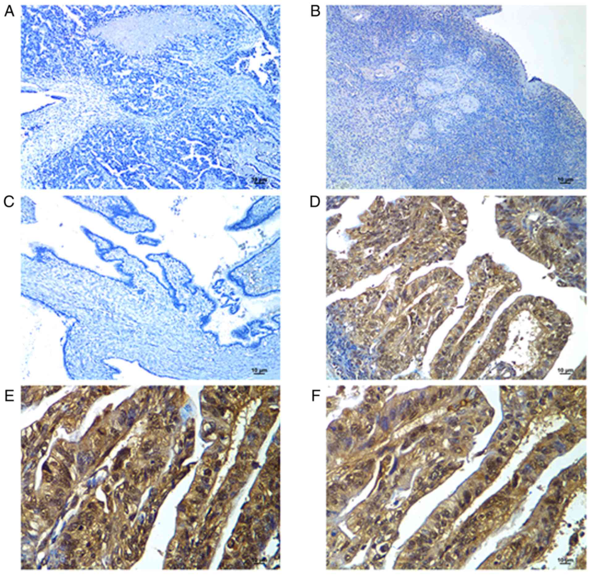 | Figure 1.Immunohistochemical staining of AGBL2
in human ovarian tissues and of IRGM and LC3A/B in epithelial
ovarian cancer tissue. (A) Negative control was obtained by
incubating tissue with normal IgG. Magnification, ×100. Scale bar,
10 µm. (B) Negative expression of AGBL2 was observed in the surface
epithelium of normal ovarian tissue. Magnification, ×100. Scale
bar, 10 µm. (C) Ovarian cystadenoma presented low AGBL2 expression
(10% of tumor cells showed moderate positive staining).
Magnification, ×100. Scale bar, 10 µm. (D) High AGBL2 expression
was detected in ovarian carcinoma tissue (case 52; >90% of
carcinoma cells showed strong positive). Magnification, ×200. Scale
bar, 10 µm. High expression of (E) IRGM and (F) LC3A/B in ovarian
carcinoma (case 52; >90% of carcinoma cells showed strong
positive staining for these two proteins). Magnification, ×400.
Scale bar, 10 µm. AGBL2, ATP/GTP binding protein like 2; IRGM,
immunity related GTPase M. |
| Table II.AGBL2 expression in normal ovarian
tissues and in benign and malignant epithelial ovarian tumor
tissues. |
Table II.
AGBL2 expression in normal ovarian
tissues and in benign and malignant epithelial ovarian tumor
tissues.
|
|
| AGBL2 protein |
|---|
|
|
|
|
|---|
| Tissue type | Number of cases,
n | Low expression, n
(%) | High expression, n
(%) |
|---|
| Normal ovaries | 30 | 30
(100) | 0
(0) |
| Cystadenomas | 35 | 32
(91) | 3
(9) |
| Borderline
tumors | 38 | 30
(79) | 8
(21) |
| Invasive
carcinomas | 165 | 103 (62) | 62 (38) |
Association of AGBL2 expression with
the clinicopathologic characteristics and survival of patients with
ovarian carcinoma
Following AGBL2 expression analysis, the
clinicopathological significance of highly expressed AGBL2 in
patients with ovarian carcinoma was analyzed. The results
demonstrated that high AGBL2 expression was positively associated
with high histological grade, advanced pathological
tumor-node-metastasis (pT/pN/pM) status and FIGO stage in patients
with ovarian carcinoma (P<0.05; Table
I). No significant association was observed between AGBL2
expression and other clinicopathological characteristics, including
the patient age and tumor histological type (P>0.05; Table I).
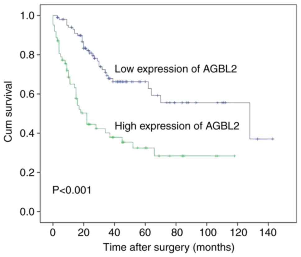
The cumulative survival curves using Kaplan-Meier
method for univariate survival analysis were calculated, and the
differences in survival times were analyzed by the log-rank test.
The results demonstrated that the prognostic predictors of patient
survival, including tumor histological grade (P=0.001), pT/pN/pM
status (P<0.01) and FIGO stage (P<0.001), had a significant
impact on patient prognosis (Table
III), which confirmed that the cohort of patients presented in
the present study was representative of the general population with
ovarian carcinoma. Subsequently, the impact of AGBL2 expression
level on the survival of patients with ovarian carcinoma was
analyzed. The results revealed that the mean survival time of
patients with ovarian carcinoma and high AGBL2 expression was of
46.4 months, which was significantly shorter than the 91.2 months
for patients with low AGBL2 expression (P<0.001; Table III; Fig.
2).
| Table III.Analysis of the clinicopathological
characteristics and AGBL2 expression for the prognosis of 165
patients with ovarian carcinoma by univariate survival analysis
(log-rank test). |
Table III.
Analysis of the clinicopathological
characteristics and AGBL2 expression for the prognosis of 165
patients with ovarian carcinoma by univariate survival analysis
(log-rank test).
| Variable | Number of cases,
n | Mean survival,
months | Median survival,
months | P-value |
|---|
| Age at surgery,
years |
|
|
| 0.949 |
|
≤51.0a | 88 | 76.6 | 64.0 |
|
|
>51.0a | 77 | 63.9 | 62.0 |
|
| Histological
type |
|
|
| 0.528 |
|
Serous | 109 | 64.6 | 52.0 |
|
|
Mucinous | 19 | 72.5 | 70.0 |
|
|
Othersc | 37 | 97.6 | NRb |
|
| Histological grade
(Silveberg) |
|
|
| 0.001 |
| G1 | 28 | 110.1 | NR |
|
| G2 | 96 | 73.3 | 64.0 |
|
| G3 | 41 | 43.6 | 28.0 |
|
| pT status |
|
|
| 0.001 |
|
pT1 | 43 | 111.8 | NR |
|
|
pT2 | 34 | 76.8 | 66.0 |
|
|
pT3 | 88 | 55.9 | 34.0 |
|
| pN status |
|
|
| <0.001 |
|
pN0 | 81 | 89.1 | 128.0 |
|
|
pN1 | 84 | 52.5 | 30.0 |
|
| pM status |
|
|
| <0.001 |
|
pMX | 140 | 85.3 | 128.0 |
|
|
pM1 | 25 | 22.1 | 13.0 |
|
| FIGO stage |
|
|
| <0.001 |
| I | 29 | 134.2 | NR |
|
| II | 20 | 107.6 | NR |
|
|
III | 91 | 60.3 | 36.0 |
|
| IV | 25 | 22.1 | 13.0 |
|
| AGBL2
expression |
|
|
| <0.001 |
| Low
expression | 103 | 91.2 | 128.0 |
|
| High
expression | 62 | 46.4 | 22.0 |
|
In order to determine whether the variables
exhibiting prognostic influence following univariate analysis were
covariates, the expression of AGBL2 and other significant
clinicopathological characteristics, including tumor histological
grade, pT/pN/pM status, and FIGO stage, were assessed with
multivariate analysis (Table IV).
Multivariate Cox regression analysis revealed that AGBL2 expression
(P=0.004), pN status (P=0.034) and FIGO stage (P<0.001) were
independent prognostic factors for poor overall survival (Table IV).
| Table IV.Multivariate analysis of the overall
survival of patients with ovarian carcinoma (Cox regression
model). |
Table IV.
Multivariate analysis of the overall
survival of patients with ovarian carcinoma (Cox regression
model).
| Variable | Relative risk | 95% confidence
interval | P-value |
|---|
| AGBL2a | 2.052 | 1.262–3.337 | 0.004 |
| pN
statusb | 1.727 | 1.042–2.861 | 0.034 |
| FIGO
stagec | 3.169 | 2.161–4.646 | 0.000 |
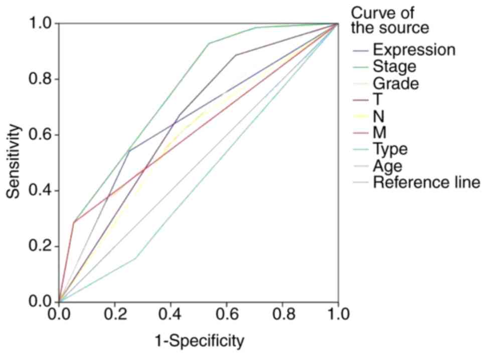
ROC can maximize both sensitivity and specificity
for the outcome and can therefore highlight the point on the curve
closest to (0.0, 1.0) for each clinicopathologic characteristic. In
order to evaluate the patient survival status, ROC curve analysis
was performed for each clinicopathologic characteristic and AGBL2
expression (area under the curve =0.645; P=0.001; Fig. 3).
Association between AGBL2, IRGM and
LC3A/B expression in patients with ovarian carcinoma
A recent study reported that AGBL2 participates in
tumorigenesis through IRGM-regulated autophagy (18). The potential association between
AGBL2 expression and IRGM and LC3A/B expression was therefore
assessed by IHC in tissues from patients with ovarian carcinoma
(Fig. 1D-F). The results
demonstrated that AGBL2 expression level was significantly
associated with IRGM (P=0.013) and LC3A/B (P=0.004) expression
level. In addition, IRGM expression level was positively associated
with LC3A/B expression level (P=0.023; Table V).
| Table V.Association between AGBL2 expression
and IRGM and LC3A/B expression in ovarian carcinoma tissues. |
Table V.
Association between AGBL2 expression
and IRGM and LC3A/B expression in ovarian carcinoma tissues.
|
|
| IRGM
expression | LC3A/B
expression |
|---|
|
|
|
|
|
|---|
| Expression
level | Number of cases,
n | Low, n (%) | High, n (%) |
P-valuea | Low, n (%) | High, n (%) |
P-valuea |
|---|
| AGBL2
expression |
|
|
| 0.013 |
|
| 0.004 |
|
Low | 103 | 35 (34) | 68 (66) |
| 65 (63) | 38 (37) |
|
|
High | 62 | 10 (16) | 52 (84) |
| 25 (40) | 37 (60) |
|
| LC3A/B
expression |
|
|
| 0.023 |
|
|
|
|
Low | 90 | 31 (34) | 59 (66) |
|
|
|
|
|
High | 75 | 14 (19) | 61 (81) |
|
|
|
|
Discussion
Epithelial ovarian cancer is the most lethal
gynecological malignancy in postmenopausal women (4). Although remarkable efforts have been
made to improve treatment modalities, ovarian carcinoma remains the
primary cause of gynecological cancer-associated mortality
(5). It is therefore crucial to
identify novel therapeutic approaches and targets to improve early
diagnosis and patient outcome.
Recent reports have verified that AGBL2 catalyzes MT
detyrosination and promotes tumorigenesis and cancer progression
(8). However, to date, the
expression status of AGBL2 and its clinicopathological significance
in ovarian carcinomas has not been elucidated (12–14). In
the present study, IHC was used to investigate whether an abnormal
AGBL2 expression could be involved in the pathogenesis of ovarian
carcinoma. By using a large cohort of ovarian tissues, including
normal, benign and borderline epithelial ovarian tumor tissues, and
malignant epithelial cancers, the results demonstrated that AGBL2
high expression level was consistent with ovarian tumor malignancy.
AGBL2 was more frequently highly expressed in ovarian carcinomas,
compared within benign and borderline tumors. Furthermore, high
AGBL2 expression was associated with high histological grade and
advanced clinical stage (pT/pN/pM and FIGO stage). These results
confirmed that AGBL2 could serve a crucial role in tumorigenesis
promotion of ovarian carcinoma. Previous studies demonstrated that
AGBL2 high expression is a prognostic marker of cancer and is
associated with poor clinical outcome in patients with
hepatocellular carcinoma and breast cancer (18,19).
Similarly, the results from this study indicated that AGBL2 high
expression could predict poor overall survival in patients with
ovarian carcinomas, and that AGBL2 may be considered as an
independent prognostic marker. Altogether, these findings suggest
that AGBL2 high expression may serve as a reliable prognosis
biomarker in patients with ovarian carcinoma. Subsequently, IHC
staining of AGBL2 may be used as an additional tool to identify
whether patients with ovarian carcinoma could have an increased
risk of tumor metastasis and/or a poor prognosis.
A recent study reported that AGBL2 stimulates
autophagy by regulating IRGM and therefore promote cancer cell
proliferation (18). The potential
association between AGBL2 expression and IRGM and LC3A/B expression
was therefore evaluated by IHC in tissues from patients with
ovarian carcinoma. The results demonstrated that AGBL2 expression
level was significantly associated IRGM and LC3A/B expression level
in ovarian carcinoma tissues. Furthermore, IRGM expression level
was positively associated with LC3A/B expression level. These
findings indicated that AGBL2 might also regulate autophagy by
modulating IRGM in ovarian carcinoma. A previous study reported
that inhibition of PI3K/AKT/mTOR and the Ras/MAPK signaling
pathways, which are involved in autophagy activation, can limit the
development of ovarian cancer (23).
The results from the present study identified a potential molecular
mechanism for the oncogenic role of AGBL2 in ovarian carcinoma
pathogenesis, and suggested that AGBL2 may enhance autophagy to
support the malignant phenotype of cancer cells. However, this
study was a retrospective study, and fresh tissue samples were only
rarely collected. The limitation of this study was the exclusive
use of IHC to determine AGBL2, IRGM and LC3A/B expression levels.
Future studies incorporating western blotting,
reverse-transcription-quantitative PCR and cell line experiments
are required for further investigation.
In conclusion, the present study was the first to
investigated AGBL2 expression in a large cohort of normal human
ovarian tissue, and in benign, borderline and malignant epithelial
ovarian tumors tissues by IHC. The results demonstrated that AGBL2
expression was upregulated in tumor tissues and may therefore serve
as a novel, reliable and independent prognostic marker in ovarian
carcinoma. In addition, these results further confirmed that
increased AGBL2 expression may enhance the aggressive behavior of
ovarian carcinoma.
Acknowledgements
Not applicable.
Funding
The present study was funded by the Nature Science
Foundation of China (grant no. 81772769) and the Research Project
in the Science and Technology Bureau in Guangzhou (grant no.
201704020125).
Availability of data and materials
The datasets used and/or analyzed during the current
study are available from the corresponding author on reasonable
request.
Authors' contributions
WPH and LLW designed and performed the experiments
and drafted and revised the manuscript. Both authors approved the
final version of the manuscript.
Ethics approval and consent to
participate
The present study was approved by the Research
Ethics Committee of the First Affiliated Hospital, Sun Yat-Sen
University (approval no. 2018-32) and all patients provided written
informed consent prior to the study.
Patient consent for publication
Not applicable.
Competing interests
The authors declare that they have no competing
interests.
References
|
1
|
Wingo PA, Ries LA, Rosenberg HM, Miller DS
and Edwards BK: Cancer incidence and mortality. 1973-1995, a report
card for the U.S. Cancer. 82:1197–1207. 1998. View Article : Google Scholar : PubMed/NCBI
|
|
2
|
Siegel RL, Miller KD and Jemal A: Cancer
statistics, 2018. CA Cancer J Clin. 68:7–30. 2018. View Article : Google Scholar : PubMed/NCBI
|
|
3
|
Coburn SB, Bray F, Sherman ME and Trabert
B: International patterns and trends in ovarian cancer incidence,
overall and by histologic subtype. Int J Cancer. 140:2451–2460.
2017. View Article : Google Scholar : PubMed/NCBI
|
|
4
|
Jelovac D and Armstrong DK: Recent
progress in the diagnosis and treatment of ovarian cancer. CA
Cancer J Clin. 61:183–203. 2011. View Article : Google Scholar : PubMed/NCBI
|
|
5
|
Oza AM, Cook AD, Pfisterer J, Embleton A,
Ledermann JA, Pujade-Lauraine E, Kristensen G, Carey MS, Beale P,
Cervantes A, et al: Standard chemotherapy with or without
bevacizumab for women with newly diagnosed ovarian cancer (ICON7):
Overall survival results of a phase 3 randomised trial. Lancet
Oncol. 16:928–936. 2015. View Article : Google Scholar : PubMed/NCBI
|
|
6
|
Coleman RL, Monk BJ, Sood AK and Herzog
TJ: Latest research and treatment of advanced-stage epithelial
ovarian cancer. Nat Rev Clin Oncol. 10:211–224. 2013. View Article : Google Scholar : PubMed/NCBI
|
|
7
|
Barnholtz-Sloan JS, Schwartz AG, Qureshi
F, Jacques S, Malone J and Munkarah AR: Ovarian cancer: Changes in
patterns at diagnosis and relative survival over the last three
decades. Am J Obstet Gynecol. 189:1120–1127. 2003. View Article : Google Scholar : PubMed/NCBI
|
|
8
|
Sahab ZJ, Hall MD, Me Sung Y,
Dakshanamurthy S, Ji Y, Kumar D and Byers SW: Tumor suppressor
RARRES1 interacts with cytoplasmic carboxypeptidase AGBL2 to
regulate the α-tubulin tyrosination cycle. Cancer Res.
71:1219–1228. 2011. View Article : Google Scholar : PubMed/NCBI
|
|
9
|
de Forges H, Bouissou A and Perez F:
Interplay between microtubule dynamics and intracellular
organization. Int J Biochem Cell Biol. 44:266–274. 2012. View Article : Google Scholar : PubMed/NCBI
|
|
10
|
Song Y and Brady ST: Post-translational
modifications of tubulin: Pathways to functional diversity of
microtubules. Trends Cell Biol. 25:125–136. 2015. View Article : Google Scholar : PubMed/NCBI
|
|
11
|
Webster DR, Gundersen GG, Bulinski JC and
Borisy GG: Differential turnover of tyrosinated and detyrosinated
microtubules. Proc Natl Acad Sci USA. 84:9040–9044. 1987.
View Article : Google Scholar : PubMed/NCBI
|
|
12
|
Kato C, Miyazaki K, Nakagawa A, Ohira M,
Nakamura Y, Ozaki T, Imai T and Nakagawara A: Low expression of
human tubulin tyrosine ligase and suppressed tubulin
tyrosination/detyrosination cycle are associated with impaired
neuronal differentiation in neuroblastomas with poor prognosis. Int
J Cancer. 112:365–375. 2004. View Article : Google Scholar : PubMed/NCBI
|
|
13
|
Mialhe A, Lafanechère L, Treilleux I,
Peloux N, Dumontet C, Brémond A, Panh MH, Payan R, Wehland J,
Margolis RL and Job D: Tubulin detyrosination is a frequent
occurrence in breast cancers of poor prognosis. Cancer Res.
61:5024–5027. 2001.PubMed/NCBI
|
|
14
|
Soucek K, Kamaid A, Phung AD, Kubala L,
Bulinski JC, Harper RW and Eiserich JP: Normal and prostate cancer
cells display distinct molecular profiles of alpha-tubulin
posttranslational modifications. Prostate. 66:954–965. 2006.
View Article : Google Scholar : PubMed/NCBI
|
|
15
|
Whipple RA, Matrone MA, Cho EH, Balzer EM,
Vitolo MI, Yoon JR, Ioffe OB, Tuttle KC, Yang J and Martin SS:
Epithelial-to-mesenchymal transition promotes tubulin
detyrosination and microtentacles that enhance endothelial
engagement. Cancer Res. 70:8127–8137. 2010. View Article : Google Scholar : PubMed/NCBI
|
|
16
|
Whipple RA, Balzer EM, Cho EH, Matrone MA,
Yoon JR and Martin SS: Vimentin filaments support extension of
tubulin-based microtentacles in detached breast tumor cells. Cancer
Res. 68:5678–5688. 2008. View Article : Google Scholar : PubMed/NCBI
|
|
17
|
Gundersen GG and Bulinski JC: Selective
stabilization of microtubules oriented toward the direction of cell
migration. Proc Natl Acad Sci USA. 85:5946–5950. 1988. View Article : Google Scholar : PubMed/NCBI
|
|
18
|
Wang LL, Jin XH, Cai MY, Li HG, Chen JW,
Wang FW, Wang CY, Hu WW, Liu F and Xie D: AGBL2 promotes cancer
cell growth through IRGM-regulated autophagy and enhanced Aurora A
activity in hepatocellular carcinoma. Cancer Lett. 414:71–80. 2018.
View Article : Google Scholar : PubMed/NCBI
|
|
19
|
Zhang H, Ren Y, Pang D and Liu C: Clinical
implications of AGBL2 expression and its inhibitor latexin in
breast cancer. World J Surg Oncol. 12:1422014. View Article : Google Scholar : PubMed/NCBI
|
|
20
|
Yang GF, Li XM and Xie D: Overexpression
of clusterin in ovarian cancer is correlated with impaired
survival. Int J Gynecol Cancer. 19:1342–1346. 2009. View Article : Google Scholar : PubMed/NCBI
|
|
21
|
He WP, Zhou J, Cai MY, Xiao XS, Liao YJ,
Kung HF, Guan XY, Xie D and Yang GF: CHD1L protein is overexpressed
in human ovarian carcinomas and is a novel predictive biomarker for
patients survival. BMC Cancer. 12:4372012. View Article : Google Scholar : PubMed/NCBI
|
|
22
|
Xu L, Li X, Cai M, Chen J, Li X, Wu WK,
Kang W, Tong J, To KF, Guan XY, et al: Increased expression of
Solute carrier family 12 member 5 via gene amplification
contributes to tumour progression and metastasis and associates
with poor survival in colorectal cancer. Gut. 65:635–646. 2016.
View Article : Google Scholar : PubMed/NCBI
|
|
23
|
Lu Z, Yang H, Sutton MN, Yang M, Clarke
CH, Liao WS and Bast RC Jr: ARHI (DIRAS3) induces autophagy in
ovarian cancer cells by downregulating the epidermal growth factor
receptor, inhibiting PI3K and Ras/MAP signaling and activating the
FOXo3a-mediated induction of Rab7. Cell Death Differ. 21:1275–1289.
2014. View Article : Google Scholar : PubMed/NCBI
|