Introduction
Neuroendocrine tumors (NETs), which account for
<1% of all malignant tumors, originate from disseminated
neuroendocrine cells. Most NETs are found in the gastrointestinal
and respiratory tracts (1).
Neuroendocrine carcinomas (NECs) in the gastrointestinal tract are
mostly found in the rectum, jejuno-ileum, and pancreas. Primary NEC
of the gallbladder is a rare disease, it makes up 1.4% of all
gastrointestinal endocrine tumors, and 0.4–4% of all malignant
tumors that occur in the gallbladder (2). In general, somatostatin receptor
scintigraphy (SRS) has a low diagnostic sensitivity for NECs and
18F fluorodeoxyglucose (FDG)-positron emission
tomography (PET) is useful in the diagnosis of tumors with a high
proliferation index and high glucose consumption.
Here, we report a case of a patient preoperatively
diagnosed with gallbladder NEC using SRS.
Case presentation
A 63-year-old man with no complaints visited a
doctor for abdominal ultrasonography (AUS) screening. AUS revealed
thickening of the gallbladder wall. The patient was therefore
admitted to our hospital for further examination. He was taking
medication for hypertension and type 2 diabetes and he had a family
history of lung cancer.
Blood analysis at the time of admission revealed
mild liver dysfunction. No elevation in the levels of tumor markers
such as carcinoembryonic antigen (CEA), cancer antigen 19-9 (CA
19-9), neurospecific enolase (NSE), or pro-gastrin-releasing
peptide was observed. There were no abnormalities in the levels of
hormones such as insulin, glucagon, or gastrin (Table I–V).
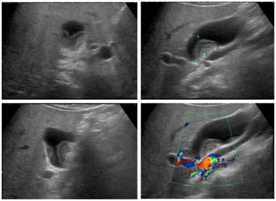
AUS showed a low-echoic mass (22×14 mm) with clear boundaries to
the liver, protruding into the lumen from the gallbladder neck
(Fig. 1).
 | Table I.Hematology. |
Table I.
Hematology.
| Hematology | Result | Unit |
|---|
| WBC | 3710 | /µl |
|
Lymph | 25.3 | % |
| Mono | 4.9 | % |
|
Eosino | 1.1 | % |
| Baso | 0.3 | % |
| Neut | 68.4 | % |
| RBC |
543×104 | /µl |
| Hb | 14.6 | g/dl |
| Hct | 44.2 | % |
| Plt |
16.3×10⁴ | /µl |
| Table V.Hormones. |
Table V.
Hormones.
| Hormones | Result | Unit |
|---|
| Insulin | 11.3 | µU/ml |
| Glucagon | 174 | pg/ml |
| Gastrin | 91 | pg/ml |
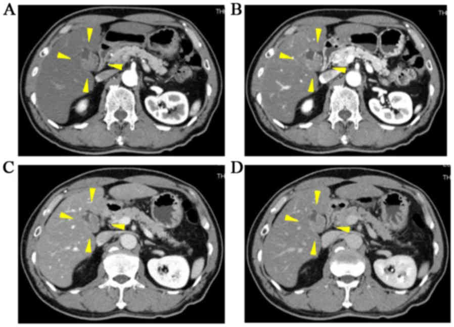
Abdominal contrast-enhanced computed tomography (CT)
showed wall thickening that appeared to extend into the lumen of
the gallbladder neck with a weak contrast effect. No obvious
out-of-wall development was observed (Fig. 2).
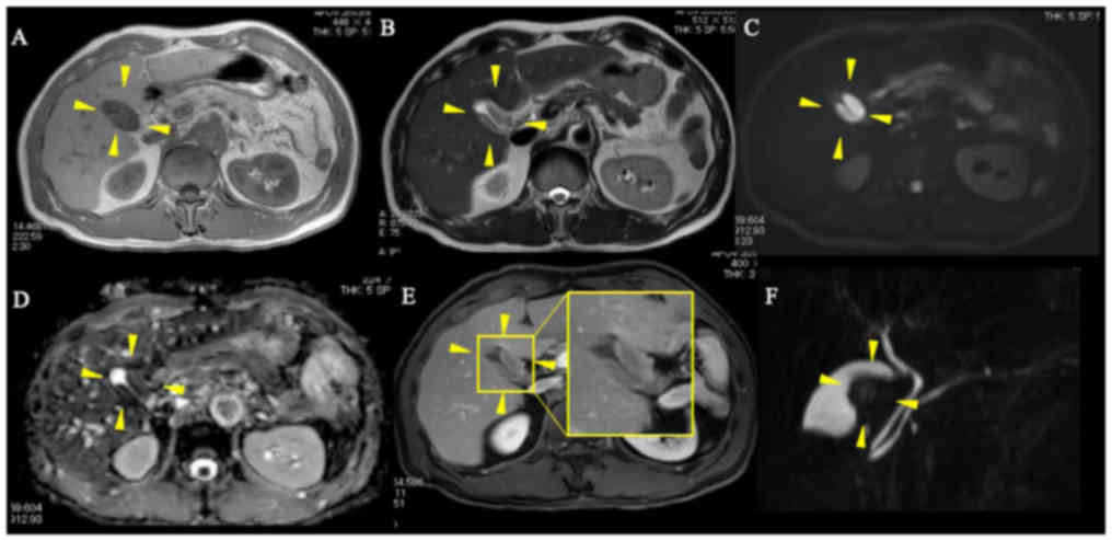
In abdominal magnetic resonance imaging (MRI), the
tumor showed a low signal intensity on T1 imaging and a high signal
intensity on T2 imaging that was slightly higher than the liver
parenchyma. An apparent high diffusion signal was observed in
diffusion-weighted imaging (DWI) and the signal declined with
apparent diffusion coefficient mapping. The mucosal surface was
preserved. These findings were indicative of a NET and a malignant
lymphoma with higher cell density than that of a typical
gallbladder cancer. Magnetic resonance cholangiopancreatography
showed a defect in the neck of the gallbladder (Fig. 3).
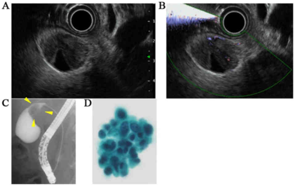
Endoscopic ultrasonography findings were similar to
those of AUS and showed a structure of submucosal tumor-like
appearance and clear boundaries to the liver. No Rokitansky-Aschoff
sinus was observed. Endoscopic retrograde cholangiopancreatography
showed a defect in the neck of the gallbladder and
pancreaticobiliary malfunction was not observed. The amylase levels
in the bile juice was not elevated. An endoscopic naso-gallbladder
drainage catheter was placed in the gallbladder, and bile cytology,
which was performed 4 times, revealed a large number of
heterogeneous cell clusters. Adenocarcinoma was suspected based on
these findings (Fig. 4).
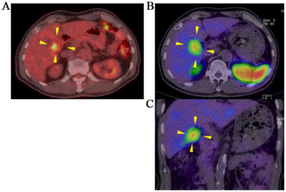
Positron emission tomography/computed tomography
(PET/CT) revealed the abnormal accumulation of 18F
fluorodeoxyglucose (FDG) in the gallbladder wall (Maximum standard
uptake value: 5.8 → 8.8), with no findings suggestive of distant
organ metastasis.
SRS was performed because gallbladder NET was also
considered as a possible diagnosis. Abnormal accumulation was
apparent in SRS and is consistent with a tumor. No other abnormal
accumulation suggestive of metastasis was observed (Fig. 5).
Gallbladder NEC was suspected based on the above
findings. However, are suspected diagnosis was stage II gallbladder
carcinoma based on the results of bile cytology. Therefore, we
performed enlarged cholecystectomy and D2 lymphadenectomy.
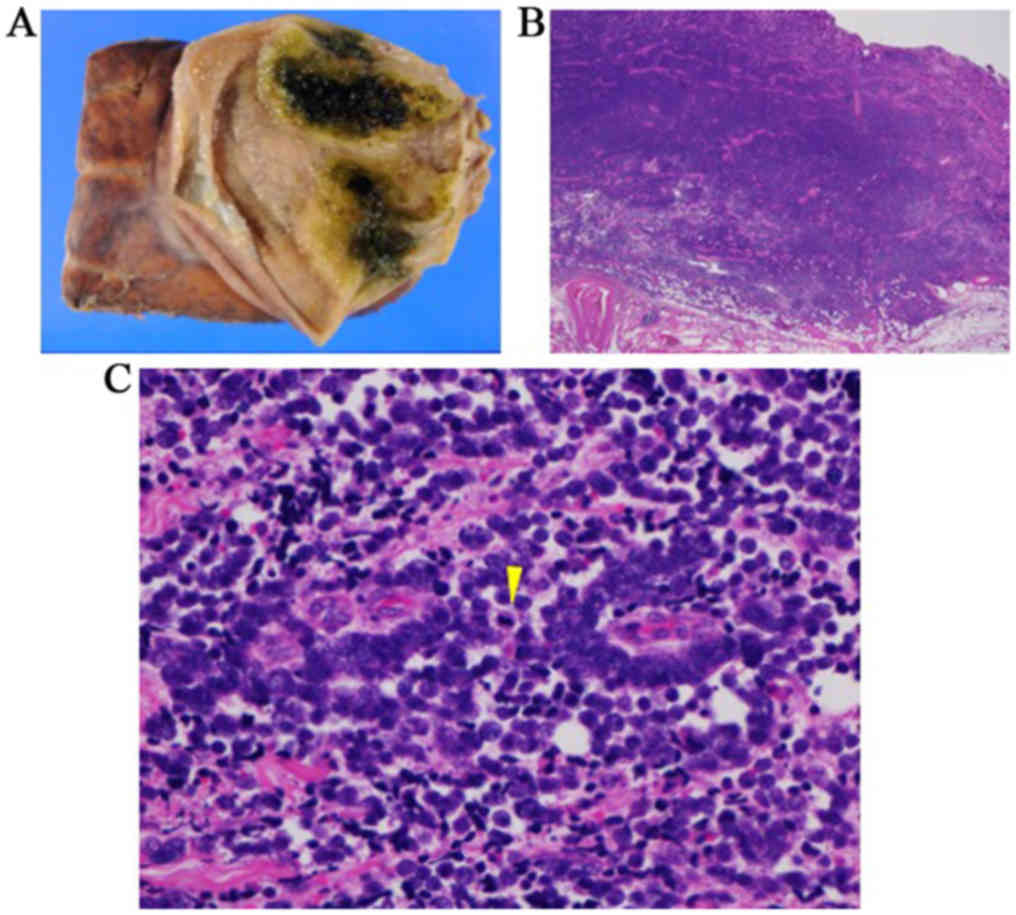
A tumor (45×35 mm) stretching from the body to the
neck of the gallbladder was found in the resected specimen.
Hematoxylin and eosin staining showed that the tumor cells had
honeycomb growth with a high nucleus-to-cytoplasm ratio, submucosal
growth in the shape of a cord, and numerous mitotic figures (20/10
high power fields; Fig. 6). In
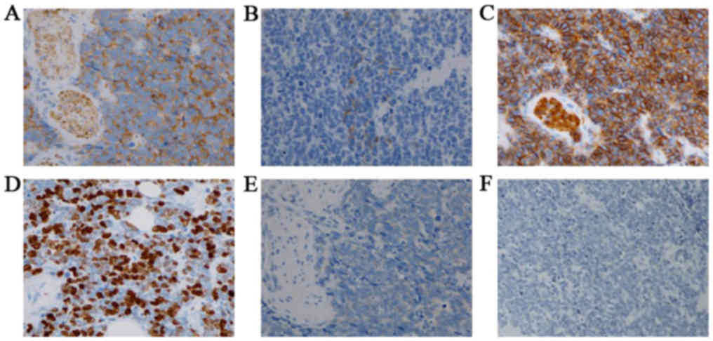
addition, Ki-67 labeling rate was >80%. The tumor cells were
positive for synaptophysin, chromogranin A, and cluster of
differentiation 56 (CD56). Immunohistochemical staining for
somatostatin receptor (SSTR) 2A was negative. Based on these
findings, the patient was diagnosed with gallbladder NEC. A small
differentiated tubular adenocarcinoma was found on the mucosal
surface; however, this made up only about 10% of the overall tumor,
which does not meet the diagnostic criteria for mixed
adenoneuroendocrine carcinoma (Fig.
7). The final pathological diagnosis was small cell NEC (pT3a,
N, M0, stage II) Gbn, 45×35 mm, nodular-infiltrating type, circ,
NEC, pT3a, int, INFb, ly1, v1, ne2, pCM0, pEM1, pR0. As multiple
liver metastases were observed on CT 4 months after the operation,
chemotherapy (CPT-11 [irinotecan] + cis-diamminedichloroplatinum
[cisplatin]) was administered according to the regimen for small
cell carcinoma of the lung. The patient is still undergoing
treatment.
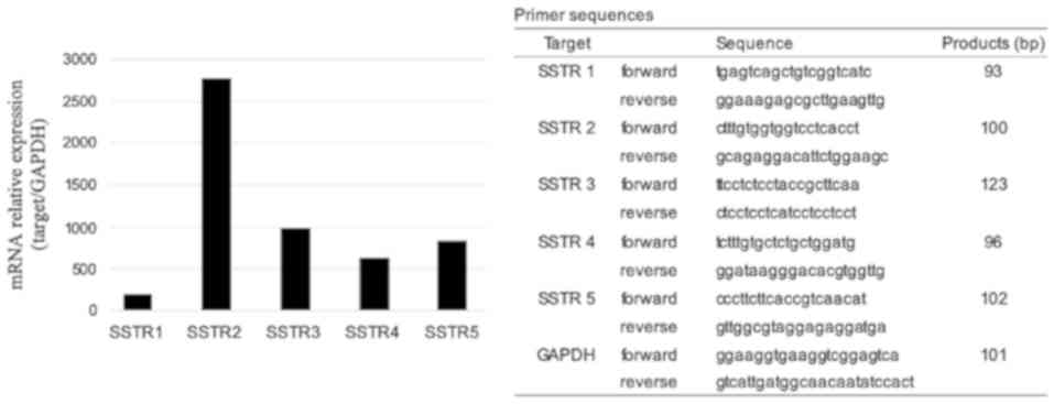
SSTR5 immunostaining was performed to elucidate why
SRS was positive when SSTR2 immunostaining was negative, but we
obtained a negative result. Additionally, we measured gene
expression using quantitative real-time polymerase chain reaction
(PCR) for all subtypes of SSTR (SSTR1-5) and compared our results
with those of earlier studies that reported on the Caco2 cell line
(3–5). The tumor was positive for all SSTR
subtypes (Fig. 8) and may express
SSTRs other than SSTR2 and SSTR5.
The study was approved by the ethics committee of
Kagoshima University Hospital (approved number 26-6).
Discussion
Here, we report a rare case of gallbladder NEC
preoperatively diagnosed using SRS. NETs occur in the
gastrointestinal tract and bronchopulmonary system. Primary NEC of
the gallbladder is a rare disease, accounting for only up to 1.4%
of all gastrointestinal endocrine tumors and 0.4–4% of all
malignant gallbladder tumors. In addition, the mean age of patients
at disease onset is 64 years, and the incidence in females is
relatively high (70%). Gallbladder NEC is characterized by rapid
development, early liver metastasis, lymph node metastasis, and
direct metastasis, and the prognosis is considered to be extremely
poor (2,6,7).
According to the 2010 World Health Organization classification of
tumors of the digestive tract, NETs are categorized into NET G1,
NET G2, and NECs according to mitotic count and Ki-67 index
(8). NEC is difficult to
preoperatively differentiate from gallbladder adenocarcinoma, and
most patients are diagnosed postoperatively by immunostaining for
markers such as NSE, CD56, chromogranin A, and synaptophysin
(9). Several hypotheses have been
proposed on the pathogenesis of gallbladder NETs. A previous report
posited that NETs are derived from undifferentiated pluripotent
cells at the site of development, which are capable of
differentiating into epithelial and neuroendocrine cells, and that
this ability to differentiate changes during cancer proliferation
and leads to a shift to small cell carcinoma (10).
No specific imaging features of gallbladder NECs
have been described (11,12). Many cases are difficult to
distinguish from typical, more common gallbladder cancers. DWI in
MRI has been reported to be useful for classifying NEC and
adenocarcinoma components. As NECs have a higher proliferative
potential than typical cancers and a high cell density, they have a
relatively low diffusion ability (13). The gallbladder NEC in our patient had
a high diffusion signal on DWI. DWI is considered potentially
useful for differentiating between benign gallbladder tumors,
typical gallbladder cancers, and gallbladder NECs (14).
SRS using 111In-pentetreotide, which
targets somatostatin receptors (SSTRs) and has a high affinity for
subtype 2A, is currently the gold standard functional imaging test
for NETs (15). For the diagnosis of
NETs, European and US guidelines recommend focal diagnosis using
SRS, diagnosis of lymph node and liver metastases, and CT and MRI.
The evaluation of SSTRs has also been reported to be useful for
predicting therapeutic response to octreotide preparations
(16). While SRS has a low
diagnostic sensitivity for poorly differentiated NECs, FDG-PET is
useful for the diagnosis of tumors with a high proliferation index.
The diagnostic sensitivity is low for NETs with low proliferation
index, slow growth rate, and low glucose consumption (17,18).
Although pathological examination revealed a poorly differentiated
small cell NET in our patient, SRS and FDG-PET results were
positive. In NETs, the lower the degree of differentiation, the
lower the rate of SRS positivity and the higher the degree of PET
accumulation. However, a comparative study reported that tumors
with proliferation (Ki-67) indices >15 and <2% had positive
SRS sensitivities of 69 and 87%, respectively. This suggests the
possibility of SRS accumulation, even in NECs (17).
SSTRs, which are receptors of the peptide hormone
somatostatin, have 5 subtypes, namely SSTR1, SSTR2, SSTR3, SSTR4,
and SSTR5. 111In-pentetreotide has an affinity for SSTR2
and SSTR5. Generally, most NETs express SSTR2, and SRS is
considered to be useful in the diagnosis of NETs (19). In NETs, the expression of SSTR2
decreases as the degree of differentiation decreases. In the case
of our patient, immunohistochemical staining was negative for
SSTR2A and SSTR5; however, real-time PCR detected the expressions
of all the subtypes (SSTR1, SSTR2, SSTR3, SSTR4, and SSTR5;
Fig. 8). SRS has a high affinity for
SSTR2, SSTR5, and SSTR3. We postulated that the real-time PCR
result, which suggested that, besides SSTR2 and SSTR5, the tumor
may also express other SSTRs such as SSTR3, may explain the
discrepancy between the loss of SSTR expression shown on
immunohistochemistry and the apparent accumulation in the tumor
shown on SRS. SRS may be useful for NEC diagnosis in some cases,
even when immunohistochemical staining for SSTRs is negative.
Several limitations associated with the present
study warrant mention. First, we performed immunohistochemical
staining for SSTR2 and SSTR5 but not for SSTR1, SSTR3, and SSTR4,
because we did not obtain antibodies against these subtypes.
Second, we detected the expressions of SSTR1, SSTR2, SSTR3, SSTR4,
and SSTR5 using real-time PCR (Fig.
8) and compared our findings with those of earlier studies that
reported on the Caco2 cell line (4,5). Owing
to the lack of tissue, we were not able to compare SSTR mRNA
expression between the tumor and the adjacent normal tissue.
In conclusion, gallbladder NEC is a rare disease and
is difficult to diagnose preoperatively. The incidence of this
disease may increase in the future, and accurate diagnostic and
effective treatment methods, which may include surgery and
chemotherapy, must be established to improve prognosis. Gallbladder
NET should be considered and SRS should be performed when
evaluation of an advanced gallbladder tumor reveals normal levels
of tumor markers, such as CEA and CA19-9, and atypical gallbladder
tumor characteristics on imaging, such as apparent high diffusion
signal on DWI.
Acknowledgements
Not applicable.
Funding
No funding was received.
Availability of data and materials
The datasets used and/or analyzed during the present
study are available from the corresponding author on reasonable
request.
Authors' contributions
YK, drafted the manuscript. ST and AI, supervised
and reviewed the contents of the manuscript and the data and image
analysis. MK, HI, SA, FS, YN, SH, SK, MH and KG contributed to
acquisition, analysis and interpretation of data, writing and
revision of the manuscript critically for important intellectual
content. All authors read and approved the final manuscript.
Ethics approval and consent to
participate
The current study was approved by the ethics
committee of Kagoshima University Hospital (approved no. 26-6).
Patient consent for publication
The consent for publication of the manuscript and
the related images was obtained from the patients.
Competing interests
The authors declare that they have no competing
interests.
References
|
1
|
Yao JC, Hassan M, Phan A, Dagohoy C, Leary
C, Mares JE, Abdalla EK, Fleming JB, Vauthey JN, Rashid A and Evans
DB: One hundred years after ‘carcinoid’: Epidemiology of and
prognostic factors for neuroendocrine tumors in 35,825 cases in the
United States. J Clin Oncol. 26:3063–3072. 2008. View Article : Google Scholar : PubMed/NCBI
|
|
2
|
Eltawil KM, Gustafsson BI, Kidd M and
Modlin IM: Neuroendocrine tumors of the gallbladder: An evaluation
and reassessment of management strategy. J Clin Gastroenterol.
44:687–695. 2010.PubMed/NCBI
|
|
3
|
Mizutani G, Nakanishi Y, Watanabe N, Honma
T, Obana Y, Seki T, Ohni S and Nemoto N: Expression of somatostatin
receptor (SSTR) subtypes (SSTR-1, 2A, 3, 4 and 5) in neuroendocrine
tumors using real-time RT-PCR method and immunohistochemistry. Acta
Histochem Cytochem. 45:167–176. 2012. View Article : Google Scholar : PubMed/NCBI
|
|
4
|
Ayiomamitis GD, Notas G, Zaravinos A,
Drygiannakis I, Georgiadou M, Sfakianaki O, Mastrodimou N, Thermos
K and Kouroumalis E: Effects of octreotide and insulin on colon
cancer cellular proliferation and correlation with hTERT activity.
Oncoscience. 1:457–467. 2014. View Article : Google Scholar : PubMed/NCBI
|
|
5
|
Colucci R, Blandizzi C, Ghisu N, Florio T
and Del Tacca M: Somatostatin inhibits colon cancer cell growth
through cyclooxygenase-2 downregulation. Br J Phasrmacol.
155:198–209. 2008. View Article : Google Scholar
|
|
6
|
Albores-Saavedra J, Batich K, Hossain S,
Henson DE and Schwartz AM: Carcinoid tumors and small-cell
carcinomas of the gallbladder and extrahepatic bile ducts: A
comparative study based on 221 cases from the Surveillance,
Epidemiology, and End Results Program. Ann Diagn Pathol.
13:378–383. 2009. View Article : Google Scholar : PubMed/NCBI
|
|
7
|
Yun SP, Shin N and Seo HI: Clinical
outcomes of small cell neuroendocrine carcinoma and adenocarcinoma
of the gallbladder. World J Gastroenterol. 21:269–275. 2015.
View Article : Google Scholar : PubMed/NCBI
|
|
8
|
Hamilton SR and Aaltonen LA: World health
organization classification of tumours. Pathology and genetics of
tumours of the digestive system. Hamilton SR and Aaltonen LA: IARC
Press; Lyon: pp. 20002000
|
|
9
|
Adachi T, Haraguchi M, Irie J, Yoshimoto
T, Uehara R, Ito S, Tokai H, Noda K, Tada N and Hirabaru M:
Gallbladder small cell carcinoma: A case report and literature
review. Surg Case Rep. 2:712016. View Article : Google Scholar : PubMed/NCBI
|
|
10
|
Horiguchi S: A case of neuroendocrine
carcinoma of gallbladder. Tando. 30:290–297. 2016.
|
|
11
|
Chen C, Wang L, Liu X, Zhang G, Zhao Y and
Geng Z: Gallbladder neuroendocrine carcinoma: Report of 10 cases
and comparision of clinicopathologic features with gallbladder
adenocarcinoma. Int J Clin Exp Pathol. 8:8218–8226. 2015.PubMed/NCBI
|
|
12
|
El Fattach H, Guerrache Y, Eveno C, Pocard
M, Kaci R, Shaar-Chneker C, Dautry R, Boudiaf M, Dohan A and Soyer
P: Primary neuroendocrine tumors of the gallbladder:
Ultrasonographic and MDCT features with pathologic correlation.
Diagn Interv Imaging. 96:499–502. 2015. View Article : Google Scholar : PubMed/NCBI
|
|
13
|
Murawaki Y, Miura M, Otani Y and Yoshida
M: A case of adenoendocrine cell carcinoma of the gallbladder
complicated by pancreaticobiliary maljunction, whereby differences
in tissue composition could be identified on diffusion-weighted
MRI. Tando. 27:200–204. 2013.
|
|
14
|
Ogawa T, Horaguchi J, Fujita N, Noda Y,
Kobayashi G, Ito K, Koshita S, Kanno Y, Masu K and Sugita R: High
b-value diffusion-weighted magnetic resonance imaging for
gallbladder lesions: Differentiation between benignity and
malignancy. J Gastroenterol. 47:1352–1360. 2012. View Article : Google Scholar : PubMed/NCBI
|
|
15
|
O'Toole D, Kianmanesh R and Caplin M:
ENETS 2016 consensus guidelines for the management of patients with
digestive neuroendocrine tumors: An update. Neuroendocrinology.
103:117–118. 2016. View Article : Google Scholar : PubMed/NCBI
|
|
16
|
van Essen M, Sundin A, Krenning EP and
Kwekkeboom DJ: Neuroendocrine tumours: The role of imaging for
diagnosis and therapy. Nat Rev Endocrinol. 10:102–114. 2014.
View Article : Google Scholar : PubMed/NCBI
|
|
17
|
Binderup T, Knigge U, Loft A, Mortensen J,
Pfeifer A, Federspiel B, Hansen CP, Højgaard L and Kjaer A:
Functional imaging of neuroendocrine tumors: A head-to-head
comparison of somatostatin receptor scintigraphy, 123I-MIBG
Scintigraphy, and 18F-FDG PET. J Nucl Med. 51:704–712. 2010.
View Article : Google Scholar : PubMed/NCBI
|
|
18
|
Kubota K, Okasaki M, Minamimoto R, Miyata
Y, Morooka M, Nakajima K and Sato T: Lesion-based analysis of
(18)F-FDG uptake and (111)In-Pentetreotide uptake by neuroendocrine
tumors. Ann Nucl Med. 28:1004–1010. 2014. View Article : Google Scholar : PubMed/NCBI
|
|
19
|
Volante M, Brizzi MP, Faggiano A, La Rosa
S, Rapa I, Ferrero A, Mansueto G, Righi L, Garancini S, Capella C,
et al: Somatostatin receptor type 2A immunohistochemistry in
neuroendocrine tumors: A proposal of scoring system correlated with
somatostatin receptor scintigraphy. Mod Pathol. 20:1172–1182. 2007.
View Article : Google Scholar : PubMed/NCBI
|