Introduction
Colorectal cancer is currently the third most
commonly occurring cancer type in males and the second most common
in females globally, with 1.4 million new cases and over 690,000
associated mortalities in 2012 (1).
Oxaliplatin is one of the most frequently used drugs for the
treatment of patients with colorectal cancer (2). Through the induction of intrastrand
adduct formation, oxaliplatin may inhibit cell cycle progression to
promote cell death in proliferative cells (3). However, de novo and acquired
oxaliplatin resistance notably reduced the treatment efficacy of
this drug in patients with colorectal cancer (4).
MicroRNAs (miRNAs) are small, non-coding,
single-stranded RNA molecules (5).
Deregulation of miRNAs has been reported to contribute to the
carcinogenesis in various types of cancer, including colorectal
cancer (6–8). Furthermore, a number of miRNAs, such as
miR-425-5p and miR-203, have been determined to be involved in
chemotherapy resistance in colorectal cancer via regulation of
their target genes (9–11). Previous studies indicated that
miR-106a was overexpressed in tumor tissues, fecal samples and
plasma of patients with colorectal cancer (12–14). A
decrease in miR-106a levels has been previously reported to predict
a reduced disease-free survival (DFS) and overall survival (OS)
times in patients with colorectal cancer (15), suggesting a dual role of miR-106a in
colorectal cancer.
Forkhead box Q1 (FOXQ1) is a member of the FOX gene
family and functions as a transcription factor (16). Accumulating evidence has indicated
that FOX proteins serve as terminal effectors for numerous
signaling pathways, including the transforming growth factor-β
signaling, Wnt, Hedgehog and mitogen-activated protein kinase
pathways (17). Previously, FOXQ1
was determined to be overexpressed in colorectal cancer and
promoted colorectal cancer progression (18,19). A
limited number of studies have indicated that FOXQ1 may be
regulated by transcriptional activation, miRNA binding and
post-translational modification under different conditions
(20–22). For example, in colorectal cancer
cells, FOXQ1 was identified as a target gene of Wnt signaling
pathway (20). However, the specific
underlying mechanism of the deregulation of FOXQ1 by miRNA in
colorectal cancer remains unknown.
In the present study, the function of miR-106a in
mediating oxaliplatin sensitivity in colorectal cancer was
investigated. Overexpression of miR-106a slightly decreased cell
growth ability and sensitized colorectal cancer cells to
oxaliplatin treatment. Reverse transcription-quantitative
polymerase chain reaction (RT-qPCR) and western blot analysis
demonstrated that miR-106a could downregulate FOXQ1 at mRNA and
protein levels in colorectal cancer cells. The dual luciferase
reporter assay confirmed that FOXQ1 was a direct target of
miR-106a. Furthermore, it was observed that there was a decrease in
miR-106a expression levels and an increase in FOXQ1 mRNA expression
levels in tumor tissues from patients with oxaliplatin-resistant
colorectal cancer, compared with patients with
oxaliplatin-sensitive colorectal cancer. In addition, a significant
association between miR-106a and FOXQ1 expression in tumor tissues
was observed. The present study identified miR-106a as a promising
target for oxaliplatin resistance in colorectal cancer.
Materials and methods
Patients
In the present study, the gene expression of 30
patients with colorectal cancer, including 17 patients (11 men and
6 women; range, 51–68 years; mean age, 59±7 years) with
oxaliplatin-sensitive colorectal cancer and 13 patients (8 males
and 5 females; range 56–69 years) with oxaliplatin-resistant
colorectal cancer were examined. All the participants received
oxaliplatin-based chemotherapy and were enrolled in Xingtai
People's Hospital (Xingtai, China) between February 2013 and March
2015. Tumor response and progression were assessed according to the
Response Evaluation Criteria in Solid Tumors version 1.1 (23). According to the outcome of therapy,
17 patients were classified as responders (complete or partial
response) and 13 patients were classified as non-responders (no
change and progressive disease). Written consents regarding
participation in the present study and use of their tissues were
obtained from all patients and the experiments were conducted with
the approval of and under the supervision of the Ethics Committee
of Xingtai People's Hospital.
Cell culture and oxaliplatin
treatment
The kidney derived 293T cell line and human
colorectal cancer cell lines, HCT116 and HT-29, which were commonly
used to study oxaliplatin sensitivity of colorectal cancer
(24–25), were obtained from the American Type
Culture Collection and used within 6 months. HT-29 cell line was
authenticated by STR profiling. The HCT116 and HT-29 cell lines
were maintained in Dulbecco's modified Eagle's medium (Gibco;
Thermo Fisher Scientific, Inc.) containing 10% fetal bovine serum
(Gibco; Thermo Fisher Scientific, Inc.) in an incubator at 37°C
with 5% CO2.
For oxaliplatin treatment, indicated concentrations
(2, 4, 6 and 8 µmol/l) of oxaliplatin (Selleck Chemicals) were
added into the culture medium of HCT116 and HT-29 cells and
sustained for 48 h at 37°C then subjected to the following
procedures.
MTT assay
Cell viability was assessed with an MTT assay.
Briefly, HCT116 and HT-29 cells were cultured at 37°C in 96-well
plates (1×10³ cells/well) and treated with oxaliplatin
for the indicated lengths of time (0, 25, 50, 75 and 100 h).
Subsequently, HCT116 and HT-29 cells were stained with thiazolyl
blue tetrazolium bromide (MTT; Sigma-Aldrich; Merck KGaA) for 2 h
at 37°C and absorbance at 570 nm was detected following purple
precipitates being dissolved with MTT detergent reagent on a
microplate reader (BioTek Instruments, Inc.). Cell viability was
calculated as the ratio of the absorbance values of treated samples
to those of controls.
RNA extraction and RT-qPCR
RNA was extracted from HCT116 and HT-29 cells and
tissues from patients using TRIzol® (Invitrogen; Thermo
Fisher Scientific, Inc.), according to the manufacturer's protocol.
Total RNA was reverse transcribed into cDNA with a PrimeScript™ RT
reagent kit (Takara Bio, Inc.), according to manufacturer's
protocol. For mRNA expression analysis of FOXQ1, the RT-qPCR was
performed on a CFX96 Real-Time PCR Detection system (Bio-Rad
Laboratories, Inc.) with SYBR® Premix Ex Taq™ II (Takara
Bio, Inc.). GAPDH was used as an endogenous reference gene.
For miRNA expression analysis, RT was performed with
a miScript II RT kit (Qiagen GmbH). Expression levels of miR-106a
were detected using miScript primer assays with a miScript SYBR
Green PCR kit (Qiagen GmbH), according to the manufacturer's
protocol. U6 was used as an endogenous reference gene. The
thermocycling conditions were as follows: 95°C for 15 min, followed
by denaturation: 95°C for 10 sec, annealing for 30 sec and
extension for 30 sec (40 cycles). The relative expression of miRNA
and mRNA were calculated using 2−ΔΔCq method (26). The primer sequences were listed as
follows: FOXQ1-Forward: 5′-CACGCAGCAAGCCATATACG-3′; FOXQ1-Reverse:
5′-CGTTGAGCGAAAGGTTGTGG-3′; MMP-2-Forward:
5′-GGCCCTGTCACTCCTGAGAT-3′; MMP-2-Reverse:
5′-GGCATCCAGGTTATCGGGGA-3′; VEGF-A-Forward:
5′-AGGGCAGAATCATCACGAAGT-3′; VEGF-A-Reverse:
5′-AGGGTCTCGATTGGATGGCA-3′; GAPDH-Forward:
5′-CTCTGATTTGGTCGTATTGGG-3′; GAPDH-Reverse:
5′-TGGAAGATGGTGATGGGATT-3′; miR-106a-RT:
5′-GTCGTATCCAGTGCGTGTCGTGGAGTCGGCAATTGCACTGGATACGACCTACCTG-3′;
miR-106a-Forward: 5′-ATCCAGTGCGTGTCGTG-3′; miR-106a-Reverse:
5′-TGCTAAAAGTGCTTACAGTG-3′; U6-Forward: 5′-CTCGCTTCGGCAGCACA-3′;
U6-Reverse: 5′-AACGCTTCACGAATTTGCGT-3′.
Western blot analysis
The FOXQ1 antibody (cat. no. SC-166265; dilution,
1:2,000) and vascular endothelial growth factor-A (VEGF-A; cat. no.
SC-365578; dilution, 1:2,000) were purchased from Santa Cruz
Biotechnology, Inc. Antibodies against matrix metallopeptidase-2
(MMP-2; cat. no. 40994; dilution, 1:1,000) was purchased from Cell
Signaling Technology, Inc. The GAPDH antibody was obtained from
Sigma-Aldrich (Merck KGaA). The secondary antibodies against mouse
and rabbit (HRP-conjugate; dilution, 1:10,000) were purchased from
ProteinTech Group, Inc. Proteins were extracted from HCT116 and
HT-29 cells using radioimmunoprecipitation assay lysis buffer
(Sigma-Aldrich; Merck KGaA). The concentration of protein lysates
was determined by bicinchoninic acid assay (BCA assay) using a BCA
protein assay kit (Thermo Fisher Scientific, Inc.), according to
the manufacturer's protocol. Western blot analysis was performed as
follows: Equal amounts (20 µg) of samples were loaded into each
lane on an 8% SDS-PAGE gel. The proteins were separated and
transferred on a polyvinylidene fluoride membrane. Following
blocking in 5% non-fat milk for 1 h at room temperature, the
membranes were incubated with primary antibodies overnight at 4°C.
Subsequently, the membranes were incubated with secondary
antibodies for 1 h at room temperature and then developed with
Enhanced Chemiluminescent Western Blotting Substrate (Pierce;
Thermo Fisher Scientific, Inc.). Images were captured with
ImageQuant LAS 4000 (GE Healthcare Life Sciences). GAPDH was used
as endogenous control.
Transfection of miR-Negative Control
(miR-NC) mimics and miR-106a mimics
miR-NC mimics and miR-106a mimics were purchased
from Shanghai GenePharma Co., Ltd. The sequences were: miR-NC
mimics, 5′-UUCUCCGAACGUGUCACGUTT-3′; miR-106a mimics,
5′-AAAAGUGCUUACAGUGCAGGUAG-3′. miR-NC mimics or miR-106a mimics
were transfected at a final concentration of 40 nM with
Lipofectamine® 2000 (Invitrogen; Thermo Fisher
Scientific, Inc.), according to the manufacturer's protocols. In
brief, miR-NC mimics or miR-106a mimics and
Lipofectamine® 2000 were mixed (to a concentration of
200 nM) in Opti-MEM medium (Thermo Fisher Scientific, Inc.) and
incubated for 20 min at 37°C. Subsequently, the mixture was added
to HCT116 and HT-29 cells in DMEM. The medium was replaced with
fresh DMEM after 6 h and sustained for 18 h before subjected to
following experiments.
Dual luciferase reporter assay
Sequencing alignment was performed using miRDB
database (www.mirdb.org). FOXQ1 3′UTR was
amplified from cDNA of 293T cells and ligated into a pGL3 plasmid
(Promega Corporation). A total of two-point mutations were
introduced into FOXQ1 3′UTR with a Site-directed mutagenesis kit
(Agilent Technologies, Inc.), in order to construct
pGL3-FOXQ1-3′UTR-Mut.
For the dual luciferase reporter assay, 293T and
HCT116 cells were plated on 24-well plates. On the following day,
cells were co-transfected with pGL3-FOXQ1-WT or
pGL3-FOXQ1-3′UTR-Mut, miR-NC mimics or miR-106a mimics and pRL-TK
plasmid (Invitrogen; Thermo Fisher Scientific, Inc.) using
Lipofectamine® 2000. The assay was performed 48 h after
with a Dual-luciferase Assay system (Promega Corporation). The
firefly luciferase activity was normalized to the Renilla
luciferase activity value.
Statistical analysis
The data were calculated with GraphPad Prism v6
(GraphPad Software, Inc.) and presented as the mean ± standard
deviation. Comparisons between two different groups were determined
with unpaired Student's t-test. P<0.05 was considered to
indicate a statistically significant difference.
Results
Overexpression of miR-106a-transfected
oxaliplatin-sensitized colorectal cancer cells
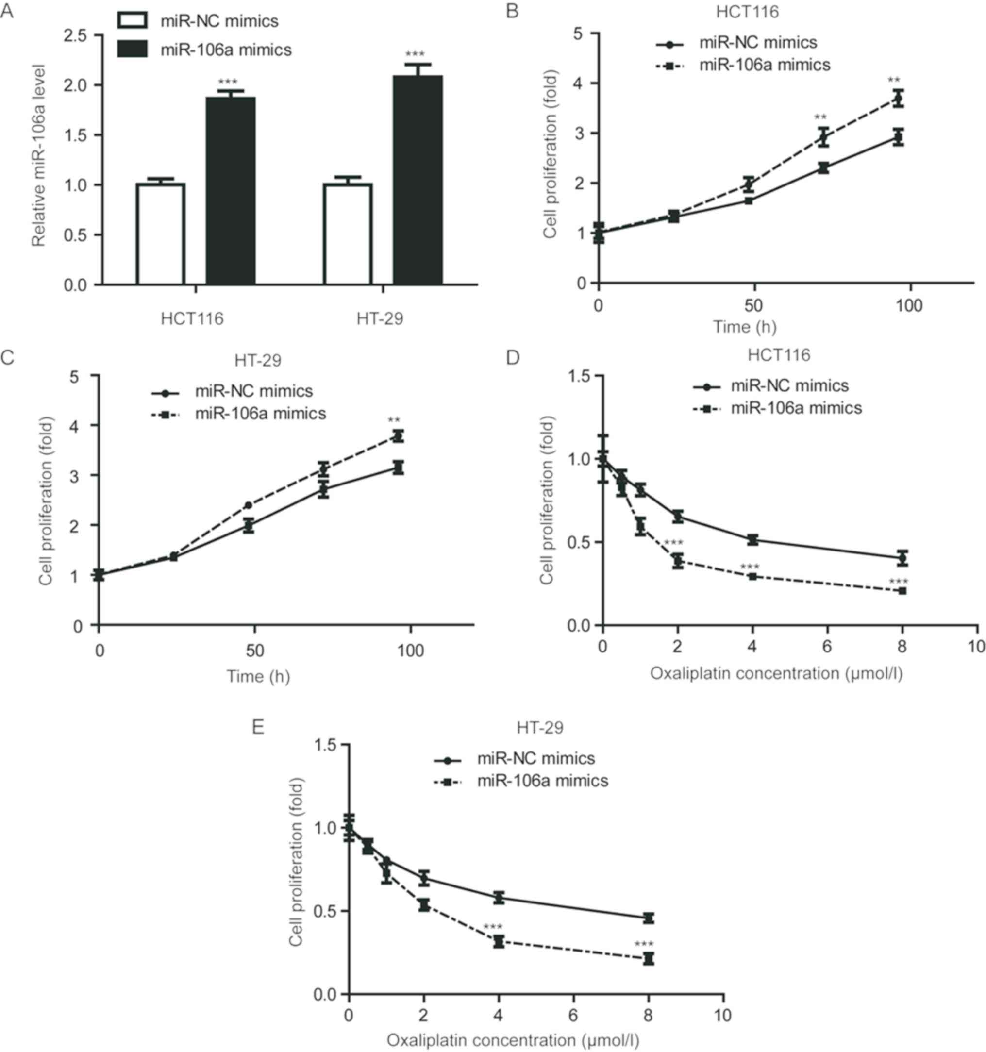
In order to evaluate the role of miR-106a in
colorectal cancer cells, the HCT116 and HT-29 cell lines were
selected. miR-106a was significantly overexpressed in these cell
lines via transfection of miR-106a mimics (P<0.001; Fig. 1A). A cell viability assay was
conducted to detect cell proliferation ability upon miR-106a
overexpression. As depicted in Fig. 1B
and C, miR-106a mimics significantly increased MTT absorbance
compared with cells transfected with miR-NC mimics (P<0.01),
indicating miR-106a overexpression promoted cell proliferation of
HCT116 and HT-29 cells. However, in cells treated with increasing
concentrations of oxaliplatin, overexpression of miR-106a
significantly reduced cell viability, compared with control groups,
in the cell lines tested (P<0.001; Fig. 1D and E). These results indicated that
miR-106a overexpression could enhance oxaliplatin sensitivity in
colorectal cancer cells.
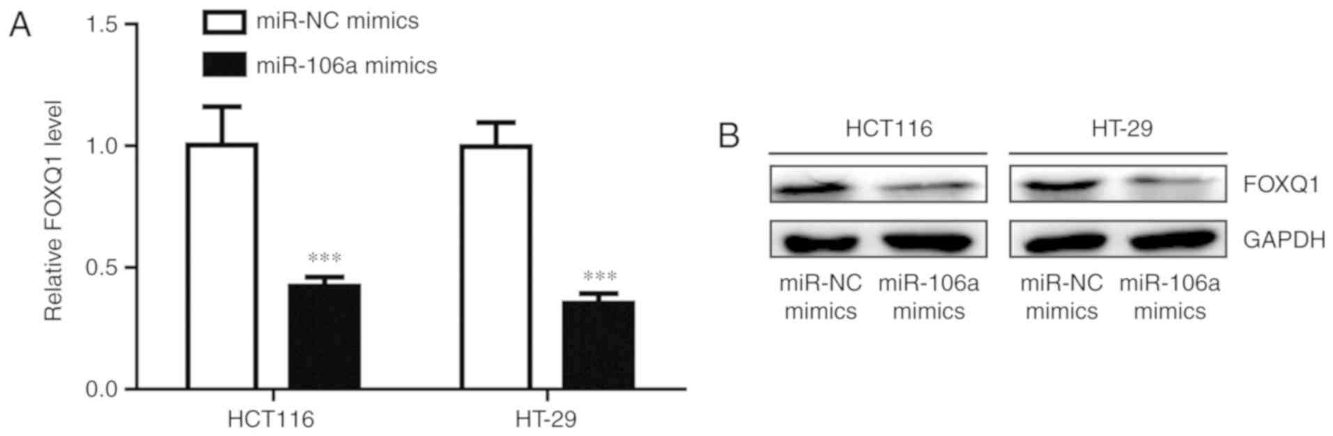
miR-106a represses FOXQ1 expression in
colorectal cancer cells
FOXQ1 is frequently overexpressed in colorectal
cancer and its expression is associated with oxaliplatin resistance
(27). Transfection of miR-106a
mimics significantly decreased FOXQ1 mRNA level in HCT116 and HT-29
cells (P<0.001; Fig. 2A). Western
blot analysis demonstrated that the protein expression level of
FOXQ1 was also reduced following miR-106 overexpression (Fig. 2B). These results indicated that
miR-106a may regulate oxaliplatin sensitivity via repression of
FOXQ1 level.
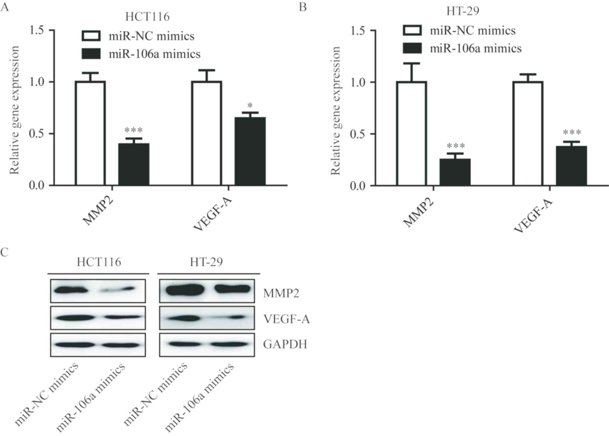
miR-106a regulates FOXQ1 target
genes
FOXQ1 is a transcriptional factor and can activate
VEGF-A and MMP-2 transcription via regulation of the Wnt signaling
pathway in colorectal cancer (27).
In addition, overexpression of miR-106a significantly reduced
VEGF-A (P<0.05) and MMP-2 (P<0.001) mRNA in HCT116 and HT-29
cells (Fig. 3A and B), and the
protein levels of VEGF-A and MMP-2 were also reduced in the two
cell lines (Fig. 3C). These results
indicated that miR-106a could inhibit FOXQ1, and repress
transcription of its target genes.
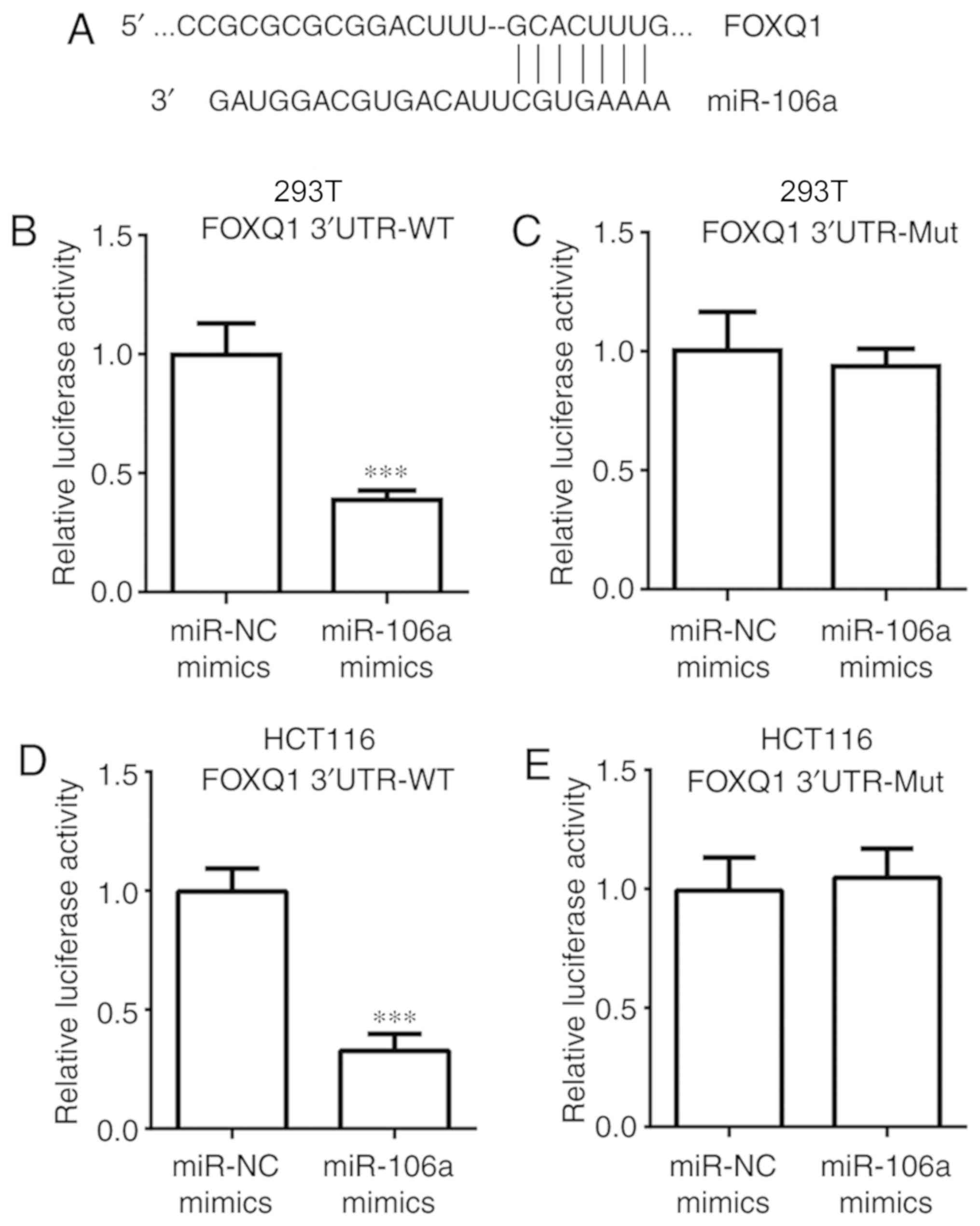
miR-106a directly inhibits FOXQ1
expression
The aim of the present study was to investigate
whether miR-106a directly regulates FOXQ1 expression. Sequence
alignment demonstrated that there were binding sites for miR-106a
in the 3′UTR of FOXQ1 mRNA (Fig.
4A). In the dual luciferase assay, it was determined that
transfection of miR-106a mimics significantly decreased relative
luciferase activity of 293T cells transfected with FOXQ1 3′UTR-WT,
but not FOXQ1 3′UTR-Mut (P<0.001; Fig. 4B and C). Similar results were
observed in HCT116 cells (P<0.001; Fig. 4D and E). These data confirmed that
miR-106a could bind to FOXQ1 mRNA and directly repress its
expression.
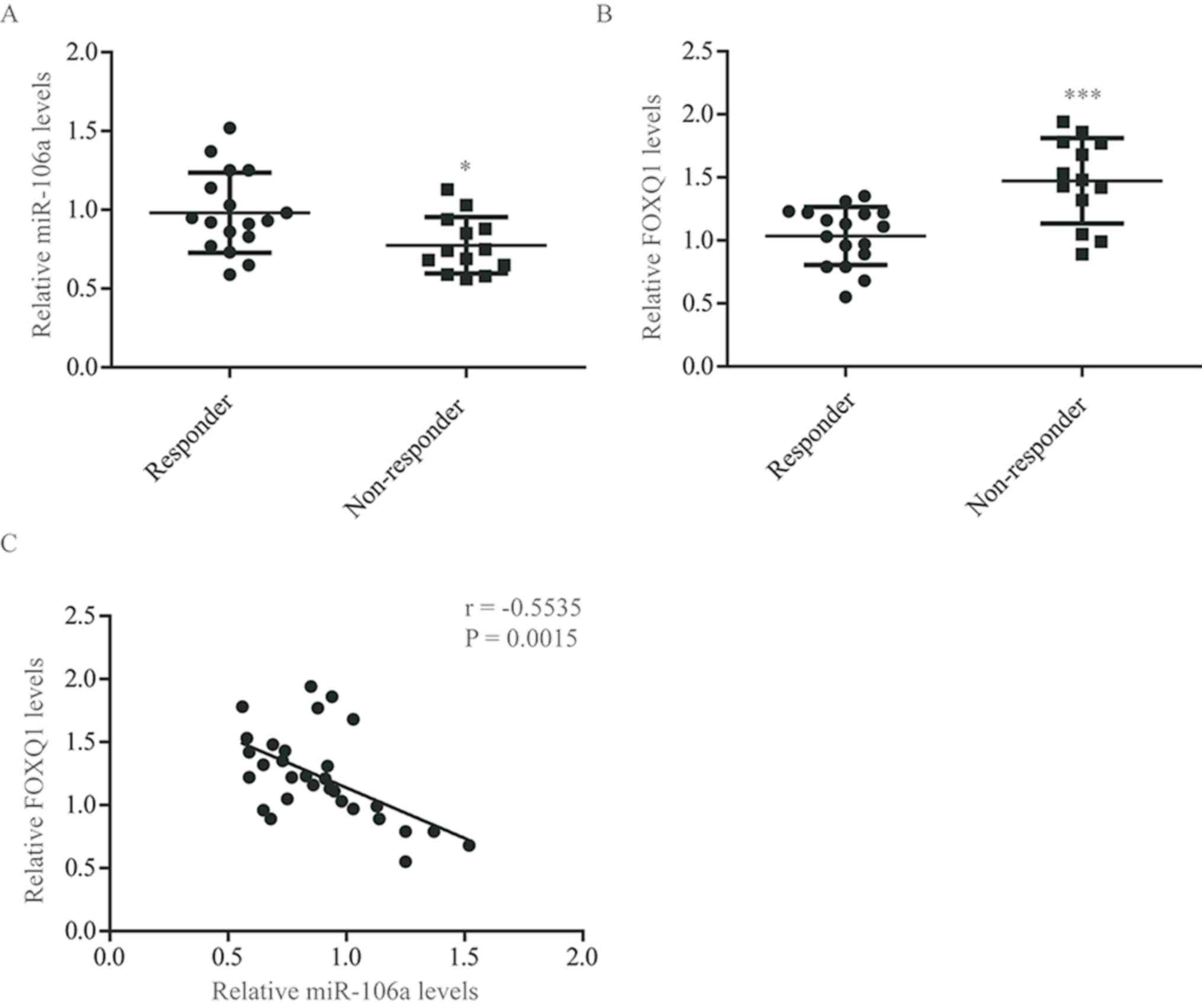
Expression of miR-106a and FOXQ1 in
tumor tissues from patients with colorectal cancer
To further evaluate the role of miR-106a and FOXQ1
in oxaliplatin sensitivity, the expression of miR-106a and FOXQ1 in
tumor tissues from patients with colorectal cancer receiving
oxaliplatin-based chemotherapy was determined. RT-qPCR demonstrated
that miR-106a expression levels (P<0.05) were significantly
decreased in tumor tissues from oxaliplatin non-responders, while
FOXQ1 (P<0.001) mRNA levels were significantly elevated
(Fig. 5A and B). Consistent with the
present in vitro data, statistical analysis indicated that
the expression of miR-106a was associated with FOXQ1 expression
(r=−0.5535; P=0.0015; Fig. 5C).
These results demonstrated that miR-106a regulates the expression
of FOXQ1 in tumor tissues of patients with colorectal cancer.
Discussion
Oxaliplatin is one of the commonly used chemotherapy
agents for patients with cancer (28). Resistance to oxaliplatin frequently
occurs in the clinical management of colorectal cancer (29). The underlying mechanism of
oxaliplatin resistance is complicated and includes dysregulation of
numerous miRNAs and proteins, including upregulation of miR-203
(10,30,31). In
the present study, miR-106a was identified as an oxaliplatin
sensitizer in colorectal cancer cells via regulation of FOXQ1
expression.
Deregulation of miRNAs was a signature and early
event in the initiation and development of colorectal cancer
(32). Previous findings
demonstrated that miR-106a was overexpressed in colorectal cancer
and predicted poor prognosis (15,33,34).
miR-106a could regulate cell proliferation, apoptosis, migration
and invasion in colon cancer cells via repression of its target
genes (12,35). In a recent meta-analysis, a high
expression of miR-106a was found to be associated with poor overall
survival of colorectal cancer patients (36). In the present study, it was validated
that miR-106a overexpression promoted colorectal cancer cell
growth. Notably, overexpression of miR-106a sensitized colorectal
cancer cells to oxaliplatin treatment. Additionally, the expression
of miR-106a was reduced in tumor tissues from non-responder
patients with colorectal cancer, compared with responders. These
data indicated a role of miR-106a in regulating oxaliplatin
sensitivity in colorectal cancer cells.
FOXQ1 is overexpressed in colorectal cancer types
(37). In colorectal cancer cells,
it was determined that miR-106a overexpression resulted in
decreased FOXQ1 level. A dual luciferase assay was conducted to
further confirm FOXQ1 as a direct target of miR-106a. As a
transcription factor, FOXQ1 could activate target gene expression
in order to regulate cell growth, migration, epithelial-mesenchymal
transition and chemoresistance in colorectal cancer cells (18,38).
Western blot analysis and RT-qPCR demonstrated that the expression
of FOXQ1 target genes were also repressed in colorectal cancer
cells with overexpressed miR-106a. Due to miR-106a repressing FOXQ1
expression, it was concluded that miR-106a may control oxaliplatin
sensitivity via the regulation of FOXQ1 in colorectal cancer.
Furthermore, FOXQ1 expression was significantly increased in
colorectal tumor tissues from non-responders, compared with
responders, and an association was indicated between FOXQ1 and
miR-106a expression levels in colorectal tumor tissues.
Collectively, these results demonstrated that miR-106a could
sensitize colorectal cancer cells to oxaliplatin.
The present results indicated that miR-106a serves
an important role in determining the sensitivity of colorectal
cancer cells to oxaliplatin treatment. The present study improved
the understanding of the role of miR-106a in colorectal cancer and
may allow for the use of miR-106a as a biomarker and target for
patients receiving chemotherapy, facilitating the development of
novel chemotherapy strategies.
The present study focused on the expression of
miR-106a in tumor tissues from oxaliplatin responder and
oxaliplatin non-responder. miR-106a expression in colorectal normal
tissues and tumor tissues and its role in regulating colorectal
cancer cell proliferation will be further evaluated in the
future.
Acknowledgements
Not applicable.
Funding
No funding was received.
Availability of data and materials
The datasets used and/or analyzed during the present
study are available from the corresponding author on reasonable
request.
Authors' contributions
ZL, YQ and SD performed the experiments, analyzed
the data and wrote the manuscript. YQ and SD participated in the
collection of patient samples. XC and ZH assisted with the western
blot analysis and data collection. ZZ conceived the idea, designed
the study and helped to draft the manuscript. All authors read and
approved the final manuscript.
Ethics approval and consent to
participate
The present study was approved by the Ethic
Committee of Xingtai People's Hospital. All subjects provided
written informed consent prior to enrollment.
Patient consent for publication
Not applicable.
Competing interests
The authors declare that they have no competing
interests.
References
|
1
|
Torre LA, Bray F, Siegel RL, Ferlay J,
Lortet-Tieulent J and Jemal A: Global cancer statistics, 2012. CA
Cancer J Clin. 65:87–108. 2015. View Article : Google Scholar : PubMed/NCBI
|
|
2
|
André T, Boni C, Mounedji-Boudiaf L,
Navarro M, Tabernero J, Hickish T, Topham C, Zaninelli M, Clingan
P, Bridgewater J, et al: Oxaliplatin, fluorouracil, and leucovorin
as adjuvant treatment for colon cancer. N Engl J Med.
350:2343–2351. 2004. View Article : Google Scholar : PubMed/NCBI
|
|
3
|
Raymond E, Faivre S, Chaney S, Woynarowski
J and Cvitkovic E: Cellular and molecular pharmacology of
oxaliplatin. Mol Cancer Ther. 1:227–235. 2002.PubMed/NCBI
|
|
4
|
Rabik CA and Dolan ME: Molecular
mechanisms of resistance and toxicity associated with platinating
agents. Cancer Treat Rev. 33:9–23. 2007. View Article : Google Scholar : PubMed/NCBI
|
|
5
|
Ambros V: microRNAs: Tiny regulators with
great potential. Cell. 107:823–826. 2001. View Article : Google Scholar : PubMed/NCBI
|
|
6
|
Ardekani AM and Naeini MM: The role of
microRNAs in human diseases. Avicenna J Med Biotechnol. 2:161–179.
2010.PubMed/NCBI
|
|
7
|
Du B, Wu D, Yang X, Wang T, Shi X, Lv Y,
Zhou Z, Liu Q and Zhang W: The expression and significance of
microRNA in different stages of colorectal cancer. Medicine
(Baltimore). 97:e96352018. View Article : Google Scholar : PubMed/NCBI
|
|
8
|
Emami SS, Akbari A, Zare AA, Agah S,
Masoodi M, Talebi A, Minaeian S, Fattahi A and Moghadamnia F:
MicroRNA expression levels and histopathological features of
colorectal cancer. J Gastrointest Cancer. 50:276–284. 2019.
View Article : Google Scholar : PubMed/NCBI
|
|
9
|
Zhang Y, Hu X, Miao X, Zhu K, Cui S, Meng
Q, Sun J and Wang T: MicroRNA-425-5p regulates chemoresistance in
colorectal cancer cells via regulation of programmed cell death 10.
J Cell Mol Med. 20:360–369. 2016. View Article : Google Scholar : PubMed/NCBI
|
|
10
|
Zhou Y, Wan G, Spizzo R, Ivan C, Mathur R,
Hu X, Ye X, Lu J, Fan F, Xia L, et al: miR-203 induces oxaliplatin
resistance in colorectal cancer cells by negatively regulating ATM
kinase. Mol Oncol. 8:83–92. 2014. View Article : Google Scholar : PubMed/NCBI
|
|
11
|
Ayers D and Vandesompele J: Influence of
microRNAs and long non-coding RNAs in cancer chemoresistance. Genes
(Basel). 8(pii): E952017. View Article : Google Scholar : PubMed/NCBI
|
|
12
|
Feng B, Dong TT, Wang LL, Zhou HM, Zhao
HC, Dong F and Zheng MH: Colorectal cancer migration and invasion
initiated by microRNA-106a. PLoS One. 7:e434522012. View Article : Google Scholar : PubMed/NCBI
|
|
13
|
Koga Y, Yamazaki N, Yamamoto Y, Yamamoto
S, Saito N, Kakugawa Y, Otake Y, Matsumoto M and Matsumura Y: Fecal
miR-106a is a useful marker for colorectal cancer patients with
false-negative results in immunochemical fecal occult blood test.
Cancer Epidemiol Biomarkers Prev. 22:1844–1852. 2013. View Article : Google Scholar : PubMed/NCBI
|
|
14
|
Liu GH, Zhou ZG, Chen R, Wang MJ, Zhou B,
Li Y and Sun XF: Serum miR-21 and miR-92a as biomarkers in the
diagnosis and prognosis of colorectal cancer. Tumour Biol.
34:2175–2181. 2013. View Article : Google Scholar : PubMed/NCBI
|
|
15
|
Diaz R, Silva J, Garcia JM, Lorenzo Y,
García V, Peña C, Rodríguez R, Muñoz C, García F, Bonilla F and
Domínguez G: Deregulated expression of miR-106a predicts survival
in human colon cancer patients. Genes Chromosomes Cance.
47:794–802. 2008. View Article : Google Scholar
|
|
16
|
Overdier DG, Porcella A and Costa RH: The
DNA-binding specificity of the hepatocyte nuclear factor 3/forkhead
domain is influenced by amino-acid residues adjacent to the
recognition helix. Mol Cell Biol. 14:2755–2766. 1994. View Article : Google Scholar : PubMed/NCBI
|
|
17
|
Zhu H: Targeting forkhead box
transcription factors FOXM1 and FOXO in leukemia (Review). Oncol
Rep. 32:1327–1334. 2014. View Article : Google Scholar : PubMed/NCBI
|
|
18
|
Kaneda H, Arao T, Tanaka K, Tamura D,
Aomatsu K, Kudo K, Sakai K, De Velasco MA, Matsumoto K, Fujita Y,
et al: FOXQ1 is overexpressed in colorectal cancer and enhances
tumorigenicity and tumor growth. Cancer Res. 70:2053–2063. 2010.
View Article : Google Scholar : PubMed/NCBI
|
|
19
|
Weng W, Okugawa Y, Toden S, Toiyama Y,
Kusunoki M and Goel A: FOXM1 and FOXQ1 are promising prognostic
biomarkers and novel targets of tumor-suppressive miR-342 in human
colorectal cancer. Clin Cancer Res. 22:4947–4957. 2016. View Article : Google Scholar : PubMed/NCBI
|
|
20
|
Christensen J, Bentz S, Sengstag T,
Shastri VP and Anderle P: FOXQ1, a novel target of the Wnt pathway
and a new marker for activation of Wnt signaling in solid tumors.
PLoS One. 8:e600512013. View Article : Google Scholar : PubMed/NCBI
|
|
21
|
Vishnubalaji R, Hamam R, Yue S, Al-Obeed
O, Kassem M, Liu FF, Aldahmash A and Alajez NM: MicroRNA-320
suppresses colorectal cancer by targeting SOX4, FOXM1, and FOXQ1.
Oncotarget. 7:35789–35802. 2016. View Article : Google Scholar : PubMed/NCBI
|
|
22
|
Calnan DR and Brunet A: The FoxO code.
Oncogene. 27:2276–2288. 2008. View Article : Google Scholar : PubMed/NCBI
|
|
23
|
Eisenhauer EA, Therasse P, Bogaerts J,
Schwartz LH, Sargent D, Ford R, Dancey J, Arbuck S, Gwyther S,
Mooney M, et al: New response evaluation criteria in solid tumours:
Revised RECIST guideline (version 1.1). Eur J Cancer. 45:228–247.
2009. View Article : Google Scholar : PubMed/NCBI
|
|
24
|
Codony-Servat J, Cuatrecasas M, Asensio E,
Montironi C, Martínez-Cardús A, Marín-Aguilera M, Horndler C,
Martínez-Balibrea E, Rubini M, Jares P, et al: Nuclear IGF-1R
predicts chemotherapy and targeted therapy resistance in metastatic
colorectal cancer. Br J Cancer. 117:1777–1786. 2017. View Article : Google Scholar : PubMed/NCBI
|
|
25
|
Ju HQ, Lu YX, Wu QN, Liu J, Zeng ZL, Mo
HY, Chen Y, Tian T, Wang Y, Kang TB, et al: Disrupting
G6PD-mediated Redox homeostasis enhances chemosensitivity in
colorectal cancer. Oncogene. 36:6282–6292. 2017. View Article : Google Scholar : PubMed/NCBI
|
|
26
|
Livak KJ and Schmittgen TD: Analysis of
relative gene expression data using real-time quantitative PCR and
the 2(-Delta Delta C(T)) method. Methods. 25:402–408. 2001.
View Article : Google Scholar : PubMed/NCBI
|
|
27
|
Peng X, Luo Z, Kang Q, Deng D, Wang Q,
Peng H, Wang S and Wei Z: FOXQ1 mediates the crosstalk between
TGF-β and Wnt signaling pathways in the progression of colorectal
cancer. Cancer Biol Ther. 16:1099–1109. 2015. View Article : Google Scholar : PubMed/NCBI
|
|
28
|
Davies JM and Goldberg RM: First-line
therapeutic strategies in metastatic colorectal cancer. Oncology
(Williston Park). 22:1470–1479. 2008.PubMed/NCBI
|
|
29
|
Hector S, Bolanowska-Higdon W, Zdanowicz
J, Hitt S and Pendyala L: In vitro studies on the mechanisms of
oxaliplatin resistance. Cancer Chemother Pharmacol. 48:398–406.
2001. View Article : Google Scholar : PubMed/NCBI
|
|
30
|
Li J, Chen Y, Zhao J, Kong F and Zhang Y:
miR-203 reverses chemoresistance in p53-mutated colon cancer cells
through downregulation of Akt2 expression. Cancer Lett. 304:52–59.
2011. View Article : Google Scholar : PubMed/NCBI
|
|
31
|
Sau A, Pellizzari Tregno F, Valentino F,
Federici G and Caccuri AM: Glutathione transferases and development
of new principles to overcome drug resistance. Arch Biochem
Biophys. 500:116–122. 2010. View Article : Google Scholar : PubMed/NCBI
|
|
32
|
Thomas J, Ohtsuka M, Pichler M and Ling H:
MicroRNAs: Clinical relevance in colorectal cancer. Int J Mol Sci.
16:28063–28076. 2015. View Article : Google Scholar : PubMed/NCBI
|
|
33
|
Bovell LC, Shanmugam C, Putcha BD,
Katkoori VR, Zhang B, Bae S, Singh KP, Grizzle WE and Manne U: The
prognostic value of microRNAs varies with patient race/ethnicity
and stage of colorectal cancer. Clin Cancer Res. 19:3955–3965.
2013. View Article : Google Scholar : PubMed/NCBI
|
|
34
|
Li J, Liu Y, Wang C, Deng T, Liang H, Wang
Y, Huang D, Fan Q, Wang X, Ning T, et al: Serum miRNA expression
profile as a prognostic biomarker of stage II/III colorectal
adenocarcinoma. Sci Rep. 5:129212015. View Article : Google Scholar : PubMed/NCBI
|
|
35
|
Qin Y, Huo Z, Song X, Chen X, Tian X and
Wang X: mir-106a regulates cell proliferation and apoptosis of
colon cancer cells through targeting the PTEN/PI3K/AKT signaling
pathway. Oncol Lett. 15:3197–3201. 2018.PubMed/NCBI
|
|
36
|
Hao H, Liu L, Zhang D, Wang C, Xia G,
Zhong F and Hu X: Diagnostic and prognostic value of miR-106a in
colorectal cancer. Oncotarget. 8:5038–5047. 2017. View Article : Google Scholar : PubMed/NCBI
|
|
37
|
Li Y, Zhang Y, Yao Z, Li S, Yin Z and Xu
M: Forkhead box Q1: A key player in the pathogenesis of tumors
(Review). Int J Oncol. 49:51–58. 2016. View Article : Google Scholar : PubMed/NCBI
|
|
38
|
Qiao Y, Jiang X, Lee ST, Karuturi RK, Hooi
SC and Yu Q: FOXQ1 regulates epithelial-mesenchymal transition in
human cancers. Cancer Res. 71:3076–3086. 2011. View Article : Google Scholar : PubMed/NCBI
|