Introduction
Uveal melanoma (UVM) is the cause of ~85% of all
ocular melanomas and is the most common primary intraocular
malignancy in adults (1). The
average annual incidence of UVM in the US is 5.1 per million
between 1973 and 2012 (95% CI, 4.2–6.1) (2). Approximately 50% of patients with UVM
develop metastatic disease (3), with
the liver being the most common initial site of metastasis.
Patients with metastatic disease are rarely candidates for curative
surgery and generally have a poor prognosis; death often occurs
within a few months of the development of metastases (4,5).
Although the incidence rate of UVM is known to be influenced by a
number of parameters, including demographic, geographic and, to a
lesser extent, hereditary factors, little is known about the
underlying mechanisms responsible for its initiation, progression
or biological heterogeneity. Consequently, there is an urgent need
to increase the understanding of the molecular and cellular biology
of UVM, which will aid in the development not only of novel
prognostic biomarkers but also of individualized treatment
regimens.
Fatty acid-binding proteins (FABPs) are a protein
family (6) that bind to hydrophobic
lipids, including various retinoids and long-chain fatty acids and
are involved in lipid metabolism (7)
by affecting lipid transport, storage, membrane incorporation and
transcriptional regulation (8,9). The
FABP5 isoform is an intracellular lipid-binding protein that
is highly expressed in macrophages and adipocytes (10). FABP5 is transcriptionally
regulated by a number of cytokines and signaling pathways,
including the phosphoinositide 3-kinase (PI3K)/AKT pathway and the
transcription factors peroxisome proliferator-activated receptor
(PPAR) β/δ and nuclear factor κ light chain enhancer of activated B
cells (NFκB) (11–13). Notably, FABP5 was
overexpressed in several tumor types and its expression level was
associated with the growth and metastasis of several cancer types,
including prostate cancer, intrahepatic cholangiocarcinoma,
colorectal cancer and cervical cancer (7,8,12,14,15).
An understanding of the regulation and function of
FABP5 in normal organ development and disease progression
may identify novel targets for UVM treatment. To investigate the
transcriptional expression of FABP5 and define its
prognostic value in patients with UVM, the present study focused on
analyzing the gene expression profiles, revealing the underlying
biological interaction networks and assessing their prognostic
value. It is postulated that the potential oncogenic activity of
FABP5 correlates with poor prognosis and might reveal its
potential therapeutic targets and the molecular pathogenesis of
UVM.
Materials and methods
Patients and transcriptional
expression profile
RNA-sequence data, from The Cancer Genome Atlas
(TCGA) database (16), including 80
patients with UVM were downloaded and analyzed. The gene expression
profile was detected experimentally using the Illumina HiSeq-2000
RNA Sequencing platform by the University of North Carolina TCGA
genome characterization center. The X-tile software (version 3.6.1)
was used to determine the cut-off value of mRNA expression of
FABP5 by assessing the biological relationships between
FABP5 mRNA expression levels and the outcome of UVM patients
(17). The differential
transcriptional expression levels of FABP5 between patients
with metastatic and non-metastatic UVM, from the GSE22138 dataset,
was acquired from Oncomine database (18).
Oncomine database
The transcriptional expression profiles of
FABP5 in patients with UVM were publicly available from the
Oncomine online database (http://www.oncomine.com), which was used to illustrate
the differential expression in patients with metastatic and
non-metastatic UVM (19). The
expression of FABP5 profiles from the Oncomine database was
obtained based on the following criteria: i) ‘Gene: FABP5’;
ii) ‘Cancer Type: Uveal Melanoma’; iii) ‘Data Type: mRNA’; iv)
Threshold Setting Condition (P<0.0001; fold change, >2; and
gene rank, top 10%); and v) Group by ‘Metastatic Event Status’.
Statistical analysis
The phenotype and expression profiles of
FABP5 in 80 patients with UVM from TCGA and Oncomine
databases were analyzed and presented. The transcriptional
expression levels of FABP5 in UVM and their association with
clinicopathological parameters (age of the patients, tumor
histology and individual cancer stages), obtained from TCGA, were
analyzed and compared among different groups visually using a
χ2 test. The differential transcriptional expression
levels of FABP5 between patients with metastatic and
non-metastatic UVM, from the GSE22138 dataset acquired from
Oncomine database (18), was
analyzed using Student's t-test. Survival comparison between
distinct mRNA expression levels groups of FABP5 was analyzed
in patients with UVM from TCGA database. Overall survival (OS),
which was evaluated from the date of first therapy to the date of
death or last follow-up, was the primary end point. The secondary
end point was progression-free survival (PFS), which was the
duration between the onset of curative treatment and the date of
progression or second-line treatment or death, whichever occurred
first. The follow-up duration was evaluated using the Kaplan-Meier
method with log-rank test and 95% CI of the separate curves.
Partial Spearman's correlation and statistical significance were
calculated for the correlation analysis between FABP5
expression levels and immune infiltration levels. The hypothetical
tests were bilateral and P<0.05 was considered to indicate a
statistically significant difference. The receiver operating
characteristic curve (ROC) was constructed by predicting the
probability of a diagnosis being of high or low integrated score of
significant hub gene expression. The area under curve (AUC)
analysis was used to assess the diagnostic ability.
Tumor immune estimation resource
(TIMER) database analysis
The correlation between FABP5 expression and
the abundance of immune infiltrates in UVM was analyzed using TIMER
(cistrome.shinyapps.io/timer/), which is an integrated resource for
the scientific analysis of immune infiltrates across multiple
cancer types (20). TIMER applies a
previously published deconvolution statistical method to infer
several tumor-infiltrating immune cells from gene expression
profiles (21). The TIMER database
includes 10,897 samples across 32 cancer types from TCGA, enabling
the evaluation of the abundance of immune infiltrates. The
correlation between FABP5 expression and the various immune
infiltrates, including CD8+ T cells, CD4+ T
cells and neutrophils, were analyzed via gene modules. The gene
expression levels against tumor purity are displayed on the
left-most panel (22). Tumor purity
is the proportion of cancer cells in the admixture. Genes highly
expressed in the microenvironment are expected to have negative
associations with tumor purity, whereas genes highly expressed in
the tumor cells are expected to have positive associations with
tumor purity.
Protein-protein interaction (PPI)
network construction and module analysis
PPIs are physical contacts of high specificity that
are established between proteins, as a result of biochemical events
steered by electrostatic forces. The PPI network is essential in
understanding cell physiology in normal and disease states and for
drug development. The Search Tool for the Retrieval of Interacting
Genes (http://string-db.org; version 10.0)
online database was used to predict the PPI network of co-regulated
hub genes and for analyzing the functional interactions between
proteins (23). An interaction with
a combined score of >0.4 was regarded as statistically
significant.
In order to detect the potential functions, the Gene
Ontology (GO) biological process (BP), cellular component (CC),
molecular function (MF) and the Kyoto Encyclopedia of Genes and
Genomes (KEGG) pathway analyses of hub genes in this module were
analyzed using the Database for Annotation, Visualization and
Integrated Discovery (http://david.ncifcrf.gov; version 6.8) online tool
(24) and subsequently visualized
using a bubble chart. P<0.05 was considered to indicate a
statistically significant difference.
Hierarchical partitioning was performed on the
transcriptional expression profiles of eleven hub genes using a
heat map. The color gradients illustrate high (blue) or low
(yellow) expression levels.
Data processing of gene set enrichment
analysis (GSEA)
GSEA was used to determine whether the differential
expression of FABP5 was associated with a particular
biological process or molecular function. TCGA database was
implemented with the GSEA method using the Category version 3.0
package (25). Student's-t-test was
performed for every separate analysis in consistent pathways and
the mean of the differentially expressed genes was calculated. A
total of 1,000 permutation tests were used to identify pathways
with significant changes. The adjusted P-values (adj. P) with
Benjamini and Hochberg (BH) false discovery rate (FDR) method by
default were utilized to correct the occurrence of false positive
results (25). The significantly
associated genes were defined with an adj. P<0.01 and
FDR<0.25. Statistical analysis and graphical plotting were
conducted using the R software (version 3.3.2).
Results
Transcriptional expression of FABP5 in
UVM based on clinicopathological parameters
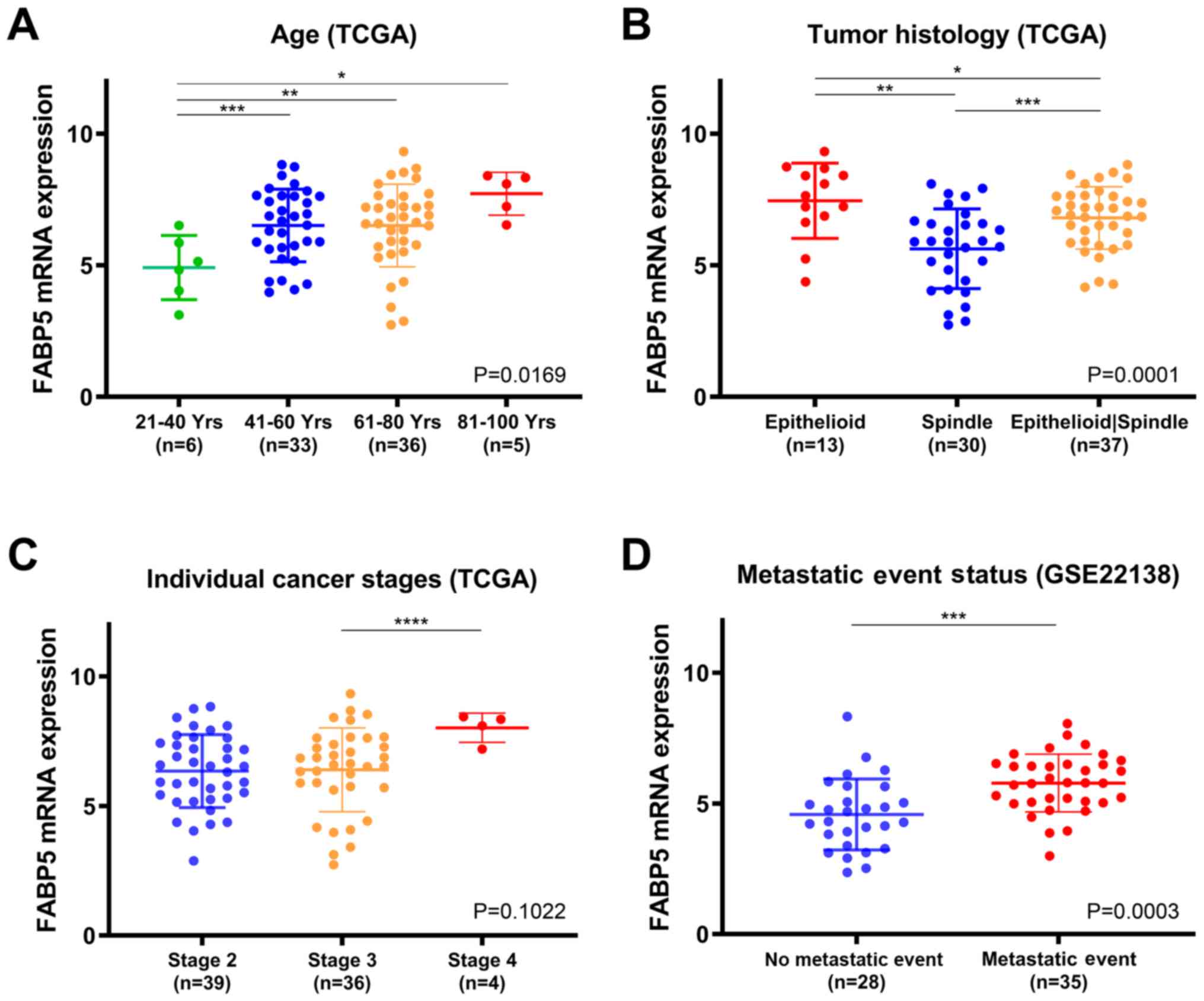
As illustrated in Fig.
1, the transcriptional expression profiles of FABP5 from
the RNA-sequence data from TCGA database and the GSE22138 dataset
were analyzed. The transcriptional expression of FABP5 in
UVM was significantly associated with the age of the patient
(P=0.0169). The lowest mRNA expression of FABP5 was detected
in the 21–40 year age group. The transcriptional expression levels
of FABP5 was found to be higher in the 81–100 (*P<0.05),
the 61–80 (**P<0.01) and the 41–60 (***P<0.001) age groups
compared with that in the 21–40 age group (Fig. 1A). In addition, the transcriptional
expression levels of FABP5 in UVM was significantly
associated with tumor histology (P=0.0001). The highest mRNA
expression levels of FABP5 was detected in epithelioid UVM,
whereas the lowest level was found in spindle cell UVM. The
transcriptional expression levesl of FABP5 was found to be
higher in the epithelioid UVM compared with that in the mixed cell
UVM (*P<0.05) and spindle cell UVM groups (**P<0.01), and
higher in the mixed cell UVM compared with that in the spindle cell
UVM (***P<0.001) group (Fig. 1B).
The transcriptional expression of FABP5 in UVM was not
significantly associated with individual cancer stages (P=0.1022;
Fig. 1C). However, patients who were
in more advanced stages tended to express higher mRNA expression
levels of FABP5. The highest mRNA expression of FABP5
was found in stage 4, which was significantly higher compared with
that in stage 3 (****P<0.0001). The transcriptional expression
of FABP5 in UVM was significantly associated with metastatic
event status in GSE22138 (P=0.0003). A higher level of FABP5
mRNA expression levels were found in patients with a metastatic
event (Fig. 1D). The baseline
clinicopathological characteristics, according to FABP5
expression status, are shown in Table
SI.
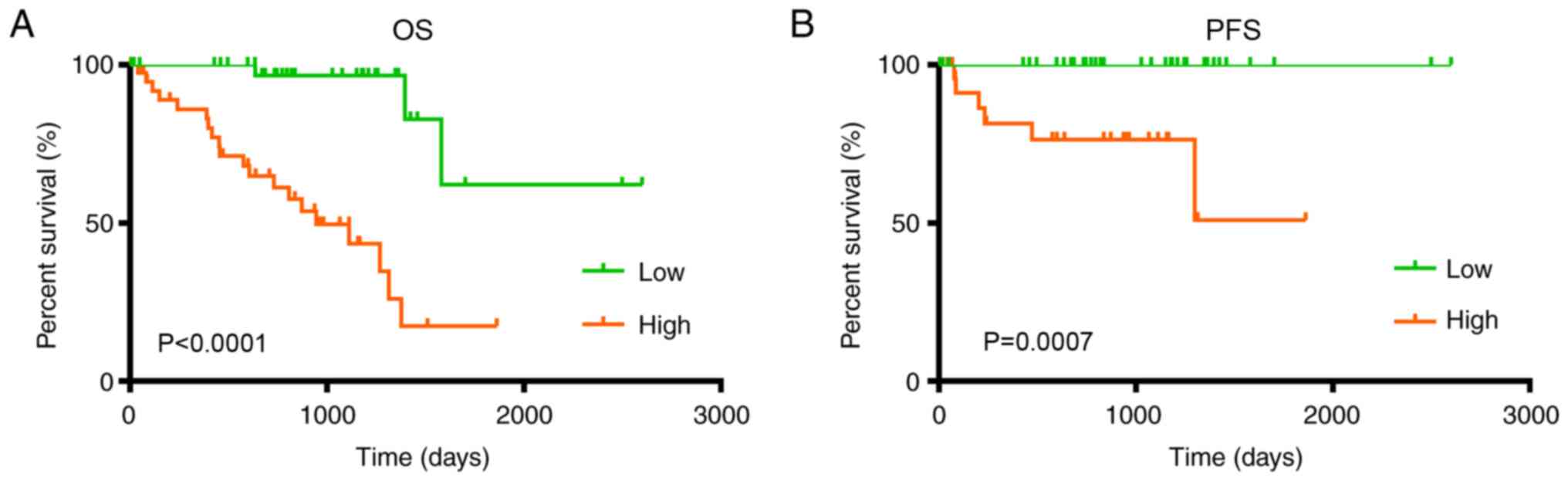
Survival outcomes of the 80 UVM
patients from TCGA
The patients have been divided according to
FABP5 expression levels, therefore overall survival is
associated with expression levels of FABP5 and patients with a high
expression levels have a significantly low overall survival time
(P<0.001; Fig. 2A). In addition,
patients with UVM and high FABP5 mRNA levels showed shorter
PFS time (P=0.0007; Fig. 2B).
Furthermore, the ROC curve was generated to validate the ability of
the logistic model to predict prognosis. The AUC index for the
integrated model was 0.867 for the OS (P=0.008) for patients with
UVM who had died (Fig. S1).
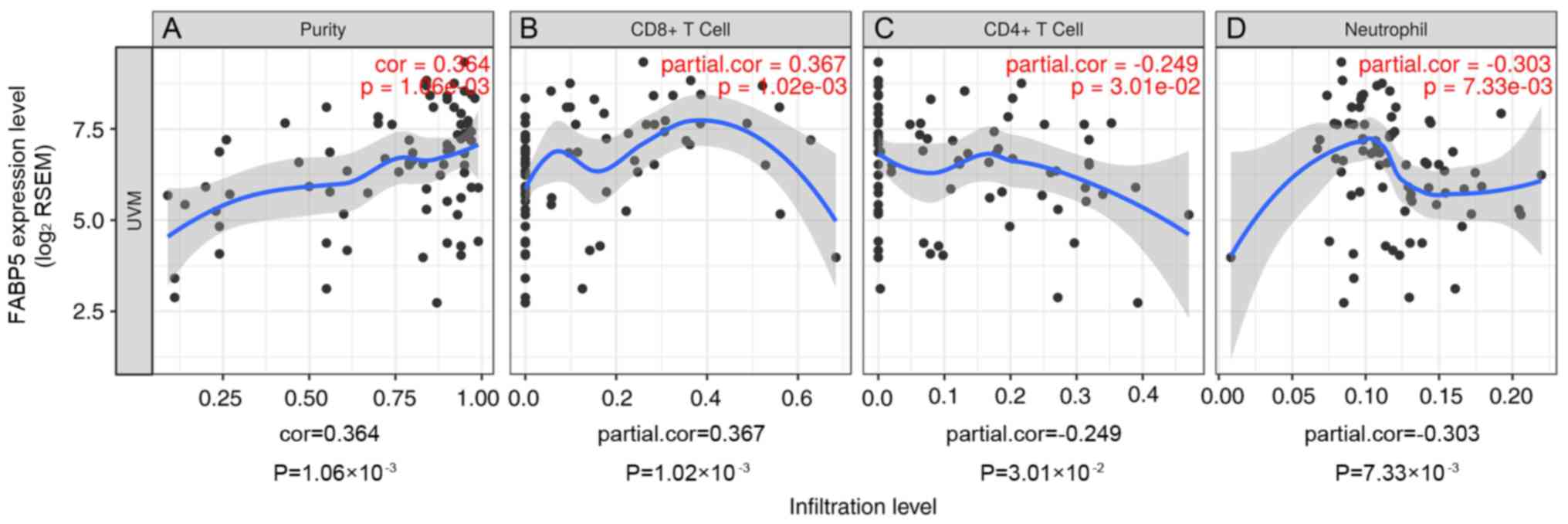
Immune infiltration level
Tumor-infiltrating lymphocytes are an independent
predictor of cancer sentinel lymph node status and survival rate
(26,27). Therefore, the correlation between
FABP5 expression and immune infiltration levels in UVM was
investigated using TIMER. The analysis demonstrated that
FABP5 expression had significant and positive correlation
with tumor purity and CD8+ T cells in UVM and
significant negative correlation with infiltrating levels of
CD4+ T cells and neutrophils in UVM (Fig. 3).
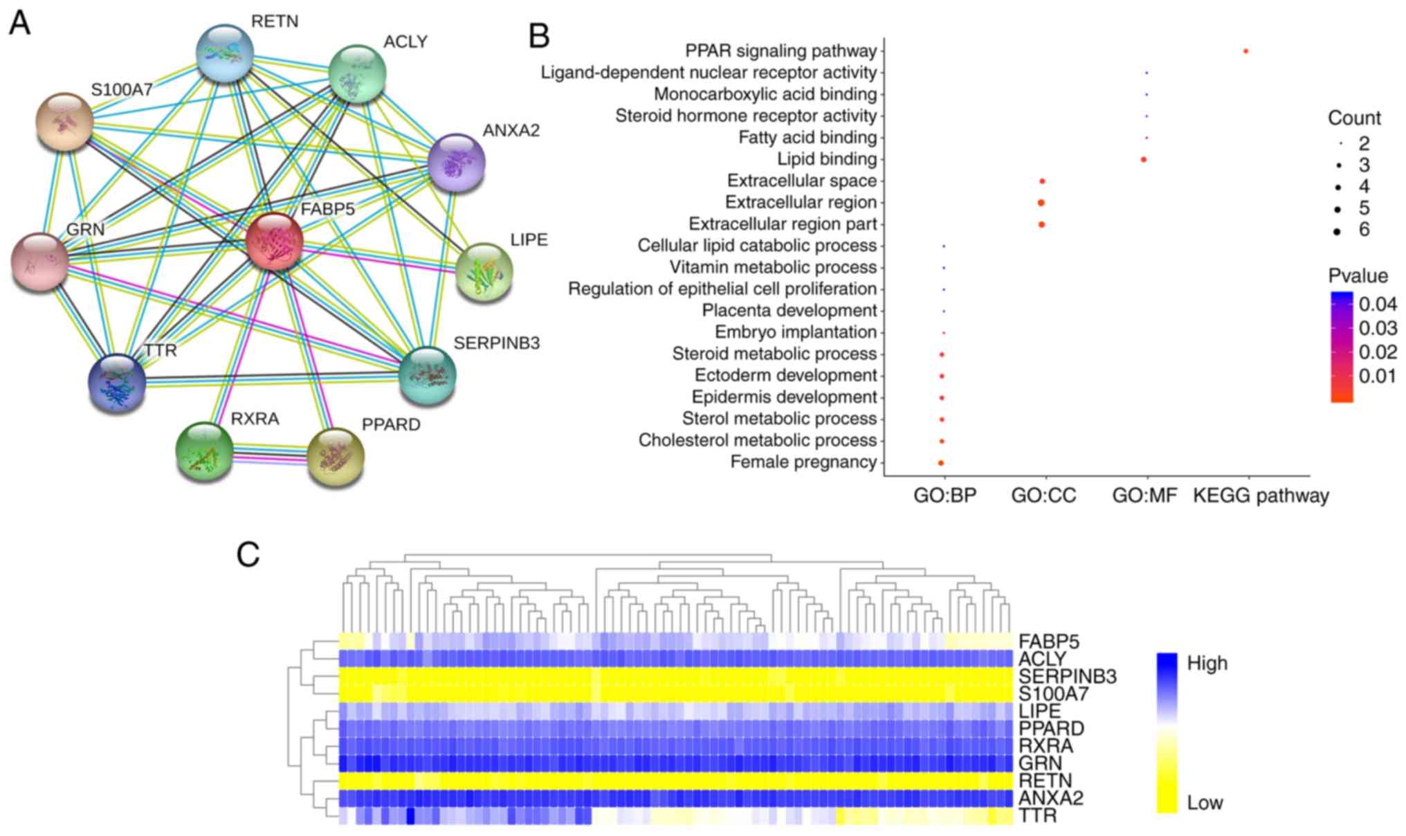
Functional annotation and predicted
signaling pathways
The PPI network of FABP5 was constructed. The
network of FABP5 and its co-expressing genes (resistin, ATP
citrate lyase, annexin A2, lipase E, Serpin family B member 3,
PPARδ, retinoid X receptor α, transthyretin, granulin precursor and
S100 calcium binding protein A7) was visualized (Fig. 4A). The PPI network derived from
active interaction sources was illustrated in detail with the
required interaction score equal to 0.400. As illustrated in
Fig. 4B, functional and pathway
enrichment analyses of a total of 11 associated genes were
performed and visualized using a bubble chart. The changes in the
BP of significant genes were significantly enriched for ‘female
pregnancy’, ‘cholesterol metabolic process’, ‘sterol metabolic
process’, ‘epidermis development’, ‘ectoderm development’ and
‘steroid metabolic process’. The changes in CC were mostly enriched
for the ‘extracellular region part’, ‘extracellular region’ and
‘extracellular space’. The GO analysis results showed that changes
in the MF of significant genes were primarily enriched in ‘lipid
binding’. The hierarchical partitioning of FABP5 and its
co-expressing genes was obtained from 80 UVM patients of TCGA
database (Fig. 4C). It represents
the levels of expression of 11 genes across 80 comparable UVM
patients from TCGA database with high expression marked in blue and
low expression marked in yellow.
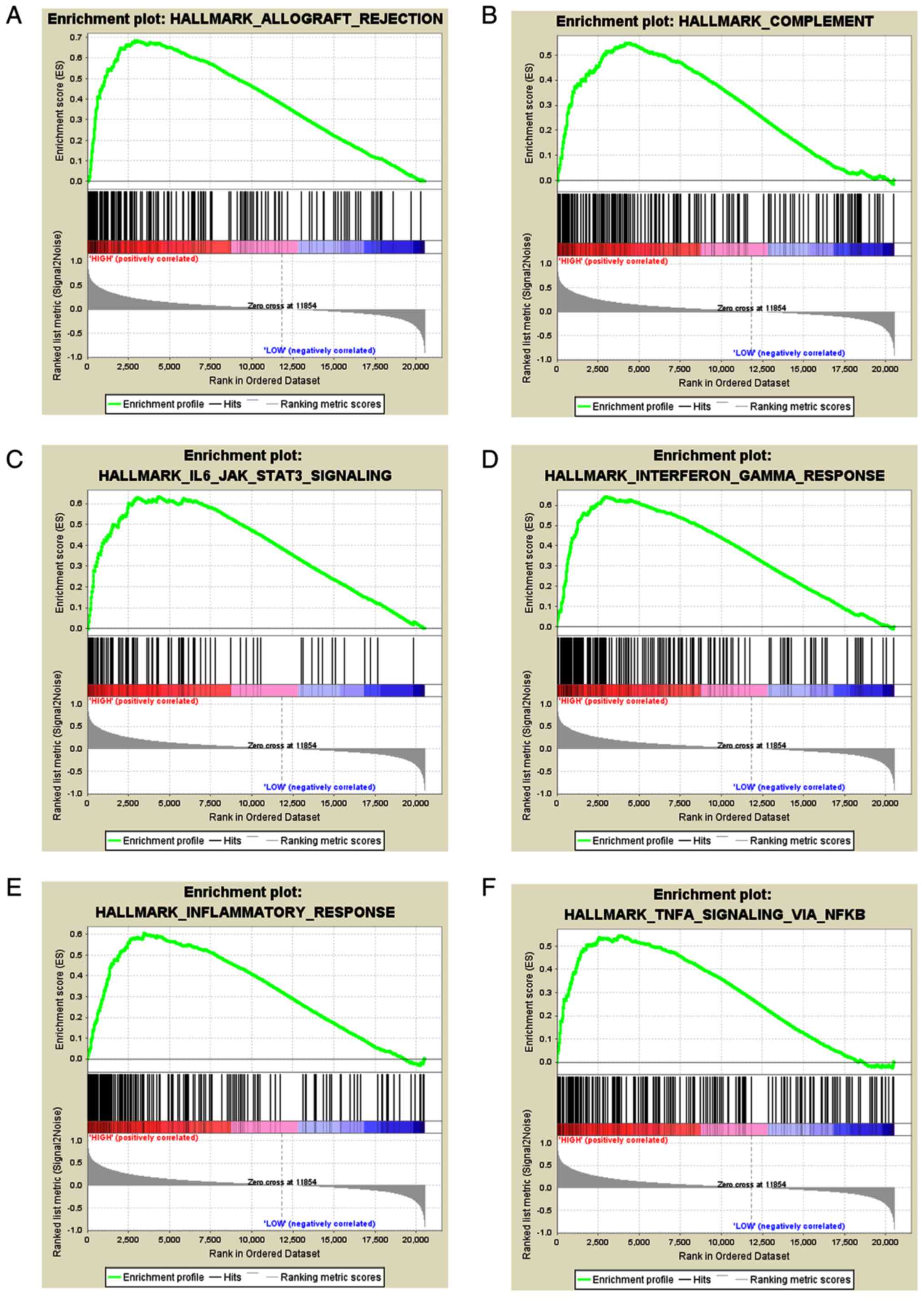
Significant genes and pathways
obtained by GSEA
A total of 100 significantly associated genes were
obtained using GSEA, including those with both positive and
negative associations. Importantly, GSEA was used to perform the
analysis of hallmark pathways that are associated with
FABP5. The results suggested the pathways that were
significantly associated with FABP5, included allograft
rejection, complement, interleukin-6/Janus kinase-STAT3 signaling,
interferon γ response, inflammatory response and tumor necrosis
factor α signaling via NFκB (Fig.
5A-F). In addition, the transcriptional expression profiles of
the 100 significant genes were analyzed using a heat map (Fig. 5G).
Discussion
Aberrant genetic and epigenetic regulation of key
metabolic pathways is known to contribute towards the development
and progression of UVM (28).
Elevated expression of the protease A disintegrin and
metalloproteinase domain 10 and the membrane transporter ATP
binding cassette subfamily B member 5 was shown to be associated
with rapid metastatic progression and worse prognosis in UVM,
suggesting that these proteins may be useful as prognostic factors
(29,30). As a major mediator of fatty acid
uptake, transport and metabolism, FABP5 may participate in
the development and aggressive behavior of cancer (7,12,31,32).
Accordingly, FABP5 is known to play an oncogenic role in
numerous types of cancer, including prostate carcinogenesis,
cervical cancer, renal cell carcinoma and hepatocellular carcinoma
(6,8,11,12),
however to the best of our knowledge the prognostic implications of
FABP5 expression in UVM are currently unknown. In order to address
this gap in knowledge; the present study investigated the
expression, potential function and prognostic value of FABP5
in UVM.
FABP5 is an intracellular carrier of
long-chain fatty acids and other bioactive lipids and also
modulates their metabolism. In addition to transporting fatty acids
within the cytoplasm, FABP5 transfers fatty acids into the
nucleus, where they activate transcription factors (33). For example, FABP5 transfers
retinoic acid to PPARδ, which contributes to cell survival and
proliferation, and FABP5 regulates the induction of
prostaglandin E synthase and inflammation via prostaglandin E2
biosynthesis and NFκB activation (34).
Previous studies have shown that FABP5
expression was associated with the malignant behavior of multiple
types of cancer. Among its oncogenic activities, FABP5
promoted cell migration, proliferation and survival by enhancing
the transcriptional activities of nuclear receptor peroxisome
proliferator-activated receptor β/δ in human breast cancer cells
(MDA-MB-231 cells), human immortalized epidermal cells (HaCaT
cells) and human colorectal adenocarcinoma cells (LS-174T cells)
(7,35–37).
FABP5 expression was associated with primary and metastatic
prostate cancer and is differentially expressed in primary and
metastatic UVM (38,39). Moreover, FABP5 was suggested
as a potential therapeutic target for prostate cancer (40). Furthermore, the expression of
FABP5 was elevated in the regional lymph nodes of patients
with vulvar carcinoma, suggesting its potential as a prognostic
marker gene for this disease (41).
FABP5 may contribute to retinoic acid resistance and
decrease the anticarcinogenic activities of retinoic acid in breast
cancer (42).
At the molecular level, elevated FABP5
expression in fibroblasts was shown to increase PPARδ activity,
cell proliferation, migration and invasion in breast cancer
(43). In human prostate cancer
cells (PC-3 cells) and human breast cancer cells (MDA-MB-231
cells), FABP5 contributes to inflammatory cytokine
production via protein kinase C and the NFκB signaling pathway in
response to elevated levels of reactive oxygen species (10). In addition to PPARβ/δ, PI3K/AKT and
NFκB activities are involved in the regulation of FABP5
activity and expression. FABP5 may increase clear cell renal
cell carcinoma cell proliferation, partly via the PI3K/AKT
signaling pathway (11). In
colorectal cancer, FABP5 promoted cell growth and metastasis
via the PPARβ/δ signaling pathway (15). In addition, FABP5 promoted the
expression of secreted proteins associated with tumor malignancy,
by activating the NFκB signaling pathway (10).
To the best of our knowledge, the present study is
the first to investigate the potential of FABP5 as a
prognostic factor of UVM. Although FABP5 has been implicated
in the development of numerous types of cancer and other human
diseases, including prostate cancer, intrahepatic
cholangiocarcinoma, colorectal cancer and cervical cancer (7,8,12,14,15),
little is known about its involvement in UVM. The present study
demonstrated that the mRNA expression levels of FABP5 was
elevated in UVM tissues, which was significantly associated with
worse clinicopathological parameters, such as shorter OS and PFS
times. Of note, another study demonstrated that patients with
spindle cell UVM tumors had longer disease-specific survival
compared with those with epithelioid and mixed tumors consisting of
epithelioid and spindle cells (44).
This association was linked to the expression levels of
FABP5 mRNA, with the highest being observed in epithelioid
UVM and the lowest in spindle cell UVM. Furthermore, younger
patients (≤20 years) with UVM at the time of diagnosis were found
to have a lower rate of metastasis compared with adults (21–60
years) and older adults (>60 years), which indicated the risk of
metastasis gradually increased with increasing age (45–47). In
the present study, the transcriptional expression of FABP5
in UVM was significantly associated with the age of the patient
suggesting it has prognostic value patients with UVM. Furthermore,
two additional major findings from the present study reveal that
FABP5 expression was positively correlated with UVM tumor
purity and CD8+ T cells whereas it was negatively
correlated with immune cell infiltration; specifically, with the
number of CD4+ T cells and neutrophils. These data
suggests that FABP5 may play a crucial role in immune cell
recruitment to and/or retention within the tumor microenvironment
in UVM.
There are several limitations to the present study.
Firstly, only FABP5 mRNA expression levels were examined as
a potential prognostic biomarker to predict OS and PFS times.
Secondly, further validation studies or prospective cohorts should
be analyzed to verify the present findings. Finally, despite
conducting bioinformatics analysis of functional annotations and
enrichment of FABP5-associated pathways, these findings were
not verified by exploring the underlying molecular mechanisms of
FABP5 signaling. Thus, further studies will be required to
understand the association between FABP5 and tumor growth in
UVM, as well as in other cancer types.
To the best of our knowledge, the present study is
the first to reveal that elevated FABP5 expression is
significantly associated with cancer progression and poor survival
in patients with UVM. Thus, FABP5 is a potential marker of
UVM, which is easily detected, thereby assisting in the selection
of monitoring and treatment strategies. The present study also
provides novel directions for further studies, in order to
elucidate the molecular pathogenesis of UVM. Such studies, together
with randomized clinical trials, will be required to understand the
precise underlying mechanisms of action of FABP5 and its
clinical application in patients with UVM.
Supplementary Material
Supporting Data
Acknowledgements
Not applicable.
Funding
The present study was supported by grants from the
National Natural Science Foundation of China (grant nos. 81202004
and 81802525).
Availability of data and material
The datasets analyzed during the current study are
available from the corresponding author on reasonable request.
Authors' contributions
XFZ designed the research and contributed towards
the analyses, interpretation and presentation of data. YX and WHX
drafted the manuscript, analyzed the data and interpreted the
results. XLY helped to perform the statistical analysis and the
literature review. HLZ co-worked on associated data collection,
data interpretation and revising the draft. All authors read and
approved the final manuscript.
Ethical approval and consent to
participate
Not applicable.
Patient consent for publication
Not applicable.
Competing interests
The authors declare that they have no competing
interests.
Glossary
Abbreviations
Abbreviations:
|
UVM
|
uveal melanoma
|
|
FABP5
|
fatty acid-binding protein 5
|
|
TCGA
|
The Cancer Genome Atlas
|
|
PFS
|
progression-free survival
|
|
OS
|
overall survival
|
|
GO
|
Gene Ontology
|
|
BP
|
biological processes
|
|
CC
|
cellular components
|
|
MF
|
molecular function
|
|
KEGG
|
Kyoto Encyclopedia of Genes and
Genomes
|
|
PPI
|
protein-protein interaction
|
|
GSEA
|
Gene Set Enrichment Analysis
|
References
|
1
|
Kaliki S and Shields CL: Uveal melanoma:
Relatively rare but deadly cancer. Eye (Lond). 31:241–257. 2017.
View Article : Google Scholar : PubMed/NCBI
|
|
2
|
Mahendraraj K, Lau CS, Lee I and
Chamberlain RS: Trends in incidence, survival, and management of
uveal melanoma: A population-based study of 7,516 patients from the
Surveillance, Epidemiology, and End Results database (1973–2012).
Clin Ophthalmol. 10:2113–2119. 2016. View Article : Google Scholar : PubMed/NCBI
|
|
3
|
Carvajal RD, Schwartz GK, Tezel T, Marr B,
Francis JH and Nathan PD: Metastatic disease from uveal melanoma:
Treatment options and future prospects. Br J Ophthalmol. 101:38–44.
2017. View Article : Google Scholar : PubMed/NCBI
|
|
4
|
Smit KN, Chang J, Derks K, Vaarwater J,
Brands T, Verdijk RM, Wiemer EAC, Mensink HW, Pothof J, de Klein A
and Kilic E: Aberrant MicroRNA expression and its implications for
uveal melanoma metastasis. Cancers (Basel). 11(pii): E8152019.
View Article : Google Scholar : PubMed/NCBI
|
|
5
|
Lorenzo D, Piulats JM, Ochoa M, Arias L,
Gutiérrez C, Català J, Cobos E, Garcia-Bru P, Dias B, Padrón-Pérez
N and Caminal JM: Clinical predictors of survival in metastatic
uveal melanoma. Jpn J Ophthalmol. 63:197–209. 2019. View Article : Google Scholar : PubMed/NCBI
|
|
6
|
Kawaguchi K, Kinameri A, Suzuki S, Senga
S, Ke Y and Fujii H: The cancer-promoting gene fatty acid-binding
protein 5 (FABP5) is epigenetically regulated during human prostate
carcinogenesis. Biochem J. 473:449–461. 2016. View Article : Google Scholar : PubMed/NCBI
|
|
7
|
Ohata T, Yokoo H, Kamiyama T, Fukai M,
Aiyama T, Hatanaka Y, Hatanaka K, Wakayama K, Orimo T, Kakisaka T,
et al: Fatty acid-binding protein 5 function in hepatocellular
carcinoma through induction of epithelial-mesenchymal transition.
Cancer Med. 6:1049–1061. 2017. View Article : Google Scholar : PubMed/NCBI
|
|
8
|
Pan L, Xiao H, Liao R, Chen Q, Peng C,
Zhang Y, Mu T and Wu Z: Fatty acid binding protein 5 promotes tumor
angiogenesis and activates the IL6/STAT3/VEGFA pathway in
hepatocellular carcinoma. Biomed Pharmacother. 106:68–76. 2018.
View Article : Google Scholar : PubMed/NCBI
|
|
9
|
Furuhashi M and Hotamisligil GS: Fatty
acid-binding proteins: Role in metabolic diseases and potential as
drug targets. Nat Rev Drug Discov. 7:489–503. 2008. View Article : Google Scholar : PubMed/NCBI
|
|
10
|
Senga S, Kobayashi N, Kawaguchi K, Ando A
and Fujii H: Fatty acid-binding protein 5 (FABP5) promotes
lipolysis of lipid droplets, de novo fatty acid (FA) synthesis and
activation of nuclear factor-kappa B (NF-κB) signaling in cancer
cells. Biochim Biophys Acta Mol Cell Biol Lipids. 1863:1057–1067.
2018. View Article : Google Scholar : PubMed/NCBI
|
|
11
|
Lv Q, Wang G, Zhang Y, Han X, Li H, Le W,
Zhang M, Ma C, Wang P and Ding Q: FABP5 regulates the proliferation
of clear cell renal cell carcinoma cells via the PI3K/AKT signaling
pathway. Int J Oncol. 54:1221–1232. 2019.PubMed/NCBI
|
|
12
|
Wang W, Chu HJ, Liang YC, Huang JM, Shang
CL, Tan H, Liu D, Zhao YH, Liu TY and Yao SZ: FABP5 correlates with
poor prognosis and promotes tumor cell growth and metastasis in
cervical cancer. Tumor Biol. 37:14873–14883. 2016. View Article : Google Scholar
|
|
13
|
Armstrong EH, Goswami D, Griffin PR, Noy N
and Ortlund EA: Structural basis for ligand regulation of the fatty
acidbinding protein 5, peroxisome proliferator-activated receptor
β/δ (FABP5-PPARβ/δ) signaling pathway. J Biol Chem.
289:14941–14954. 2014. View Article : Google Scholar : PubMed/NCBI
|
|
14
|
Jeong CY, Hah YS, Cho BI, Lee SM, Joo YT,
Jung EJ, Jeong SH, Lee YJ, Choi SK, Ha WS, et al: Fatty
acid-binding protein 5 promotes cell proliferation and invasion in
human intrahepatic cholangiocarcinoma. Oncol Rep. 28:1283–1292.
2012. View Article : Google Scholar : PubMed/NCBI
|
|
15
|
Kawaguchi K, Senga S, Kubota C, Kawamura
Y, Ke Y and Fujii H: High expression of fatty acid-binding protein
5 promotes cell growth and metastatic potential of colorectal
cancer cells. FEBS Open Bio. 6:190–199. 2016. View Article : Google Scholar : PubMed/NCBI
|
|
16
|
Tomczak K, Czerwińska P and Wiznerowicz M:
The cancer genome atlas (TCGA): An immeasurable source of
knowledge. Contemp Oncol (Pozn). 19:A68–A77. 2015.PubMed/NCBI
|
|
17
|
Camp RL, Dolled-filhart M and Rimm DL:
X-tile: A new bio-informatics tool for biomarker assessment and
outcome-based cut-point optimization. Clin Cancer Res.
10:7252–7259. 2004. View Article : Google Scholar : PubMed/NCBI
|
|
18
|
Laurent C, Valet F, Planque N, Silveri L,
Maacha S, Anezo O, Hupe P, Plancher C, Reyes C, Albaud B, et al:
High PTP4A3 phosphatase expression correlates with metastatic risk
in uveal melanoma patients. Cancer Res. 71:666–674. 2011.
View Article : Google Scholar : PubMed/NCBI
|
|
19
|
Rhodes DR, Yu J, Shanker K, Deshpande N,
Varambally R, Ghosh D, Barrette T, Pandey A and Chinnaiyan AM:
ONCOMINE: A cancer microarray database and integrated data-mining
platform. Neoplasia. 6:1–6. 2004. View Article : Google Scholar : PubMed/NCBI
|
|
20
|
Li T, Fan J, Wang B, Traugh N, Chen Q, Liu
JS, Li B and Liu XS: TIMER: A web server for comprehensive analysis
of tumor-infiltrating immune cells. Cancer Res. 77:e108–e110. 2017.
View Article : Google Scholar : PubMed/NCBI
|
|
21
|
Li B, Severson E, Pignon JC, Zhao H, Li T,
Novak J, Jiang P, Shen H, Aster JC, Rodig S, et al: Comprehensive
analyses of tumor immunity: Implications for cancer immunotherapy.
Genome Biol. 17:1742016. View Article : Google Scholar : PubMed/NCBI
|
|
22
|
Aran D, Sirota M and Butte AJ: Systematic
pan-cancer analysis of tumour purity. Nat Commun. 6:89712015.
View Article : Google Scholar : PubMed/NCBI
|
|
23
|
Franceschini A, Szklarczyk D, Frankild S,
Kuhn M, Simonovic M, Roth A, Lin J, Minguez P, Bork P, von Mering C
and Jensen LJ: STRING v9.1: Protein-protein interaction networks,
with increased coverage and integration. Nucleic Acids Res.
41:D808–D815. 2013. View Article : Google Scholar : PubMed/NCBI
|
|
24
|
Huang DW, Sherman BT, Tan Q, Collins JR,
Alvord WG, Roayaei J, Stephens R, Baseler MW, Lane HC and Lempicki
RA: The DAVID gene functional classification tool: A novel
biological module-centric algorithm to functionally analyze large
gene lists. Genome Biol. 8:R1832007. View Article : Google Scholar : PubMed/NCBI
|
|
25
|
Subramanian A, Tamayo P, Mootha VK,
Mukherjee S, Ebert BL, Gillette MA, Paulovich A, Pomeroy SL, Golub
TR, Lander ES and Mesirov JP: Gene set enrichment analysis: A
knowledge-based approach for interpreting genome-wide expression
profiles. Proc Natl Acad Sci USA. 102:15545–15550. 2005. View Article : Google Scholar : PubMed/NCBI
|
|
26
|
Dunn GP, Dunn IF and Curry WT: Focus on
TILs: Prognostic significance of tumor infiltrating lymphocytes in
human glioma. Cancer Immun. 7:122007.PubMed/NCBI
|
|
27
|
Azimi F, Scolyer RA, Rumcheva P, Moncrieff
M, Murali R, McCarthy SW, Saw RP and Thompson JF:
Tumor-infiltrating lymphocyte grade is an independent predictor of
sentinel lymph node status and survival in patients with cutaneous
melanoma. J Clin Oncol. 30:2678–2683. 2012. View Article : Google Scholar : PubMed/NCBI
|
|
28
|
Vichitvejpaisal P, Dalvin LA, Mazloumi M,
Ewens KG, Ganguly A and Shields CL: Genetic analysis of uveal
melanoma in 658 patients using the cancer genome atlas
classification of uveal melanoma as A, B, C, and D. Ophthalmology.
126:1445–1453. 2019. View Article : Google Scholar : PubMed/NCBI
|
|
29
|
Caltabiano R, Puzzo L, Barresi V, Ieni A,
Loreto C, Musumeci G, Castrogiovanni P, Ragusa M, Foti P, Russo A,
et al: ADAM 10 expression in primary uveal melanoma as prognostic
factor for risk of metastasis. Pathol Res Pract. 212:980–987. 2016.
View Article : Google Scholar : PubMed/NCBI
|
|
30
|
Broggi G, Musumeci G, Puzzo L, Russo A,
Reibaldi M, Ragusa M, Longo A and Caltabiano R: Immunohistochemical
expression of ABCB5 as a potential prognostic factor in uveal
melanoma. Appl Sci. 9:13162019. View Article : Google Scholar
|
|
31
|
Pan J, Dai Q, Zhang T and Li C: Palmitate
acid promotes gastric cancer metastasis via FABP5/SP1/UCA1 pathway.
Cancer Cell Int. 19:692019. View Article : Google Scholar : PubMed/NCBI
|
|
32
|
Ju J, Wang N, Wang J, Wu F, Ge J and Chen
F: 4-Amino-2-trifluoromethyl-phenyl retinate inhibits
proliferation, invasion, and migration of breast cancer cells by
independently regulating CRABP2 and FABP5. Drug Des Devel Ther.
12:997–1008. 2018. View Article : Google Scholar : PubMed/NCBI
|
|
33
|
Kaczocha M, Vivieca S, Sun J, Glaser ST
and Deutsch DG: Fatty acid-binding proteins transport
N-acylethanolamines to nuclear receptors and are targets of
endocannabinoid transport inhibitors. J Biol Chem. 287:3415–3424.
2012. View Article : Google Scholar : PubMed/NCBI
|
|
34
|
Siegenthaler G, Hotz R, Chatellard-Gruaz
D, Didierjean L, Hellman U and Saurat JH: Purification and
characterization of the human epidermal fatty acid-binding protein:
Localization during epidermal cell differentiation in vivo and in
vitro. Biochem J. 302:363–371. 1994. View Article : Google Scholar : PubMed/NCBI
|
|
35
|
Di-Poï N, Michalik L, Tan NS, Desvergne B
and Wahli W: The anti-apoptotic role of PPARbeta contributes to
efficient skin wound healing. J Steroid Biochem Mol Biol.
85:257–265. 2003. View Article : Google Scholar : PubMed/NCBI
|
|
36
|
Adhikary T, Brandt DT, Kaddatz K, Stockert
J, Naruhn S, Meissner W, Finkernagel F, Obert J, Lieber S, Scharfe
M, et al: Inverse PPARβ/δ agonists suppress oncogenic signaling to
the ANGPTL4 gene and inhibit cancer cell invasion. Oncogene.
32:5241–5252. 2013. View Article : Google Scholar : PubMed/NCBI
|
|
37
|
Wang D, Wang H, Guo Y, Ning W, Katkuri S,
Wahli W, Desvergne B, Dey SK and DuBois RN: Crosstalk between
peroxisome proliferator-activated receptor delta and VEGF
stimulates cancer progression. Proc Natl Acad Sci USA.
103:19069–19074. 2006. View Article : Google Scholar : PubMed/NCBI
|
|
38
|
Alshalalfa M, Bismar TA and Alhajj R:
Detecting cancer outlier genes with potential rearrangement using
gene expression data and biological networks. Adv Bioinformatics.
2012:3735062012. View Article : Google Scholar : PubMed/NCBI
|
|
39
|
Xu Y, Han W, Xu W, Wang Y, Yang XL, Nie
HL, Yao J, Shen GL and Zhang XF: Identification of differentially
expressed genes and functional annotations associated with
metastases ofthe uveal melanoma. J Cell Biochem. 120:19202–19214.
2019. View Article : Google Scholar : PubMed/NCBI
|
|
40
|
Morgan EA, Forootan SS, Adamson J, Foster
CS, Fujii H, Igarashi M, Beesley C, Smith PH and Ke Y: Expression
of cutaneous fatty acid-binding protein (C-FABP) in prostate
cancer: Potential prognostic marker and target for
tumourigenicity-suppression. Int J Oncol. 32:767–775.
2008.PubMed/NCBI
|
|
41
|
Kowalewska M, Radziszewski J, Goryca K,
Bujko M, Oczko-Wojciechowska M, Jarzab M, Siedlecki JA and
Bidzinski M: Estimation of groin recurrence risk in patients with
squamous cell vulvar carcinoma by the assessment of marker gene
expression in the lymph nodes. BMC Cancer. 12:2232012. View Article : Google Scholar : PubMed/NCBI
|
|
42
|
Schug TT, Berry DC, Toshkov IA, Cheng L,
Nikitin AY and Noy N: Overcoming retinoic acid-resistance of
mammary carcinomas by diverting retinoic acid from PPARbeta/delta
to RAR. Proc Natl Acad Sci USA. 105:7546–7551. 2008. View Article : Google Scholar : PubMed/NCBI
|
|
43
|
Levi L, Lobo G, Doud MK, Von Lintig J,
Seachrist D, Tochtrop GP and Noy N: Genetic ablation of the fatty
acid-binding protein FABP5 suppresses HER2-induced mammary
tumorigenesis. Cancer Res. 73:4770–4780. 2013. View Article : Google Scholar : PubMed/NCBI
|
|
44
|
Andreoli MT, Mieler WF and Leiderman YI:
Epidemiological trends in uveal melanoma. Br J Ophthalmol.
99:1550–1553. 2015. View Article : Google Scholar : PubMed/NCBI
|
|
45
|
Kaliki S, Shields CL and Shields JA: Uveal
melanoma: Estimating prognosis. Indian J Ophthalmol. 63:93–102.
2015. View Article : Google Scholar : PubMed/NCBI
|
|
46
|
Shields CL, Kaliki S, Furuta M, Mashayekhi
A and Shields JA: Clinical spectrum and prognosis of uveal melanoma
based on age at presentation in 8,033 cases. Retina. 32:1363–1372.
2012. View Article : Google Scholar : PubMed/NCBI
|
|
47
|
Kaliki S, Shields CL, Mashayekhi A, Ganesh
A, Furuta M and Shields JA: Influence of age on prognosis of young
patients with uveal melanoma: A matched retrospective cohort study.
Eur J Ophthalmol. 23:208–216. 2013. View Article : Google Scholar : PubMed/NCBI
|