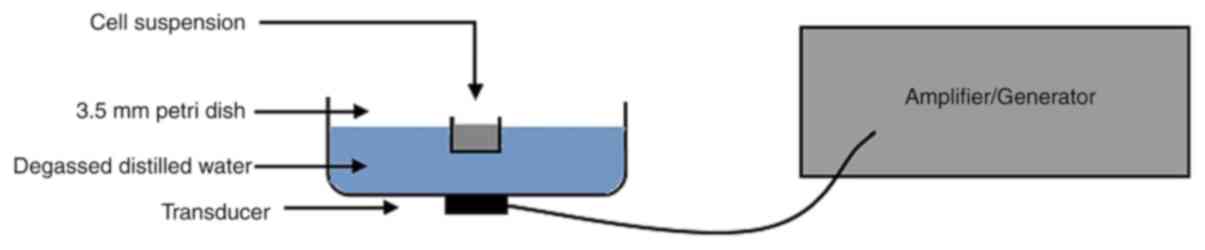
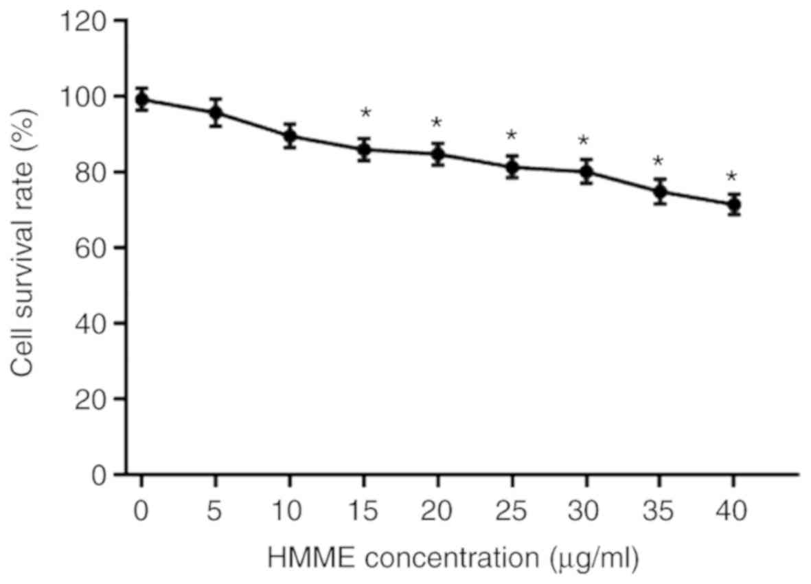
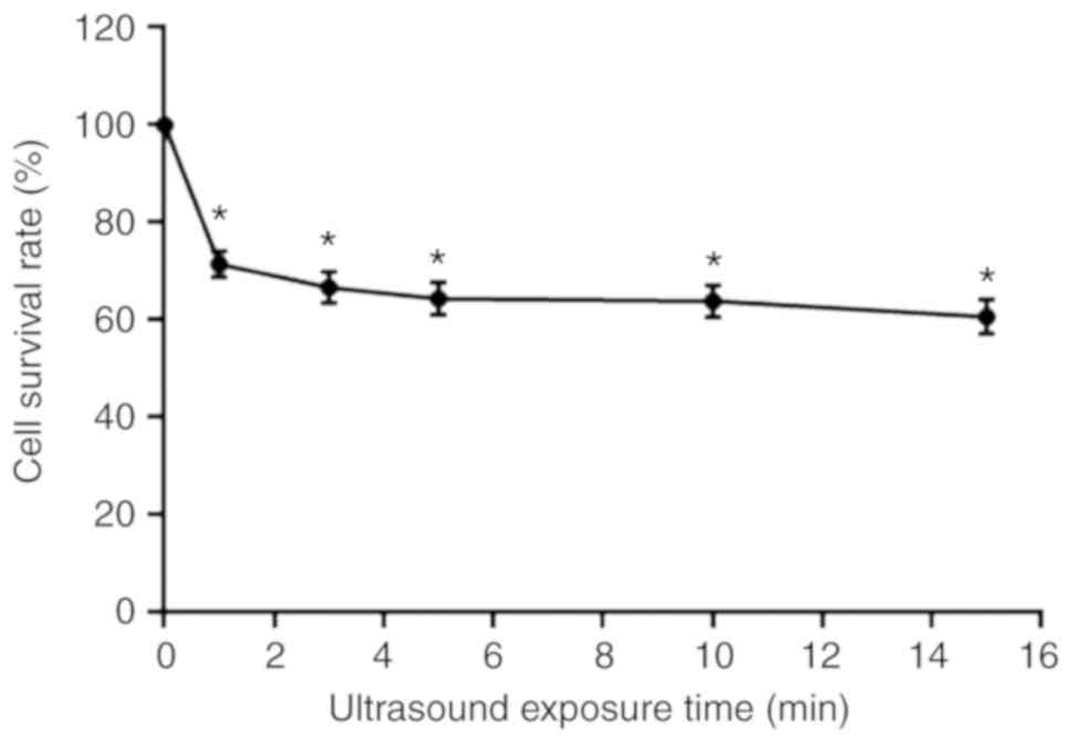
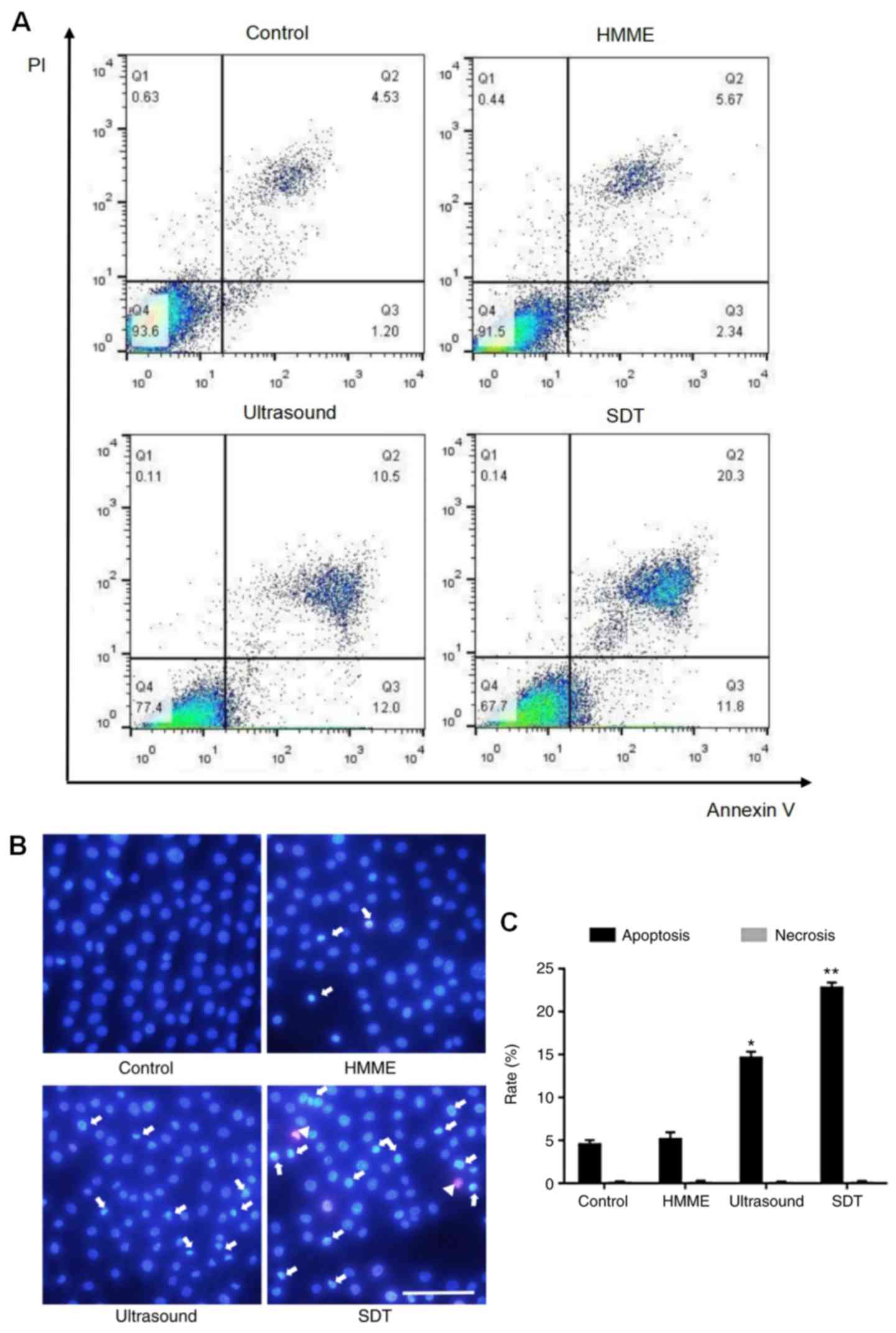
|
1
|
Bray F, Ferlay J, Soerjomataram I, Siegel
RL, Torre LA and Jemal A: Global cancer statistics 2018: GLOBOCAN
estimates of incidence and mortality worldwide for 36 cancers in
185 countries. CA Cancer J Clin. 68:394–424. 2018. View Article : Google Scholar : PubMed/NCBI
|
|
2
|
Pérez-Sayáns M, Suárez-Peñaranda JM,
Gayoso-Diz P, Barros-Angueira F, Gándara-Rey JM and García-García
A: The role of p21Waf1/CIP1 as a Cip/Kip type cell-cycle regulator
in oral squamous cell carcinoma (Review). Med Oral Patol Oral Cir
Bucal. 18:e219–e225. 2013. View Article : Google Scholar : PubMed/NCBI
|
|
3
|
Somasundaram RT, Kaur J, Leong I,
MacMillan C, Witterick IJ, Walfish PG and Ralhan R: Subcellular
differential expression of Ep-ICD in oral dysplasia and cancer is
associated with disease progression and prognosis. BMC Cancer.
16:4862016. View Article : Google Scholar : PubMed/NCBI
|
|
4
|
Taghavi N and Yazdi I: Prognostic factors
of survival rate in oral squamous cell carcinoma: Clinical,
histologic, genetic and molecular concepts. Arch Iran Med.
18:314–319. 2015.PubMed/NCBI
|
|
5
|
Kerker FA, Adler W, Brunner K, Moest T,
Wurm MC, Nkenke E, Neukam FW and von Wilmowsky C: Anatomical
locations in the oral cavity where surgical resections of oral
squamous cell carcinomas are associated with a close or positive
margin-a retrospective study. Clin Oral Investig. 22:1625–1630.
2018. View Article : Google Scholar : PubMed/NCBI
|
|
6
|
Su X, Wang P, Wang X, Cao B, Li L and Liu
Q: Apoptosis of U937 cells induced by hematoporphyrin monomethyl
ether-mediated sonodynamic action. Cancer Biother Radiopharm.
28:207–217. 2013. View Article : Google Scholar : PubMed/NCBI
|
|
7
|
Zhuang D, Hou C, Bi L, Han J, Hao Y, Cao W
and Zhou Q: Sonodynamic effects of hematoporphyrin monomethyl ether
on staphylococcus aureus in vitro. FEMS Microbiol Lett.
361:174–180. 2014. View Article : Google Scholar : PubMed/NCBI
|
|
8
|
Yang Y, Liu Y, Chen X, Gong J, Huang Z,
Wang W, Shi Y, Wang Y, Yao J, Shen Z, et al: 5-aminolevulinic
acid-mediated sonodynamic therapy alleviates atherosclerosis via
enhancing efferocytosis and facilitating a shift in the Th1/Th2
balance toward Th2 polarization. Cell Physiol Biochem. 47:83–96.
2018. View Article : Google Scholar : PubMed/NCBI
|
|
9
|
Bilmin K, Kujawska T, Secomski W, Nowicki
A and Grieb P: 5-Aminolevulinic acid-mediated sonosensitization of
rat RG2 glioma cells in vitro. Folia Neuropathol. 54:234–240. 2016.
View Article : Google Scholar : PubMed/NCBI
|
|
10
|
Hao D, Song Y, Che Z and Liu Q: Calcium
overload and in vitro apoptosis of the C6 glioma cells mediated by
sonodynamic therapy (hematoporphyrin monomethyl ether and
ultrasound). Cell Biochem Biophys. 70:1445–1452. 2014. View Article : Google Scholar : PubMed/NCBI
|
|
11
|
Su X, Wang P, Yang S, Zhang K, Liu Q and
Wang X: Sonodynamic therapy induces the interplay between apoptosis
and autophagy in K562 cells through ROS. Int J Biochem Cell Biol.
60:82–92. 2015. View Article : Google Scholar : PubMed/NCBI
|
|
12
|
Li Y, Zhou Q, Deng Z, Pan M, Liu X, Wu J,
Yan F and Zheng H: IR-780 Dye as a sonosensitizer for sonodynamic
therapy of breast tumor. Sci Rep. 6:259682016. View Article : Google Scholar : PubMed/NCBI
|
|
13
|
Bullon P, Cordero MD, Quiles JL, Morillo
JM, del Carmen Ramirez-Tortosa M and Battino M: Mitochondrial
dysfunction promoted by Porphyromonas gingivalis lipopolysaccharide
as a possible link between cardiovascular disease and
periodontitis. Free Radic Biol Med. 50:1336–1343. 2011. View Article : Google Scholar : PubMed/NCBI
|
|
14
|
Wang X, Jia Y, Wang P, Liu Q and Zheng H:
Current status and future perspectives of sonodynamic therapy in
glioma treatment. Ultrason Sonochem. 37:592–599. 2017. View Article : Google Scholar : PubMed/NCBI
|
|
15
|
Sun X, Xu H, Shen J, Guo S, Shi S, Dan J,
Tian F and Tian Y and Tian Y: Real-time detection of intracellular
reactive oxygen species and mitochondrial membrane potential in
THP-1 macrophages during ultrasonic irradiation for optimal
sonodynamic therapy. Ultrason Sonochem. 22:7–14. 2015. View Article : Google Scholar : PubMed/NCBI
|
|
16
|
Li Y, Wang P, Zhao P, Zhu S, Wang X and
Liu Q: Apoptosis induced by sonodynamic treatment by protoporphyrin
IX on MDA-MB-231 cells. Ultrasonics. 52:490–496. 2012. View Article : Google Scholar : PubMed/NCBI
|
|
17
|
Liu ZR, Chen SQ, Zou YW, Wu XY, Li HY,
Wang XQ, Shi Y and Niu HX: Hypochlorite modified albumins promote
cell death in the tubule interstitium in rats via mitochondrial
damage in obstructive nephropathy and the protective effects of
antioxidant peptides. Free Radic Res. 52:616–628. 2018. View Article : Google Scholar : PubMed/NCBI
|
|
18
|
Li N, Sun M, Wang Y, Lv Y, Hu Z, Cao W,
Zheng J and Jiao X: Effect of cell cycle phase on the sensitivity
of SAS cells to sonodynamic therapy using low-intensity ultrasound
combined with 5-aminolevulinic acid in vitro. Mol Med Rep.
12:3177–3183. 2015. View Article : Google Scholar : PubMed/NCBI
|
|
19
|
Lv Y, Zheng J, Zhou Q, Jia L, Wang C, Liu
N, Zhao H, Ji H, Li B and Cao W: Antiproliferative and
apoptosis-inducing effect of exo-protoporphyrin IX based
sonodynamic therapy on human oral squamous cell carcinoma. Sci Rep.
7:409672017. View Article : Google Scholar : PubMed/NCBI
|
|
20
|
Sisto M, Lisi S, D'Amore M, Mitolo V and
Scagliusi P: Anti-Ro and anti-La autoantibodies induce TNF-alpha
production by human salivary gland cells: An in vitro study.
Reumatismo. 59:221–226. 2007.PubMed/NCBI
|
|
21
|
Li W, Fei JF, Yang Q, Li BL, Lin C, Yue Q
and Meng QG: Acute toxic effects of sonodynamic therapy on
hypertrophic scar fibroblasts of rabbit ears. Genet Mol Res.
14:4203–4214. 2015. View Article : Google Scholar : PubMed/NCBI
|
|
22
|
Xie R, Xu T, Zhu J, Wei X, Zhu W, Li L,
Wang Y, Han Y, Zhou J and Bai Y: The combination of glycolytic
inhibitor 2-deoxyglucose and microbubbles increases the effect of
5-aminolevulinic acid-sonodynamic therapy in liver cancer cells.
Ultrasound Med Biol. 43:2640–2650. 2017. View Article : Google Scholar : PubMed/NCBI
|
|
23
|
Xiong W, Wang P, Hu J, Jia Y, Wu L, Chen
X, Liu Q and Wang X: A new sensitizer DVDMS combined with multiple
focused ultrasound treatments: An effective antitumor strategy. Sci
Rep. 5:174852015. View Article : Google Scholar : PubMed/NCBI
|
|
24
|
Sun H, Ge W, Gao X, Wang S, Jiang S, Hu Y,
Yu M and Hu S: Apoptosis-promoting effects of hematoporphyrin
monomethyl ether-sonodynamic therapy (HMME-SDT) on endometrial
cancer. PLoS One. 10:e01379802015. View Article : Google Scholar : PubMed/NCBI
|
|
25
|
Wang F, Gao Q, Guo S, Cheng J, Sun X, Li
Q, Wang T, Zhang Z, Cao W and Tian Y: The sonodynamic effect of
curcumin on THP-1 cell-derived macrophages. BioMed Res Int.
2013:7372642013.PubMed/NCBI
|
|
26
|
Qin P, Han T, Yu ACH and Xu L: Mechanistic
understanding the bioeffects of ultrasound-driven microbubbles to
enhance macromolecule delivery. J Control Release. 272:169–181.
2018. View Article : Google Scholar : PubMed/NCBI
|
|
27
|
Li J, Yue W, Huang Z, Chen ZQ, Zhan Q, Ren
FB, Liu JY and Fu SB: Calcium overload induces C6 rat glioma cell
apoptosis in sonodynamic therapy. Int J Radiat Biol. 87:1061–1066.
2011. View Article : Google Scholar : PubMed/NCBI
|
|
28
|
Tian J, Gan Y, Pan C, Zhang M, Wang X,
Tang X and Peng X: Nerol-induced apoptosis associated with the
generation of ROS and Ca2+ overload in saprotrophic
fungus Aspergillus flavus. Appl Microbiol Biotechnol.
102:6659–6672. 2018. View Article : Google Scholar : PubMed/NCBI
|
|
29
|
Tran TA, Roger S, Le Guennec JY, Tranquart
F and Bouakaz A: Effect of ultrasound-activated microbubbles on the
cell electrophysiological properties. Ultrasound Med Biol.
33:158–163. 2007. View Article : Google Scholar : PubMed/NCBI
|
|
30
|
Lentacker I, De Cock I, Deckers R, De
Smedt SC and Moonen CT: Understanding ultrasound induced
sonoporation: Definitions and underlying mechanisms. Adv Drug Deliv
Rev. 72:49–64. 2014. View Article : Google Scholar : PubMed/NCBI
|