Introduction
Ovarian cancer is a common gynecological malignancy
that has the highest mortality rate among all cancer types
affecting women worldwide, with >200,000 new cases each year
(1). Ovarian cancers are
histologically defined as type I or type II. Type I refers to a
relatively low histological grade, including endometrioid, mucinous
and clear-cell carcinomas, whereas type II refers to a higher
histological grade, including serous carcinoma and carcinosarcoma
(2). Epithelial ovarian cancer (EOC)
is the main type of ovarian cancer and represents >90% cases. In
addition, ~90% of patients dying from EOC suffer from type II EOC
(3). Treatment for ovarian cancer
usually includes a combination of surgery, radiation therapy and
chemotherapy (4). However, the
outcome of patients depends also on their clinicopathological
characteristics, including the subtype of ovarian cancer and the
presence of other medical conditions (5). In addition to the aforementioned
conventional treatments, targeted therapy, also known as
molecularly targeted therapy, is one of the major treatment
options, which only targets cancer cells (6). Targeted therapy may therefore be
considered as a promising cure for patients with ovarian cancer in
the near future.
Semaphorins are members of a family of
membrane-bound and secreted molecules, which were originally
identified as evolutionarily conserved axon-guidance cues in the
human neural circuitry (7,8). The semaphorin family is divided into
eight classes, which consist of >30 genes, while the number of
semaphorins is still rising. The neuropilin and plexin gene
families encode the main semaphorin receptors (9). It has been widely reported that
semaphorins are highly expressed in the human nervous system. For
example, previous studies demonstrated that certain semaphorin
members, including semaphorins 6B and 5B, are involved in the
progression of various types of cancer, including gastric cancer
(10) and renal cell carcinoma
(11). These semaphorins promote the
progression and angiogenesis of tumor cells via numerous
mechanisms, including the modulation of tumor angiogenesis
(10,11). Furthermore, certain semaphorins,
including class 3 semaphorins, have been reported to inhibit tumor
progression, whereas others, inducing semaphoring 4D, were
demonstrated to promote tumor progression (9). To the best of our knowledge, there is
no study about the expression of semaphorin-4C (Sema4C) in EOC.
Therefore, the present study investigated the
expression of Sema4C in EOC and determined its association with the
clinicopathological characteristics of patients with EOC.
Materials and methods
Patients and tumor samples
EOC cancer tissues were obtained from patients who
were surgically treated at the Department of Oncology of Yantaishan
Hospital (Yantai, China) between January 2013 and January 2018.
Cancer tissues were obtained within 30 min of the resection, placed
in liquid nitrogen and stored at −80°C. In total, 74 cases of EOC,
20 cases of ovarian epithelial benign tumor, 20 cases of ovarian
borderline epithelial tumor and 15 cases of normal ovarian tissues
were collected. The age distribution of patients with EOC was 29–67
years (mean age, 51.2±7.6 years). The age distribution of patients
with ovarian epithelial benign tumor was 30–68 years (mean age,
47.2±7.7 years). The age distribution of patients with ovarian
borderline epithelial tumor was 28–61 years (mean age, 43.6±6.8
years). The age distribution of patients with normal ovarian
tissues was 30–63 years (mean age, 45.6±7.4 years). The patients
had no heart-, liver-, lung-, kidney- or other important
organ-related diseases, and had no history of chemotherapy,
radiotherapy or other treatment prior to surgery. Patients with
other malignant tumors were also excluded.
The 74 cases with EOC were graded according to the
World Health Organization (WHO) standards for histopathological
clinical staging (12) as follows: A
total of 44 patients had stages I and II EOC, whereas 30 patients
had stage III EOC. However, according to the Union for
International Cancer Control (UICC) standards (13), 21 cases were in stages I–II, whereas
53 cases were in stages III–IV. In total, 55 patients out of the 74
cases were >50 years old, and 49 out of the 74 patients
presented with ascites.
EOC cancer tissues were obtained from patients who
were surgically treated at the Department of Oncology of Yantaishan
Hospital (Yantai, China) between January 2013 and January 2018 and
who were diagnosed with EOC. The tissues previously mentioned were
part of these tissues. In total, 111 EOC tissues were collected and
embedded in paraffin before analyzing Sema4C protein expression.
According to the WHO standards for histopathological clinical
staging, 69 cases were in stages I and II, whereas 44 cases were in
stage III. However, according to the UICC standards, 29 cases were
in stages I–II, whereas 82 cases were in stages III–IV. In total,
84 patients were >50 years old and 75 patients presented with
ascites. The clinical data of all the patients were complete, and
the pathological data were provided by a physician in-chief from
the Pathology department of Yantaishan Hospital.
Reverse transcription-quantitative
polymerase chain reaction (RT-qPCR)
Total RNA was extracted from tissues using
TRIzol® reagent (Invitrogen; Thermo Fisher Scientific,
Inc.), and RNA purity was determined by calculating the 260/280
ratio of optical densities using a nucleic acid-protein detector
(DU-640; Beckman Coulter, Inc.). The result was between 1.8 and
2.0, which indicated sufficient RNA purity. cDNA was synthesized
using an Eppendorf PCR Mastercycler (Eppendorf) according to the
manufacturer's instructions, whereas qPCR was performed using
SYBR-Green Master Mix (Applied Biosystems; Thermo Fisher
Scientific, Inc.) and the GeneAmp 5700 Sequence Detector (Applied
Biosystems; Thermo Fisher Scientific, Inc.). PCRs were performed as
follows: 94°C, melting under pre-denaturation for 5 min; 94°C for
additional 30 sec, 72°C for 45 sec and 62°C for 30 sec (all steps
were repeated for 35 cycles); and maintenance at 72°C for 10 min.
The primer sequences for Sema4C were synthesized by Shanghai
GenePharma Co., Ltd. and were as follows: Sema4C, forward,
5′-ACCTTGTGCCGCGTAAGACAG-3′ and reverse,
5′-CGTCAGCGTCAGTGTCAGGAA-3′; and β-actin, forward,
5′-CCTGGGCATGGAGTCCTGTG-3′ and reverse, 5′-AGGGGCCGGACTCGTCATAC-3′.
The relative expressions level of Sema4C was normalized to the
endogenous control β-actin and was expressed as 2−ΔΔCq
(14).
Immunohistochemistry (IHC)
staining
Tissue sections from paraffin-embedded cancer
tissues were incubated at 60°C for 30 min, dewaxed using xylene and
rehydrated using a decreasing ethanol gradient (100, 95, 75 and
50%, 5 min each time). Sections were washed three times for 5 min
with PBS. Sections were incubated in 3% H2O2
dissolved in 80% methanol at room temperature for 10 min to
inactivate endogenous peroxidase. Tissues were heated at 95°C for
20 min and blocked with 5% bovine serum albumin (cat. no. B2064;
Sigma-Aldrich; Merck KGaA) for 20 min. Tissues were then incubated
with rabbit polyclonal human primary antibody against Sema4C
(1:400; cat. no. PA5-52788; Thermo Fisher Scientific, Inc.) at 4°C
overnight, and incubated with goat anti-rabbit IgG secondary
antibody (1:1,000; MH1732; Thermo Fisher Scientific, Inc.) at 37°C
for 20–30 min. Signals were visualized using 3′-diaminobenzidine
staining (cat. no. TA-060-QHDX; Thermo Fisher Scientific, Inc.) at
37°C for 5–10 min and hematoxylin counterstained at 37°C for 30
sec-1 min. Differentiation was induced by hydrochloric acid and
ethanol dehydration (80, 95 and 100%, 5 min each time). For each
slice, images of 10 sections were acquired under an optical
microscope (BX45-72H05; Olympus Corporation; magnification, ×100)
to count positively stained cells. A percentage of positively
stained cells >30% was considered as a positive staining.
Statistical analysis
SPSS version 13.0 statistical software (SPSS Inc.)
was used to statistically analyze the data. The results were
expressed as the means ± standard deviation. The t-test was used
for comparisons between two datasets, whereas one-way analysis of
variance followed by least-significant difference post hoc test was
used for comparisons among multiple datasets. P<0.05 was
considered to indicate a statistically significant difference. The
Sema4C protein expression levels were compared between groups using
χ2 test. The correction between Sema4C mRNA level and
Sema4C protein expression were analyzed by Pearson's correlation
analysis.
Results
Sema4C is upregulated in EOC
tissues
The results from RT-qPCR demonstrated that Sema4C
expression level was significantly higher in malignant tissues
compared with that in borderline, benign and normal tissues
(P<0.001; Table I). In addition,
the 74 cases of EOC were divided into four groups as follows:
Serous carcinoma, mucinous adenocarcinoma, endometrial cancer
uterus and clear cell carcinoma (Table
I). These results indicated that Sema4C was highly expressed in
all cancer tissues, but its expression level was not associated
with the histological type (P>0.05).
 | Table I.Sema4C mRNA expression in different
ovarian tissues. |
Table I.
Sema4C mRNA expression in different
ovarian tissues.
| Variable | Number | Sema4C mRNA
(x ±
SD) | T-value | P-value |
|---|
| Category |
|
Malignant | 74 | 0.0505±0.0308 | 34.193 | <0.001 |
|
Borderline | 20 | 0.0074±0.0113 |
|
|
|
Benign | 20 | 0.0067±0.0082 |
|
|
|
Normal | 15 | 0.0059±0.0072 |
|
|
| Histological
type |
| Serous
carcinoma | 24 | 0.0440±0.0212 | 1.012 | >0.05 |
| Mucinous
adenocarcinoma | 21 | 0.0595±0.0444 |
|
|
|
Endometrial cancer uterus | 15 | 0.0497±0.0295 |
|
|
| Clear
cell carcinoma | 14 | 0.0501±0.0282 |
|
|
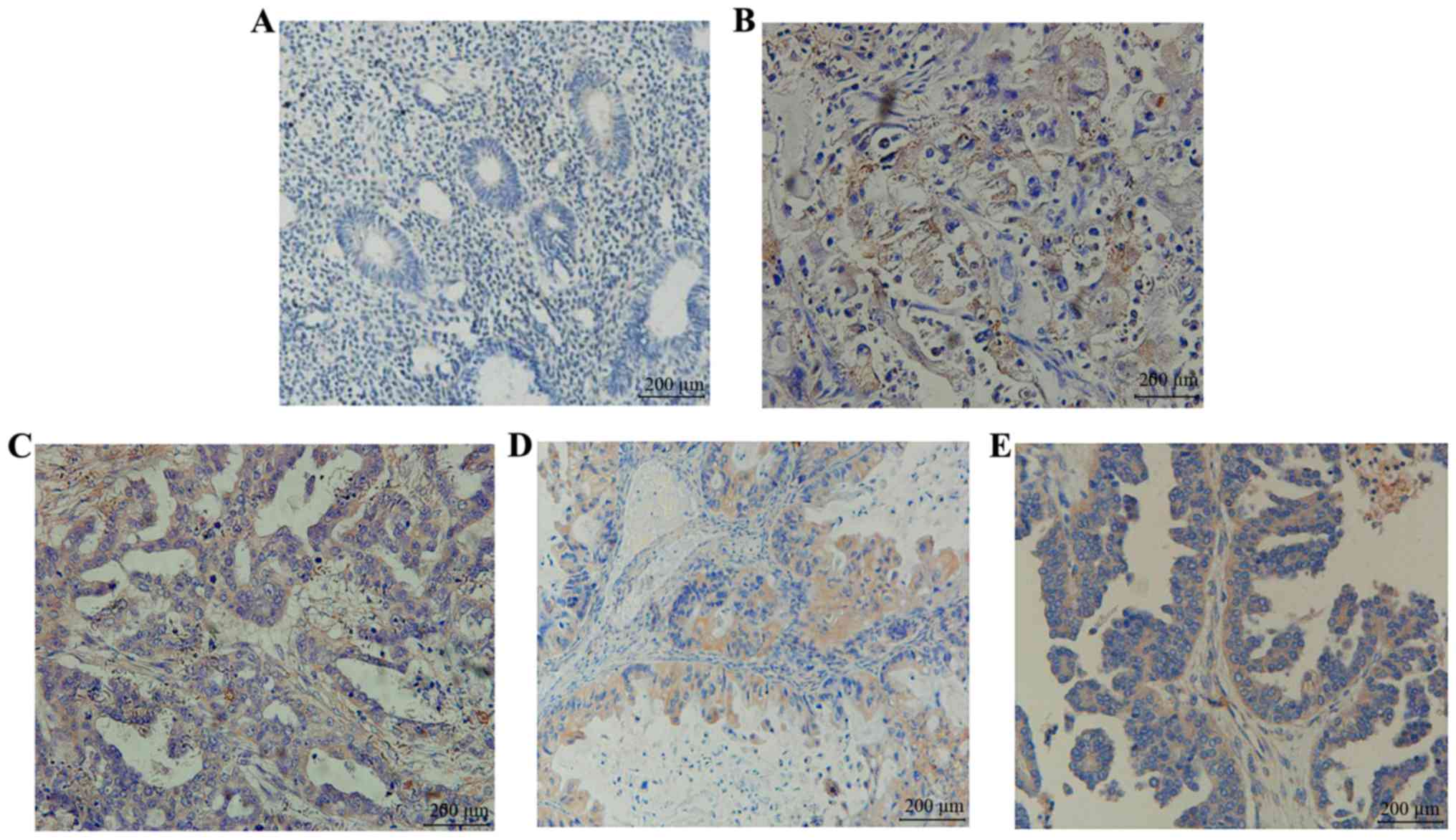
To examine the expression of Sema4C protein in
ovarian cancer, IHC was used. The results demonstrated that Sema4C
protein was hardly expressed in the normal ovarian tissue (Fig. 1A), whereas it was highly expressed in
EOC tissues (Fig. 1B-E). These
findings confirmed that Sema4C mRNA and protein expression were
highly expressed in EOC tissues.
Sema4C expression is associated with
differentiation and clinical stage of EOC tissues
The association between Sema4C mRNA expression and
numerous factors was analyzed, including cancer differentiation
level, clinical stage, EOC ascites and age of the patients at
disease onset. The results demonstrated that Sema4C mRNA expression
was significantly higher in the medium/high differentiation group
compared with that in the low differentiation group (P=0.011;
Table II). Furthermore, Sema4C mRNA
expression in stages III and IV of ovarian cancer was significantly
higher than that in stages I and II (P=0.014). These results
indicated that Sema4C mRNA expression in EOC was associated with
tissue differentiation, FIGO stage and ascites.
| Table II.Association between Sema4C mRNA
expression and clinicopathological features of patients with
epithelial ovarian cancer. |
Table II.
Association between Sema4C mRNA
expression and clinicopathological features of patients with
epithelial ovarian cancer.
| Variable | Number | Sema4C mRNA
(x ±
SD) | T-value | P-value |
|---|
| Differentiation |
| Low | 44 | 0.0431±0.0238 | 2.598 | 0.011 |
|
Medium/high | 30 | 0.0614±0.0367 |
|
|
| FIGO stage |
| I–II | 21 | 0.0367±0.0236 | 2.527 | 0.014 |
|
III–IV | 53 | 0.0560±0.0318 |
|
|
| Ascites |
| Yes | 49 | 0.0536±0.0331 | 1.213 | 0.229 |
| No | 25 | 0.0445±0.0253 |
|
|
| Age, years |
|
<50 | 19 | 0.0467±0.0279 | −6.332 | 0.529 |
| ≥50 | 55 | 0.0519±0.0319 |
|
|
Furthermore, the association between Sema4C protein
and clinicopathological factors was investigated, including
histological type, ascites, age, differentiation and FIGO stage
(Table III). The results
demonstrated that Sema4C protein positive expression in the
medium/highly-differentiated group (75.0%) was significantly higher
compared with that in the low differentiation group (52.2%;
P=0.016). In addition, Sema4C protein positive expression in
tissues at clinical stages III–IV (68.3%) was also significantly
increased compared with that at clinical stages I–II (41.4%;
P=0.011). These findings indicated that Sema4C protein expression
was associated with the differentiation and FIGO stage of EOC, but
not with histological type, ascites and age (P>0.05).
| Table III.Association between semaphoring-4C
protein expression and clinicopathological characteristics of
patients with ovarian cancer. |
Table III.
Association between semaphoring-4C
protein expression and clinicopathological characteristics of
patients with ovarian cancer.
| Variable | Positive, n (%) | Negative, n (%) | χ2 | P-value |
|---|
| Histological
type |
| Serous
carcinoma | 16 (53.3) | 14 (46.7) | 2.907 | 0.406 |
| Mucinous
adenocarcinoma | 22 (73.3) | 8
(26.7) |
|
|
|
Endometrial cancer uterus | 17 (56.7) | 13 (43.3) |
|
|
| Clear
cell carcinoma | 13 (61.9) | 8
(38.1) |
|
|
| Ascites |
| Yes | 49 (65.3) | 26 (34.7) | 1.616 | 0.204 |
| No | 19 (52.8) | 17 (47.2) |
|
|
| Age, years |
|
<50 | 14 (56.0) | 11 (44.0) | 0.376 | 0.540 |
|
≥50 | 54 (62.8) | 32 (37.2) |
|
|
|
Differentiation |
|
Low | 35 (52.2) | 32 (47.8) | 5.798 | 0.016 |
|
Medium/high | 33 (75.0) | 11 (25.0) |
|
|
| FIGO stage |
|
I–II | 12 (41.4) | 17 (58.6) | 6.539 | 0.011 |
|
III–IV | 56 (68.3) | 26 (31.7) |
|
|
Sema4C is upregulated in late-stage
EOC
The positive expression rate of Sema4C in EOC
tissues was 61.3% (Table IV),
whereas the values for borderline ovarian epithelial tumor, benign
tumor and normal ovarian tissues were 26.7, 16.7 and 10.0%,
respectively. The results of χ2 test demonstrated that
the positive expression rate of Sema4C in EOC tissues was
significantly higher compared with that in the other three types of
tissue (P<0.01; Table IV).
| Table IV.Expression of semaphorin-4C protein
in different categories of ovarian tissue. |
Table IV.
Expression of semaphorin-4C protein
in different categories of ovarian tissue.
| Category | Positive, n
(%) | Negative, n
(%) | χ2 | P-value |
|---|
| Malignant | 68 (61.3) | 43 (38.7) | 40.367 | <0.01 |
| Borderline | 8
(26.7) | 22 (38.7) |
|
|
| Benign | 5
(16.7) | 25 (83.3) |
|
|
| Normal | 3
(10.0) | 27 (90.0) |
|
|
The results from Pearson's correlation analysis
(Table V) revealed that Sema4C mRNA
expression and Sema4C protein expression in EOC tissues were
positively correlated (P<0.01). The regression equation was
Y=−1.50814+1.052126X, with a correlation coefficient of
R2=0.955 (P<0.01). Furthermore, the results from
Pearson's correlation analysis revealed that Sema4C mRNA expression
and Sema4C positive expression rate were positively correlated with
tumor malignancy and clinical stage.
| Table V.Pearson's correlation analysis
between the mRNA and protein expression of Sema4C. |
Table V.
Pearson's correlation analysis
between the mRNA and protein expression of Sema4C.
| Sema4C
expression | Positive, n
(%) | Negative, n
(%) | R2 | P-value |
|---|
| mRNA | 44 (59.46) | 30 (40.54) | 0.955 | <0.001 |
| Protein | 68 (61.26) | 43 (38.74) |
|
|
Discussion
Semaphorins were originally reported as serving
crucial role in nervous system (10,15,16).
Over the past decade, semaphorins have been thought to be involved
in numerous developmental processes, including cell migration and
invasion (15–18). In particular, Sema3B and Sema3F were
successfully identified as modulators of tumor progression
(17,18). In addition to these two semaphorins,
semaphorins 6B has been characterized as regulators of tumor
progression (10). To the best of
our knowledge, the present study was the first to confirm that both
Sema4C mRNA and protein expression were highly expressed in EOC
tissues. Furthermore, Sema4C mRNA expression in EOC was associated
with tissue differentiation, FIGO stage and ascites. Sema4C protein
expression was also found to be upregulated in late-stage EOC.
Class 4 semaphorins are single-pass transmembrane
proteins that usually exert clear influences on tumor progression.
For example, Sema4D was demonstrated to be upregulated in several
types of cancer, including head and neck, cervical, colon,
prostate, lung and breast cancer (19). In addition, Sema4C is expressed at a
relatively low rate (3.3%), or not at all in normal ovarian
tissues, which was similar to the findings of the present study. In
the present study, Sema4C protein was expressed at a low rate
(3.0%) in normal ovary. In addition, Sema4C protein was positively
expressed in EOC (56.0%), and was mostly located in the cytoplasm
and/or cell membrane.
A previous study reported that Sema4C stimulates the
production of angiogenin and colony-stimulating factor-1 in breast
cancer cells by activating the NF-κB signaling pathway (20). Furthermore, Gurrapu et al
(21) reported that Sema4C/PlexinB2
signaling pathway was essential for breast carcinoma cell
proliferation, suggesting that it might be considered as a novel
potential therapeutic target. In addition, it was reported that
elevated Sema4C expression enables indolent luminal-type tumors to
become resistant to estrogen deprivation, invasive and metastatic
in vivo. The present study reported that Sema4C was highly
expressed in ovarian epithelial cancer tissues; however, the
underlying mechanisms remain clear. The role of Sema4C in the
stimulation of ovarian epithelial cancer growth requires therefore
further investigation.
In conclusion, the present study demonstrated that
Sema4C was highly expressed in EOC tissues, and that Sema4C mRNA
and protein expression were associated with tumor malignancy and
clinical stage. These findings suggested that high Sema4C
expression in EOC tissues may be associated with poor prognosis in
patients with EOC.
Acknowledgements
Not applicable.
Funding
The present study was funded by the Key Research and
Development Plan in Shandong Province (grant no.
2018GSF118054).
Availability of data and materials
The datasets used and/or analyzed during the current
study are available from the corresponding author on reasonable
request.
Authors' contributions
SYH, SH and JW participated in the design of the
study. SYH, SH, JZ and ZZ carried out RT-qPCR and IHC experiments
and performed statistical analysis. SYH drafted the manuscript. All
authors read and approved the final version of the manuscript.
Ethics approval and consent to
participate
The present study was approved by the Ethics
Committee of the Yantaishan Hospital and all patients provided
written informed consent (clinical trial no. ChiCTR1900020785).
Patient consent for publication
Not applicable.
Competing interests
The authors declare that they have no competing
interests.
References
|
1
|
Funston G, Van Melle M, Bain ML, Jensen H,
Helsper C, Emery J, Crosbie EJ, Thompson M, Hamilton W and Walter
FM: Variation in the initial assessment and investigation for
ovarian cancer in symptomatic women: A systematic review of
international guidelines. BMC Cancer. 19:10282019. View Article : Google Scholar : PubMed/NCBI
|
|
2
|
Previs R, Leath CA 3rd, Coleman RL, Herzog
TJ, Krivak TC, Brower SL, Tian C and Secord AA: Evaluation of in
vitro chemoresponse profiles in women with type I and type II
epithelial ovarian cancers: An observational study ancillary
analysis. Gynecol Oncol. 138:267–271. 2015. View Article : Google Scholar : PubMed/NCBI
|
|
3
|
Nhokaew W, Kleebkaow P, Chaisuriya N and
Kietpeerakool C: Programmed Death ligand (PD-L1) expression in
epithelial ovarian cancer: A comparison of I and type II tumors.
Aaian Pac J Cancer Prev. 20:1161–1169. 2019. View Article : Google Scholar
|
|
4
|
Modugno F and Edwards RP: Ovarian cancer:
Prevention, detection, and treatment of the disease and its
recurrence. Molecular mechanisms and personalized medicine meeting
report. Int J Gynecol Cancer. 22:S45–S57. 2012. View Article : Google Scholar : PubMed/NCBI
|
|
5
|
Webb PM and Jordan SJ: Epidemiology of
epithelial ovarian cancer. Best Pract Res Clin Obstet Gynaecol.
41:3–14. 2017. View Article : Google Scholar : PubMed/NCBI
|
|
6
|
Grunewald T and Ledermann JA: Targeted
therapies for ovarian cancer. Best Pract Res Clin Obstet Gynaecol.
41:139–152. 2017. View Article : Google Scholar : PubMed/NCBI
|
|
7
|
Alto LT and Terman JR: Semaphorins and
their signaling mechanism. Methods Mol Biol. 1493:1–25. 2017.
View Article : Google Scholar : PubMed/NCBI
|
|
8
|
Wei L, Li H, Tamagnone L and You H:
Semaphorins and their receptors in hematological malignancies.
Front Oncol. 9:3822019. View Article : Google Scholar : PubMed/NCBI
|
|
9
|
Neufeld G, Mumblat Y, Smolkin T, Toledano
S, Nir-Zvi I, Ziv K and Kessler O: The semaphorins and their
receptors as modulators of tumor progression. Drug Resist Updat.
29:1–12. 2016. View Article : Google Scholar : PubMed/NCBI
|
|
10
|
Ge C, Li Q, Wang L and Xu X: The role of
axon guidance factor semaphorin 6B in the invasion and metastasis
of gastric cancer. J Int Med Res. 41:284–292. 2013. View Article : Google Scholar : PubMed/NCBI
|
|
11
|
Nenfeld G, Mumblat Y, Smolkin T, Toledano
S, Nir-Zvi I, Ziv K and Kessler O: The role of the semaphorins in
cancer. Cell Adh Migr. 10:652–674. 2016. View Article : Google Scholar : PubMed/NCBI
|
|
12
|
Word Health Organization (WHO) (2010), .
Treatment of tuberculosis: Guidelines. Fourth Edition.
|
|
13
|
Wittekind C and Sobin LH: TNM
Classification of Malignant Tumors. 5th. Wiley-Liss; New York, NY:
1997
|
|
14
|
Livak KJ and Schmittgen TD: Analysis of
relative gene expression data using real-time quantitative PCR and
the 2(-Delta Delta C(T)) method. Methods. 25:402–408. 2001.
View Article : Google Scholar : PubMed/NCBI
|
|
15
|
Vadasz Z, Rubinstein J, Bejar J, Sheffer H
and Halachmi S: Overexpression of semaphorin 3A in patients with
urothelial cancer. Urol Oncol. 36:161 e1–161 e6. 2018. View Article : Google Scholar
|
|
16
|
Schaeffer J, Tannahill D, Cioni JM,
Rowlands D and Keynes R: Identification of the extracellular matrix
protein Fibulin-2 as a regulator of spinal nerve organization. Dev
Biol. 442:101–114. 2018. View Article : Google Scholar : PubMed/NCBI
|
|
17
|
Kaitu'u-Lino TJ, Hastie R, Cannon P,
Binder NK, Lee S, Stock O, Hannan NJ and Tong S: Placental SEMA3B
expression is not altered in severe early onset preeclampsia.
Placenta. 35:1102–1105. 2014. View Article : Google Scholar : PubMed/NCBI
|
|
18
|
Rao J, Zhou ZH, Yang J, Shi Y, Xu SL, Wang
B, Ping YF, Chen L, Cui YH, Zhang X, et al: Semaphorin-3F
suppresses the stemness of colorectal cancer cells by inactivating
Rac1. Cancer Lett. 358:76–84. 2015. View Article : Google Scholar : PubMed/NCBI
|
|
19
|
Cao Z, Yu D, Fu S, Zhang G, Pan Y, Bao M,
Tu J, Shang B, Guo P, Yang P and Zhou Q: Lycorine hydrochloride
selectively inhibits human ovarian cancer cell proliferation and
tumor neovascularization with very low toxicity. Toxicol Lett.
218:174–185. 2013. View Article : Google Scholar : PubMed/NCBI
|
|
20
|
Yang J, Zeng Z, Qiao L, Jiang X, Ma J,
Wang J, Ye S, Ma Q, Wei J, Wu M, et al: Semaphorin 4C promotes
macrophage recruitment and angiogenesis in breast cancer. Mol
Cancer Res. 17:2015–2028. 2019. View Article : Google Scholar : PubMed/NCBI
|
|
21
|
Gurrapu S, Pupo E, Franzolin G, Lanzetti L
and Tamagonoe L: Sema4C/PlexinB2 signaling controls breast cancer
cell growth, hormonal dependence and tumorigenic potential. Cell
Death Differ. 25:1259–1275. 2018. View Article : Google Scholar : PubMed/NCBI
|