Introduction
Carcinoma of the cervix remains a common cause of
cancer-associated mortality in women in developing countries,
including Thailand. Almost all cases of cervical cancer are caused
by persistent infection with the high-risk (oncogenic type) human
papilloma virus (HPV), where types 16 and 18 are responsible for
75% of cervical cancer cases, and types 31 and 45 are responsible
for 10% of cases (1). Generally, the
majority of women with the early-stages of cervical cancer are
asymptomatic, and patients become aware of symptoms when the
disease has advanced. Therefore, preventative measures, including
vaccination against HPVs and early detection of abnormal
cytological changes through screening tests are recommended. In
developed countries, widespread use of the typical screening
procedure, the Papanicolaou (Pap) smear, enables early detection of
cervical cancer, and thereby has markedly decreased the incidence
and mortality rates of cervical cancer where these tests are
frequently adopted (2). However, the
disadvantages of the Pap test and other screening programs, such as
visual inspection with acetic acid, HPV DNA testing and
liquid-based cytology include discomfort, the invasive and
sensitive nature of the tests, and low levels of sensitivity and
specificity (3). Therefore,
developing a simpler, less invasive and more accurate method for
cervical cancer diagnosis may further improve early detection
rates.
Serum is an attractive biomaterial for diagnosis due
to the minimally invasive means of sample collection. However, a
significant drawback of analyzing serum proteins is the dynamic
concentration range of different proteins in serum samples. The 10
most abundant serum proteins constitute >85% of total serum
proteins, whereas the proteins of interest are likely present at
extremely low concentrations. A variety of high-abundant protein
depletion methods have been reported, for example, affinity
purification, organic precipitation, solid phase extraction and
immunodepletion. In the present study, the Human 14 Multiple
Affinity Removal System (MARS-14) immunoaffinity column was used to
remove the 14 most abundant serum proteins. Depletion of these
highly abundant proteins allows for improved detection of the
potential pathogenic factors and biomarkers.
Serum biomarkers are valuable tools for non-invasive
diagnosis, disease staging, prognosis, prediction of therapy
response and detection of disease recurrence. Elevated levels of
various serum biomarkers, including cancer antigen (CA)15-3,
CA19-9, CA125, serum fragments of cytokeratin (CYFRA 21–1),
carcinoma embryonic antigen, squamous cell carcinoma (SCC), tissue
polypeptide antigen, tissue polypeptide specific antigen and
immunosuppressive acidic protein have been detected in patients
with cervical cancer (4–7). Some of these biomarkers, such as SCC
and CYFRA 21-1 have been extensively studies in regard to their
potential role as prognostic indicators and predictors of therapy
response during follow-up (8,9).
However, the biomarkers currently available have limitations in
their clinical applications, such as low sensitivity and lack of
specificity. Identification of new and clinically useful biomarkers
for the improved detection and treatment of patients with cervical
cancer remains a challenge.
The purpose of the present study was to identify
candidate serum biomarkers for detection of cervical cancer. Serum
samples from normal control individual and patients with cervical
cancer diagnosed with different stages of the disease were studied
using MARS-14 immunochromatography, followed by SDS-PAGE, in-gel
digestion and liquid chromatography with tandem mass spectrometry
(LC-MS/MS) analysis. The results of the present study may result in
the identification of non-invasive, sensitive and specific
biomarkers useful for clinical diagnosis and detection of cervical
cancer.
Materials and methods
Patients
Blood samples were collected from 16 healthy females
who voluntarily registered into the screening program and 31
patients with cervical cancer who were diagnosed at the Chulabhorn
Hospital (Bangkok, Thailand) between July 2014 and April 2015.
Patients provided written informed consent and the study was
approved by the Ethical Review Board of the Chulabhorn Hospital
(approval no. 31/2554). Normal cervical status was defined as
patients without diagnosis of cervical cancer. Patients with
pathologically confirmed invasive carcinoma of the cervix were
considered as cervical cancer cases. Exclusion criteria were as
follows: Absence of cervix invasion; previous HPV vaccination;
prior HPV infection; presence of any other type of cancer, with the
exception of cervical cancer, during the last 5 years; or loss to
follow-up throughout the program. The International Federation of
Gynecology and Obstetrics staging system was used for staging in
the present study (10).
Serum samples
Blood samples were maintained at room temperature
for 30 min; serum was then obtained by centrifuging the clotted
blood at 3,000 × g for 5 min at 4°C. Immediately following
centrifugation, a protease inhibitor cocktail (Sigma-Aldrich; Merck
KGaA) was added to the serum at a ratio of 1:100 (inhibitor:
Serum). Subsequently, serum samples were stored at −80°C in
aliquots of 50 µl until further analysis. As certain proteins
reflect the inflammatory state, which is typically observed in
cervical cancer, controls with inflammatory conditions were
included in the present study. Among the 47 participants, 16
healthy subjects were divided into two groups: HPV-negative and
HPV-positive (Table I), whereas the
31 patients with cervical cancer were grouped into four groups
based on cancer stage (stage I–IV), as summarized in Table II. The clinical features of healthy
controls and patients with cervical cancer including age, cancer
stage, HPV genotype and pathology information are presented in
Tables I and II.
 | Table I.Characteristics of normal control
samples. |
Table I.
Characteristics of normal control
samples.
| Sample no. | Age | BMI | HPV genotype |
|---|
| N1 | 51 | 20.55 | Negative |
| N2 | 32 | 20.45 | Negative |
| N3 | 54 | 18.32 | Negative |
| N4 | 60 | 27.83 | Negative |
| N5 | 55 | 26.08 | Negative |
| N6 | 42 | 23.49 | Negative |
| N7 | 53 | 29.31 | Negative |
| N8 | 34 | 22.63 | 16 |
| N9 | 45 | 21.06 | N/A |
| N10 | 45 | 21.01 | 52 |
| N11 | 57 | 20.07 | 51 |
| N12 | 61 | 16.60 | 52, 59, 72 |
| N13 | 38 | 22.19 | 52 |
| N14 | 50 | 24.17 | 39 |
| N15 | 60 | 26.60 | 52, 62, 71, 72 |
| N16 | 39 | 22.07 | 59 |
| Table II.Clinicopathological data of samples
from patients with cervical cancer. |
Table II.
Clinicopathological data of samples
from patients with cervical cancer.
| Sample no. | Age | BMI | CA stage | HPV genotype | Pathology
information |
|---|
| C1 | 38 | 19.63 | IB1 | Negative | Adenocarcinoma in
situ |
| C2 | 41 | 21.23 | IB1 | 18 | Adenocarcinoma |
| C3 | 44 | 26.37 | IB1 | 33 | Non-keratinizing
squamous cell carcinoma, moderately differentiated, metastasis |
| C4 | 55 | 25.89 | IB1 | 16 | Keratinizing
squamous cell carcinoma |
| C5 | 57 | 24.89 | IB2 | 53 | Keratinizing
squamous cell carcinoma, poorly differentiated |
| C6 | 51 | 22.56 | IIA | 16 | Squamous cell
carcinoma |
| C7 | 50 | 21.90 | IIA | 68 | Mucinous
adenocarcinoma, moderately-poorly differentiated, metastasis |
| C8 | 56 | 29.50 | IIA1 | Negative | Papillary squamous
cell carcinoma |
| C9 | 55 | 25.80 | IIB | Negative | Large cell
non-keratinizing squamous cell carcinoma, poorly
differentiated |
| C10 | 59 | 29.09 | IIB | 18 | Adenocarcinoma,
endocervical like, metastasis |
| C11 | 62 | 27.40 | IIB | 68 | Squamous cell
carcinoma |
| C12 | 58 | 28.39 | IIB | 52 | Squamous cell
carcinoma, metastasis |
| C13 | 51 | 22.79 | IIB | 59 | Keratinizing
squamous cell carcinoma |
| C14 | 54 | 21.80 | IIB | 16 | Endometrioid
adenocarcinoma |
| C15 | 33 | 18.67 | IIB | 16 | Large cell
keratinizing squamous cell carcinoma |
| C16 | 80 | 22.60 | IIB | 58 | Non-keratinizing
squamous cell carcinoma |
| C17 | 60 | 22.09 | IIB | 16 | Squamous cell
carcinoma |
| C18 | 64 | 20.53 | IIB | 18 | Non-keratinizing
squamous cell carcinoma, moderately-poorly differentiated |
| C19 | 50 | 23.92 | IIB | 33, LR11 | Keratinizing
squamous cell carcinoma, moderately differentiated |
| C20 | 62 | 23.51 | IIB | 18 | Squamous cell
carcinoma |
| C21 | 64 | 31.23 | IIB | Negative | Endometrial
adenocarcinoma, moderately differentiated, metastasis |
| C22 | 61 | 27.04 | IIB | LR 72 | Endocervical
adenocarcinoma, poorly differentiated, metastasis |
| C23 | 49 | 23.88 | IIIB | Negative | Carcinoma,
malignant neoplasm of exocervix |
| C24 | 50 | 13.28 | IIIB | Negative | Keratinizing
squamous cell carcinoma |
| C25 | 42 | 23.21 | IIIB | 16 | Non-keratinizing
squamous cell carcinoma, moderately differentiated |
| C26 | 61 | 28.07 | IIIB | 16 | Non-keratinizing
squamous cell carcinoma, well-moderately differentiated |
| C27 | 58 | 23.13 | IIIB | 31 | Non-keratinizing
squamous cell carcinoma, moderately differentiated, metastasis |
| C28 | 49 | 17.94 | IVA | 52, 33 | Non-keratinizing
squamous cell carcinoma, moderately differentiated |
| C29 | 49 | 25.76 | IVB | 18 | Squamous cell
carcinoma, poorly differentiated |
| C30 | 59 | 22.98 | IVB | 52 | Non-keratinizing
squamous cell carcinoma, moderately differentiated, metastasis |
| C31 | 43 | 18.27 | IVB | 18, 51 | Adenocarcinoma,
metastasis |
Cytology examination and HPV
genotyping
Endocervical sampling was performed by gynecological
oncologists at the Chulabhorn Hospital using a cytobrush, and
samples were used for both liquid-based cytology and HPV DNA
testing. All cervical cytology slides were interpreted using the
Bethesda 2001 system by qualified pathologists at Chulabhorn
Hospital (11). Linear array HPV
testing (Roche Molecular Diagnostics) was used according to the
manufacturer's protocol for the identification of 37 HPV genotypes,
including 12 high-risk (genotypes
16/18/31/33/35/39/45/51/52/56/58/59), 8 probable high-risk
(genotypes 26/53/66/67/68/70/73/82) and 17 low-risk genotypes
(genotypes
6/11/40/42/54/55/61/62/64/69/71/72/81/83/84/IS39/CP6108).
Immunoaffinity chromatography using
MARS™
MARS-14 columns (4.6×100 mm), purchased from Agilent
Technologies, Inc., were used to deplete the 14 most abundant
proteins from pooled serum samples. Albumin, immunoglobulin (Ig)G,
antitrypsin, IgA, transferrin, haptoglobin, fibrinogen,
α2-macroglobin, α1-acid glycoprotein, IgM, apolipoprotein A-I,
apolipoprotein A-II, complement C3 and transthyretin (TTR) were
depleted at room temperature using an Agilent 1260 Infinity high
performance liquid chromatography (HPLC) system. The serum samples
were prepared by adding four volumes of buffer A
(‘Equil/load/wash’; Agilent Technologies, Inc.) and filtered to
remove particulates using a 0.22 µm Spin-X cartridge tube filter
(Corning Life Sciences). The filter was centrifuged at 16,000 × g
for 1 min at 4°C and filtrate was then ready for HPLC analysis. The
HPLC system was set up by equilibrating the MARS-14 column with
100% buffer A at a flow rate of 0.125 ml/min for 10 min. A total of
six groups of pooled serum samples were used: Normal HPV-negative
(N1-N7), normal HPV-positive (N8-N16), cervical cancer stage I
(C1-C5), stage II (C6-C22), stage III (C23-C27) and stage IV
(C28-C31). A total of 80 µl of each pooled serum was loaded onto
the column and the system was run for 18 min at the same flow rate,
then run for 2 min with a 1 ml/min flow rate. The system was
changed to 100% buffer B (elution) for 7 min and back to 100% of
buffer A for column equilibration at a flow rate of 0.125 ml/min
for 11 min. Detection was performed at a wavelength of 280 nm. The
flow-through and bound fractions were collected and the salt buffer
in each fraction was removed using a Spin-X UF concentrator (5 kDa
cutoff; Corning Life Sciences) with fixed angle centrifugation at
15,000 × g for 30 min at 4°C. Protein content in the samples were
determined using a Bradford assay and stored at −80°C for further
use.
SDS-PAGE and in-gel digestion
A total of 5 µg protein in bound and flow-through
fractions from six sample groups (normal HPV-negative controls,
normal HPV-positive controls, and patients with cervical cancer
with stage I, II, III or IV) were loaded on a 10% SDS gel and
resolved using SDS-PAGE. Electrophoresis was performed in a Hoefer
system (Hoefer Inc.) at 10 mA for 15 min. Following
electrophoresis, the gels were stained with 0.1% Coomassie
brilliant blue R-250 (SERVA Electrophoresis GmbH) in 40% methanol
and 10% acetic acid. An entire gel lane was cut into 5 similar
slices and destained with 0.1 M NH4HCO3 in
50% acetonitrile (ACN). The gel pieces were dried completely using
a SpeedVac Evaporator (Labconco Corporation) and the solvent was
discarded. The pieces were then reduced in 50 µl 0.1 M
NH4HCO3, 10 mM DTT and 1 mM EDTA and
incubated for 45 min at 60°C. Alkylation was performed using 100 mM
iodoacetamide in 0.1 M NH4HCO3 and incubated
for 30 min at room temperature in the dark. The solution was
removed, the gel pieces were dried and digested with trypsin
(Promega Corporation). Following incubation at 37°C overnight, 1%
trifluoroacetic acid was added to stop the reaction and the
peptides were extracted using 5% formic acid in 50% ACN. All
solutions containing peptides were pooled and dried using SpeedVac.
Samples were purified by C18 ZipTip® (EMD
Millipore) and stored at −20°C until they were used for
analysis.
LC-MS/MS analysis
The nanoACQUITY UltraPerformance Liquid
Chromatography System (Waters Corporation) coupled with amaZon
speed ion trap mass spectrometry (Bruker Corporation) with a
CaptiveSpray ion source was used for peptide sample analysis. The
trypsin digested samples were injected onto the C18 Nano ACQUITY
BEH column, 75 µm × 200 mm (Waters Corporation). Separation was
performed at 300 nl/min with a 70 min linear gradient of 1–50% ACN
at 40°C. The mass spectrometer was operated in positive ion mode
with a spray voltage of 1,500 V, dry temperature of 150°C, without
nebulizer gas and mass range between 400–1,400 m/z. The parameter
was optimized at 922 m/z with an ion charge count target of
400,000. The raw LC-MS/MS data were processed using Bruker Compass
version 1.4 (Bruker Corporation). DataAnalysis™ version 4.0 (Bruker
Corporation) created Mascot compatible files (.mgf) for performing
Mascot database searches. The Swiss-Prot database was used with the
following parameters: 2+ and 3+ ions, peptide tolerance 0.5 Da,
fragment tolerance 0.5 Da and the missed cleavages: 1 (12).
Data analysis
All LC-MS/MS identified proteins in bound and
flow-through fractions with a score ≥35 were categorized based on
the molecular function, biological process and cellular component
identification, and classification was performed using Protein
Analysis Through Evolutionary Relationships (PANTHER) Gene Ontology
Classification System (13). The
identified proteins in bound and flow-through fractions were
further subjected to Venn diagram analysis (14) to examine the unique and overlapping
proteins between three sample groups: Normal HPV-negative; normal
HPV-positive; and cervical cancer samples. The performance of a
single biomarker or panels of biomarkers were used to distinguish
between two clinical groups and analyzed by plotting the receiver
operating characteristic (ROC) curves and calculating the area
under the ROC curve (AUC) using GraphPad Prism v.5 (GraphPad
Software, Inc.). Sensitivity and specificity were selected at the
highest likelihood ratios. The probabilities of the panel
biomarkers were calculated using binary logistic regression in SPSS
(version 11.5; IBM Corp.) prior to analysis using ROC curves.
Western blot analysis
Expression of proteins of interest in pooled serum
samples from healthy controls and patients with cervical cancer was
determined using traditional western blot analysis. Total protein
from serum was measured using a Bradford assay and 25 µg of protein
were separated by 12.5% SDS-PAGE and transferred onto PVDF
membranes (GE Healthcare). Membranes were blocked in 3% (w/v) BSA
(Sigma-Aldrich; Merck KGaA) in TBS containing 0.1% Tween-20 (TBS-T)
for 1 h at room temperature, the membrane was probed with the
following primary antibodies: Rabbit anti-vitamin D binding protein
(VDBP; 1:2,500; cat. no. ab81307; Abcam), rabbit anti-transthyretin
(TTR; 1:2,500; cat. no. ab75815; Abcam), rabbit anti-α1 antitrypsin
(A1AT; 1:200; cat. no. ab167414; Abcam), rabbit
anti-pyrroline-5-carboxylate reductase 2 (PYCR2; 1:500; cat. no.
ab103535; Abcam), rabbit anti-multimerin-1 (MMRN1; 1:1,000; cat.
no. ab130585; Abcam) and mouse anti-apolipoprotein A-I (ApoA-I;
1:5,000; cat. no. 3350; Cell Signaling Technology, Inc.) at 4°C
overnight. The membranes were subsequently washed with TBS-T and
incubated with an appropriate secondary antibody conjugated with
horseradish peroxidase (1:5,000; cat. no. P0217 (Rabbit) and P0260
(Mouse); Dako; Agilent Technologies, Inc.) for 1 h at room
temperature. After three washes with TBS-T, protein bands were
visualized using a WesternBright ECL detection kit (Advansta
Inc.).
Capillary western blot analysis
Capillary electrophoresis was performed to
investigate the differentially expressed proteins in serum from
each patient individually using the Wes Simple Western Analysis
(ProteinSimple). The assays were performed according to the
manufacturer's protocol with 5 µg total protein. Samples were mixed
with SDS-containing sample buffer, fluorescent molecular weight
markers and 400 mM of DTT. The sample mixture was heated at 95°C
for 5 min and run on 12–230 kDa assay capillaries using the default
parameters. The proteins were probed with the following primary
antibodies: Rabbit anti-VDBP (1:50); rabbit anti-TTR (1:10,000);
rabbit anti-A1AT (1:75); rabbit anti-PYCR2 (1:5); rabbit anti-MMRN1
(1:100); and mouse anti-ApoA-I (1:25,000) and visualized using
HRP-conjugated rabbit or mouse secondary antibodies [no dilution;
cat. no. 042-206 (rabbit), 042-205 (mouse)] and luminol-peroxide
mixture (ProteinSimple). Chemiluminescence images were captured and
data was analyzed using Compass (ProteinSimple). The relative
quantities of each target protein band was determined based on peak
area and normalized against the intensity of total protein
content.
Statistical analysis
Automated western blot data were analyzed using
GraphPad Prism (version 5). Data are presented as mean ± standard
deviation. To determine differences between multiple groups, a
one-way analysis of variance with a post-hoc Tukey's test was
performed. P<0.05 was considered to indicate a statistically
significant difference.
Results
Protein identification using
LC-MS/MS
A total of six groups of pooled serum samples
[normal HPV-negative (N1-N7); normal HPV-positive (N8-N16);
cervical cancer stage I (C1-C5); stage II (C6-C22); stage III
(C23-C27) and stage IV (C28-C31)] were fractionated using a MARS-14
column. The flow-through (appearing from 8–13 min) and bound
(appearing from 22–23 min) fractions were collected. All 12
fractions (6 flow-through and 6-bound) were separated using
SDS-PAGE. A total of 60 gel bands were trypsinized and identified
by LC-MS/MS. Protein identification revealed a total of 724 and 967
proteins in the bound and the flow-through fractions, respectively.
Keratins were removed from the protein lists and the remaining
proteins with a score ≥35 were selected for further study.
Classification, functional analysis
and Venn diagram analysis of the identified proteins
All LC-MS/MS identified proteins with a score ≥35 in
bound (341 proteins) and flow-through fractions (563 proteins) are
summarized in Tables SI and
SII and were further analyzed for
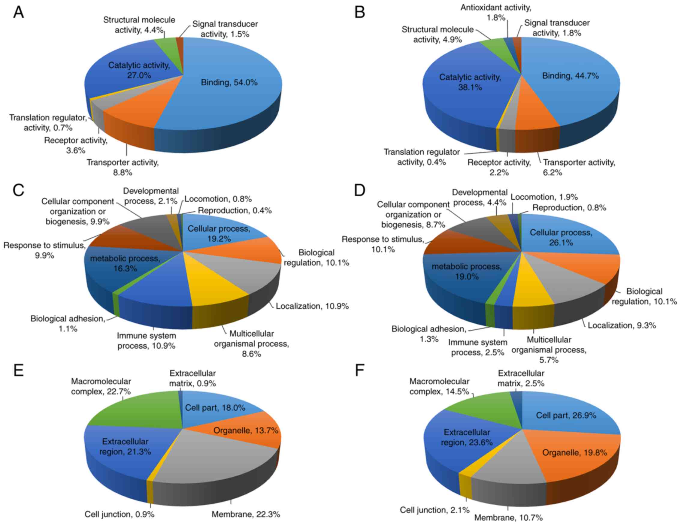
Gene Ontology using the PANTHER classification system. The proteins
were categorized according to their ‘molecular function’,
‘biological process’ and ‘cellular compartment’, as demonstrated in
Fig. 1. For proteins in the bound
fraction, the molecular functions were classified into 7 groups,
with the majority of proteins being involved in binding (54.0%),
followed by catalytic activity (27.0%) (Fig. 1A). The biological process analysis
classified these proteins into 12 groups and the majority of
proteins were involved in cellular process (19.2%), followed by
metabolic process (16.3%) (Fig. 1C).
Gene Ontology analysis for cellular compartments classified these
proteins into 7 groups, and the macromolecular complex was the most
common cellular compartment (22.7%), followed by membrane (22.3%)
and extracellular region (21.3%) (Fig.
1E). For the flow-through fraction, the molecular functions
were classified into 8 groups, with the most abundant proteins
involved in binding (44.7%), followed by catalytic activity (38.1%)
(Fig. 1B). Similar to the bound
fraction, the biological processes of these proteins were
classified into 12 groups with the most commonly identified being
cellular process (26.1%), followed by metabolic process (19.0%)
(Fig. 1D). The proteins were
classified according to the cellular compartments into 7 groups,
and were primarily involved in cell part (26.9%), extracellular
region (23.6%) and organelle (19.8%) (Fig. 1F).
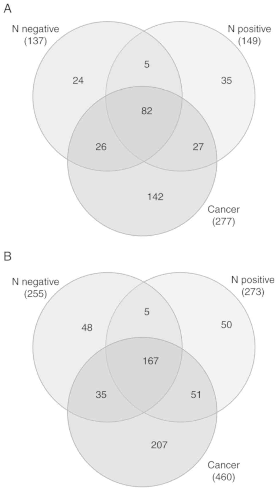
Venn diagram analysis demonstrated the unique and
overlapping proteins between controls and patient samples in the
bound and flow-through fractions (Fig.
2A and B). Overlapping regions indicated the proteins common or
partially common to each sample group. The number of unique or
overlapping proteins are indicated in Fig. 2. A total of 82 and 167 common
proteins between the 3 datasets in bound and flow-through fractions
were identified, respectively. There were 24, 35 and 142 unique
proteins in the normal HPV-negative, normal HPV-positive and
patients with cervical cancer groups, respectively, in the bound
fraction. For the flow-through fraction, 48, 50 and 207 proteins
were uniquely identified in the normal HPV-negative, normal
HPV-positive and patients with cervical cancer groups,
respectively. Among the 207 proteins uniquely identified in the
flow-through fraction of patients with cervical cancer, 2 proteins
that have been previously unreported in cervical cancer, PYCR2 and
isoform-2 of MMRN1, were selected for further evaluation as
cervical cancer biomarkers. VDBP, identified in the flow-through
fraction of both normal controls and patients with cervical cancer,
has been extensively studied in various types of cancer, but the
association between VDBP levels and the risk of cancer remains
controversial; therefore, VDBP was selected for further study.
Furthermore, certain MARS-14 targeted acute phase proteins, such as
A1AT, TTR and ApoA-I, have been associated with distinct stages in
various types of cancer. Acute phase proteins may serve as
diagnostic biomarkers as they reflect a systemic inflammatory
response often observed in a number of types of cancer, including
cervical cancer. The changes in the levels of A1AT, TTR and ApoA-I
were therefore determined in the validation set of serum
samples.
A1AT, PYCR2, TTR, ApoA-I, VDBP and
isoform-2 of MMRN1 are differentially expressed in serum from
patients with cervical cancer
Traditional western blot analysis was performed on
the six groups of pooled serum samples from healthy controls and
patients with cervical cancer. The results indicated that numerous
proteins exhibited differential serum expression levels between the
groups. The expression of A1AT and PYCR2 was elevated in serum from
patients with cervical cancer and the increased expression was
associated with advanced cancer stages. Another 4 proteins, VDBP,
TTR, ApoA-I and isoform-2 of MMRN1, were demonstrated to be
downregulated in serum in patients with cervical cancer compared
with the normal controls (Fig.
3).
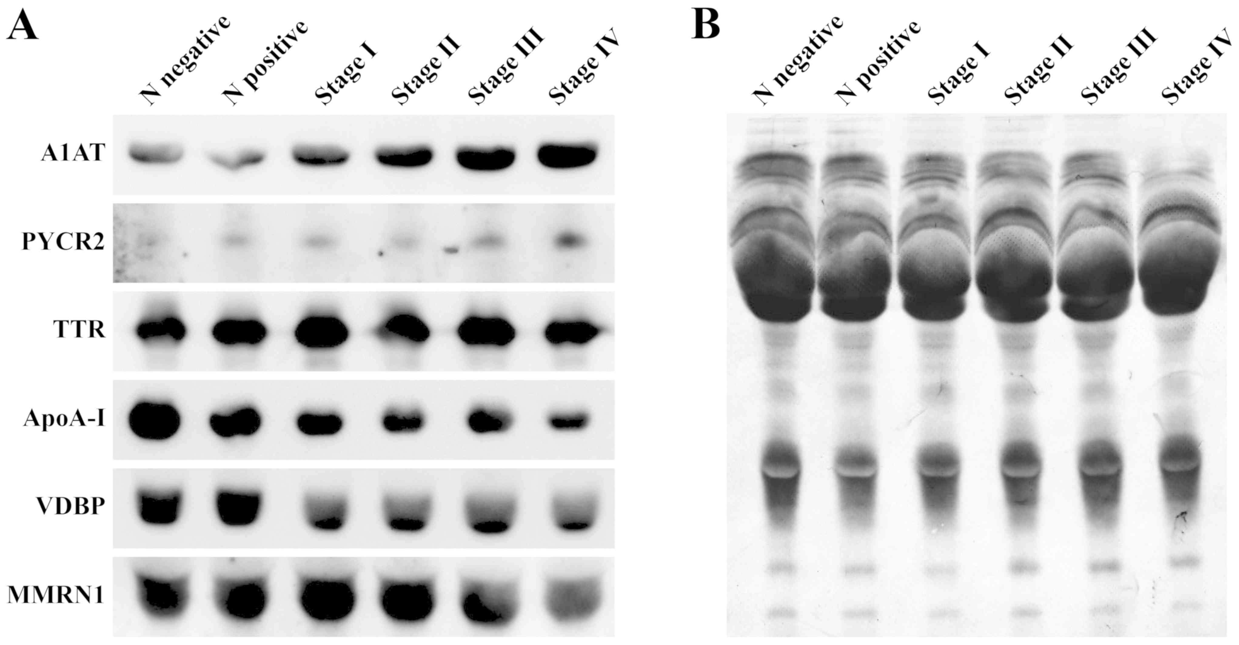 | Figure 3.Western blot analysis. (A) Levels of
A1AT, VDBP, TTR, ApoA-I, MMRN1 and PYCR2 in pooled serum samples
from N negative, N positive and stage I–IV cervical cancer
patients. (B) A Coomassie-stained western blot membrane indicating
the total protein profile as a loading control. N negative, Normal
controls with HPV negative; N positive, normal controls with HPV
positive; A1AT, α-1-antitrypsin; PYCR2, pyrroline-5-carboxylate
reductase 2; TTR, transthyretin; ApoA-I, apolipoprotein A-I; VDBP,
vitamin D binding protein; MMRN1, multimerin-1. |
Quantitative western blot analysis was used to
further validate the diagnostic potential of these proteins in each
individual case. As presented in Fig.
4, serum levels of A1AT were significantly increased in the
stage III–IV group compared with the normal HPV-negative control
group (P<0.01), the normal HPV-positive control group
(P<0.001) and stage I–II group (P<0.01). The results also
demonstrated significantly increased expression levels of PYCR2 in
the stage III–IV group compared with the normal HPV-positive
control group (P<0.01) and the stage I–II group (P<0.01). The
expression of TTR was significantly decreased in the stage III–IV
group compared with the normal HPV-negative control group
(P<0.05) and the stage I–II group (P<0.05). The ApoA-I levels
were significantly downregulated in the stage III–IV group
(P<0.01) and the stage I–II group (P<0.01) compared with the
normal HPV-negative control group. The VDBP expression levels were
significantly downregulated in the serum from the normal
HPV-positive control group (P<0.05), the stage I–II group
(P<0.01) and the stage III–IV group (P<0.05) compared with
the normal HPV-negative control group. In addition to being
significantly decreased in serum from the stage III–IV group
compared with the normal HPV-negative control group (P<0.01),
the expression levels of isoform-2 MMRN1 were also decreased in
serum from the stage I–II group (P<0.001) and the normal
HPV-positive control group (P<0.001).
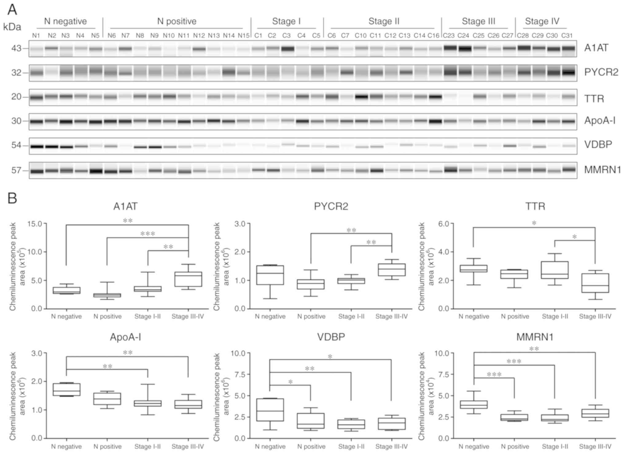 | Figure 4.Quantitative measurement of protein
expression levels using capillary electrophoresis. (A)
Electropherogram of the A1AT, PYCR2, TTR, ApoA-I, VDBP and
isoform-2 of MMRN1 levels in serum samples from the HPV negative
normal controls (N1-N5), HPV positive normal controls (N6-N15), and
patients with stage I (C1-C5), stage II (C6, C7, C10-C14 and C16),
stage III (C23-C27) and stage IV (C28-C31) cervical cancer. (B) Box
plots demonstrating the distribution of protein expression in serum
samples from the normal and stage I–IV cervical patients.
*P<0.05, **P<0.01 and ***P<0.001. A1AT, α-1-antitrypsin;
PYCR2, pyrroline-5-carboxylate reductase 2; TTR, transthyretin;
ApoA-I, apolipoprotein A-I; VDBP, vitamin D binding protein; MMRN1,
multimerin-1. |
A panel of 6 candidate biomarkers
improves detection
ROC curves were constructed to assess the
performance of candidate biomarkers to discriminate between sample
groups. The diagnostic accuracy of numerous combinations of A1AT,
PYCR2, TTR, ApoA-I, VDBP and MMRN1 were also examined using a
binary logistic regression model (Table
SIII). Individually, the AUC values ranged from 0.539 (VDBP,
for discriminating between early-stage vs. late stage cervical
cancer groups) to 0.982 (MMRN1, for discriminating between normal
HPV-negative vs. HPV-positive controls). Notably, a combination of
6 biomarkers yielded an increased predictive accuracy with the
highest AUC value of 1.000 (sensitivity of 100% and specificity of
85.7%), which was increased compared with that of any individual
biomarker, with the exception of MMRN1, for discriminating between
the HPV-negative and HPV-positive normal controls. The best models
for the differential diagnosis of the normal HPV-negative controls,
normal HPV-positive controls, and early-stage and late stage
cervical cancer samples are presented in Fig. 5.
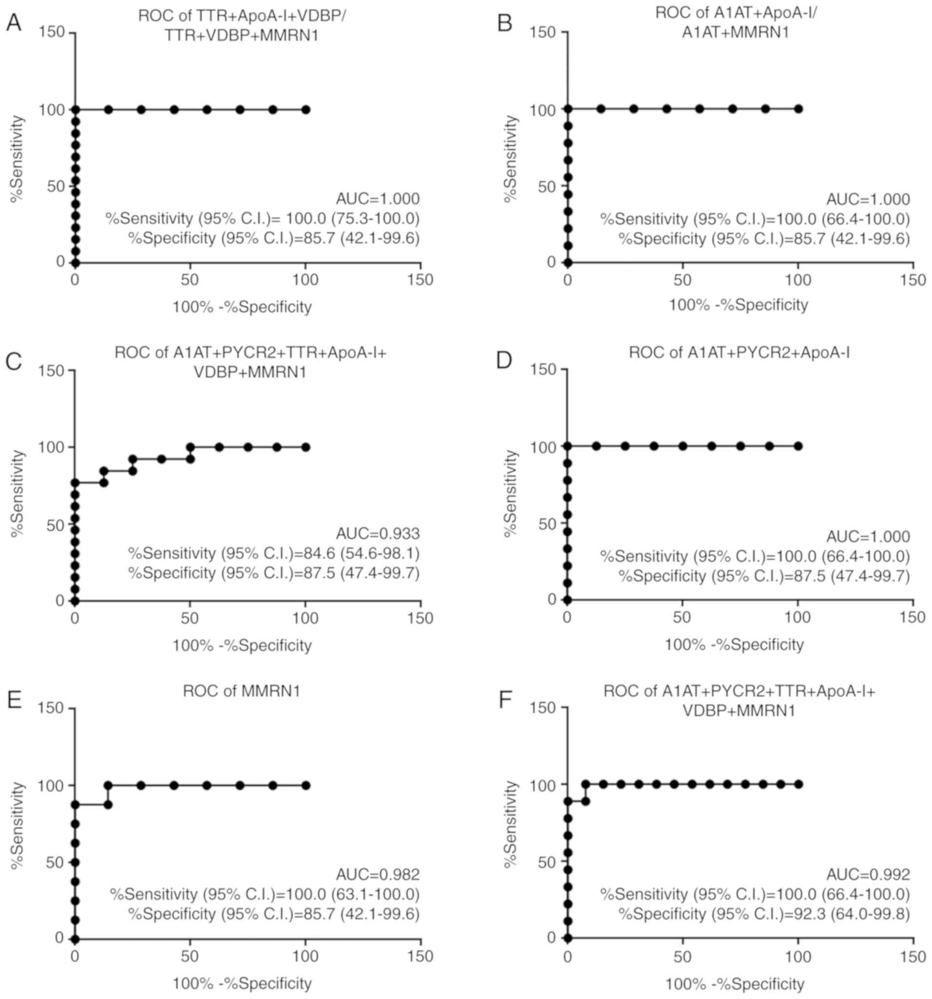 | Figure 5.Performance of the best marker models
for each comparison. AUC, sensitivity with 95% CI and specificity
with 95% CI are stated. (A) Comparison of normal controls with
HPV-negative vs. early-stage (N- vs. E). (B) Comparison of normal
controls with HPV-negative vs. late stage (N- vs. L). (C)
Comparison of normal controls with HPV-positive vs. early-stage (N+
vs. E). (D) Comparison of normal controls with HPV-positive vs.
late stage (N+ vs. L). (E) Comparison of normal controls with
HPV-negative vs. normal controls with HPV-positive (N- vs. N+). (F)
Comparison of early-stage vs. late stage cervical cancer (E vs. L).
ROC, receiver operating characteristic; AUC, area under the ROC
curve; CI, confidence interval; A1AT, α-1-antitrypsin; PYCR2,
pyrroline-5-carboxylate reductase 2; TTR, transthyretin; ApoA-I,
apolipoprotein A-I; VDBP, vitamin D binding protein; MMRN1,
multimerin-1. |
Discussion
Cervical cancer, unlike most other types of cancer,
is a potentially preventable disease with the use of vaccination
and screening tests to detect precancerous lesions. The 5-year
survival rate of patients with cervical cancer at advanced-stage is
only 15–35% (15); however, it is
>90% when detected in the early, treatable stages (16). Therefore, early detection is
important for decreasing the incidence and mortality rates of
cervical cancer. The identification of non-invasive biomarkers for
detection of cervical cancer, therefore, is a high priority. In the
present study, the western blot analysis results indicated the
differential abundance of 6 proteins between healthy controls and
patients with cervical cancer, and among different stages of
cancer, which were A1AT, PYCR2, TTR, ApoA-I, VDBP and MMRN1. The
significantly different expression profiles of these proteins in
each individual case was successfully validated by quantitative
examination using capillary electrophoresis.
Human A1AT, a serine protease inhibitory, is a serum
glycoprotein which serves a central role in tissue protection
through its inhibitory effects on enzymes in inflammatory cells,
including trypsin, chymotrypsin, neutrophil elastase and thrombin.
A1AT is primarily expressed in the liver; however, previous studies
have indicated that a number of different types of cancer cells,
including lung, prostate, thyroid, pancreatic and breast cancer
cells, are capable of expression and secretion of A1AT (17,18). The
results of the present study revealed that A1AT expression levels
were significantly upregulated in patients with stage III–IV
cervical cancer, and that it could be used to differentiate between
patients with advanced-stage cervical cancer and those with stage
I–II cancer and non-cancerous healthy controls. Upregulation of
A1AT serum levels has also been observed in different types of
malignancies, such as hepatocellular carcinoma, lung, prostate,
breast, pancreatic and colorectal cancer (17,19,20). In
addition, the positive association between upregulation of A1AT
expression levels with cancer staging has been demonstrated in
different types of cancer (21,22).
El-Akawi et al (23)
demonstrated the potential use of A1AT blood levels as an indicator
of lung and prostate cancer treatment.
PYCR2 is a mitochondrial enzyme and a member of the
PYCR family of proteins, which catalyzes the final step of proline
synthesis by conversion of pyrroline-5-carboxylate (P5C) to
proline. Unlike PYCR1, another isozyme in the PYCR family that has
been demonstrated to be one of the most frequently overexpressed
enzymes in cancer (24) and
correlated with invasiveness and poor outcomes of breast cancer
(25), relatively less is known
about the role of PYCR2 in cancer. A few studies have revealed that
PYCR2 is essential for cell proliferation (26,27). In
the present study, the expression levels of PYCR2 were increased
significantly in patients with advanced-stage cervical cancer,
compared with patients with early-stage cervical cancer and with
HPV positive normal controls. This was consistent with a previous
study demonstrating that the upregulated levels of PYCR2 were
observed in a panel of melanoma cell lines, but not in normal
melanocytes (28). In addition, it
has been demonstrated that the expression of PYCR2 is induced by
two frequently constitutively expressed oncogenic transcription
factors, c-MYC and PI3K (27,29). To
the best of our knowledge, the results of the present study are the
first to demonstrate the association between PYCR2 and cervical
cancer.
TTR, also known as vitamin A, is a homo-tetrameric
protein primarily synthesized by the liver and choroid plexus in
the brain, and is involved in the transport of thyroid hormone
thyroxine and retinol. TTR has been previously used in nutritional
indexes, due to the association between its plasma levels and the
nutritional supply. TTR levels were identified to be decreased in
the sera of various cancer types, such as lung, prostate, gastric,
ovarian, pancreatic cancer, cholangiocarcinoma and chronic myeloid
leukemia (30–36); therefore, TTR may be considered a
potential biomarker. Consistent with these studies, the results
from the present study indicated significantly decreased TTR levels
in the sera of patients with advanced-stage cervical cancer,
compared with the HPV negative normal controls and patients with
early-stage cervical cancer. In addition, the TTR levels
demonstrated an inverse correlation with tumor volume in gastric
and ovarian cancer (32,37), were associated with prostate cancer
stage (31) and serve as a
therapy-associated biomarker in patients with chronic myeloid
leukemia (36). It has been well
established that the liver increases the production of positive
acute phase reactants and decreases the production of negative
acute phase reactants in response to insults, including trauma in
the body, inflammation, infection and malignancy. As A1AT is a
positive acute phase reactant and TTR is a negative acute phase
reactant, the results of the present study, which demonstrated an
increase in serum A1AT levels concomitant with a reduction in TTR
levels, are in concordance with these established trends.
ApoA-I, the major structural protein of high-density
lipoprotein, is predominantly synthesized in the liver (70%) and
the small intestine (30%). ApoA-I is considered atheroprotective as
it participates in transporting excess cholesterol from tissues to
the liver for excretion, a process called reverse cholesterol
transport. Previous studies have indicated that ApoA-I also
demonstrated antioxidant, anti-inflammatory and anti-apoptotic
effects (38–40). Recent studies have reported that
ApoA-I inhibits cell proliferation in vitro and suppresses
tumor growth and metastasis in vivo in animal models
expressing ApoA-I (41,42). Similar to TTR, ApoA-I is a negative
acute phase reactant and serum ApoA-I levels are inversely
associated with systemic inflammation, advanced cancer stage and
poor patient survival (43). The
results of the present study, which were consistent with previous
studies, demonstrated that ApoA-I expression levels were
significantly decreased in patients with cervical cancer, at both
early and advanced-stages of the disease, compared with the HPV
negative normal controls. Previous studies have identified that
decreased serum ApoA-I levels are associated with a number of
different types of cancer, including ovarian, breast, pancreatic,
colorectal and gastric cancer (34,44–46).
VDBP, also known as Gc-globulin, belongs to the
multigene family of proteins that includes albumin and
α-fetoprotein. VDBP is synthesized and secreted predominantly by
the liver and is the major protein involved in transporting vitamin
D and its metabolites to target tissues. In addition to its role in
vitamin D transport, other important biological functions of VDBP
include extracellular G-actin scavenging, macrophage activation
(immuno-modulatory function) and neutrophil chemotaxis
(anti-inflammatory function) (47,48).
Previous studies have suggested an association between low levels
of VDBP and the risk of several types of cancer, including multiple
myeloma, pancreatic, prostate and kidney cancer (49–51).
These previous data were supported by the inverse association
between serum VDBP levels and cervical cancer observed in the
present study. Significantly decreased serum levels of VDBP were
observed in patients with cervical cancer, in both patients with
early and advanced-stage cancer, and also in HPV positive normal
controls compared with HPV negative normal controls. However,
controversial results have been observed regarding the association
between VDBP levels and the risk of certain types of cancer,
including colorectal and bladder cancer (52,53).
MMRN1, also known as elastin microfibril interface
located protein 4, is a soluble homopolymeric glycoprotein
identified in α granules in platelets and endothelial cells. MMRN1
may serve as a carrier protein for platelet factor V, and serve as
an adhesion protein via integrin receptors and the extracellular
matrix. MMRN1 has been reported as an adverse marker in pediatric
acute myeloid leukemia (AML). An association between MMRN1
expression and clinical outcomes indicated that high MMRN1
expression levels were correlated with shorter overall survival,
event-free survival and a higher relapse risk of AML (54). The 5-year survival rate of patients
with high MMRN1 expression is ~27% and low MMRN1 expression levels
are observed in 66% of patients with stomach cancer (55). Similarly, 5-year survival rates of 65
and 80% was reported for high and low levels of MMRN1 expression,
respectively for renal cancer (56),
indicating the prognostic value of MMRN1 in stomach and renal
cancer. MMRN1 has 2 isoforms, each measuring 138 and 58 kDa, due to
alternative splicing. Isoform-1 of MMRN-1 has been identified as a
potential serum biomarker for multiple myeloma and lung cancer
(49) and urinary biomarker for
cervical cancer (57). In the
present study, serum levels of isoform-2 of MMRN-1, as confirmed by
mass spectrometry, were decreased in the serum of HPV positive
normal controls and in patients with early and late stage cervical
cancer. To the best of our knowledge, the present is the first
study to demonstrate that isoform-2 of multimerin-1 was associated
with cervical cancer.
In conclusion, 6 candidate biomarkers, A1AT, PYCR2,
TTR, ApoA-I, VDBP and isoform-2 of MMRN1, were identified to be
differentially expressed between serum samples from healthy
controls and patients with cervical cancer. A1AT and PYCR2 levels
were significantly increased, whereas the levels of TTR, ApoA-I,
VDBP and isoform-2 of MMRN1 were significantly decreased in
patients with cervical cancer. To the best of our knowledge, PYCR2
and isoform-2 of MMRN1 were identified to be potentially involved
in cervical cancer for the first time. Furthermore, ROC curve
analysis supported the potential of use of these candidate
biomarkers to distinguish between patients with cervical cancer and
healthy controls, and for differentiating between patients with
early-stage and advanced-stage cervical cancer. Additionally, a
panel of 6 candidate biomarkers was identified to perform better
compared with the majority of the individual biomarkers alone for
prediction and discrimination. Therefore, the results of this pilot
study, using relatively a small sample size, suggested that these
candidate biomarkers may potentially be used for detection of
cervical cancer and that combining the biomarkers increased the
sensitivity and specificity. Further validation will be required
using an independent sample set with a larger cohort, including
patients with precancerous lesions, in order to evaluate the
potential of these candidate biomarkers as prognostic indicators of
cervical cancer.
Supplementary Material
Supporting Data
Acknowledgements
Not applicable.
Funding
The present study was supported by the Chulabhorn
Research Institute (grant no. BC 2008-02) and Chulabhorn Royal
Academy (grant no. 31/2554).
Availability of data and materials
The datasets used and/or analyzed during the current
study are available from the corresponding author on reasonable
request.
Authors' contributions
SK and PS performed the experiments, including
immunoaffinity chromatography, sample preparation for mass
spectrometry, classical and automated western blot analysis and
data analysis. CS and SK wrote the manuscript. DC performed the
mass spectrometry. PS and DC helped in preparing the manuscript. CS
and CW designed the study, developed the methodology and
interpreted the results obtained. KS performed statistical and
bioinformatics analyses. NS performed the clinical data collection,
provided the HPV genotyping data and contributed to ethical
information in the materials and methods of manuscript. NK
performed the patient recruitment, surgery and treatment on
enrolled subjects. KW performed serum sample collection and data
analysis. TS performed the histological examination and provided
pathology information. JS supervised the quality of design and gave
final approval of the manuscript prior to submission. JS, CA and
NMP provided technical assistance and were involved in the study
design. All authors read and approved the final manuscript.
Ethics approval and consent to
participate
The present study was approved by the Ethical Review
Board of the Chulabhorn Hospital (no. 31/2554), and written
informed consent was obtained from the patients.
Patient consent for publication
Patients provided written informed consent.
Competing interests
The authors declare that they have no competing
interests.
References
|
1
|
Oldham RK and Dillman RO: Principles of
Cancer Biotherapy. Springer Science and Business Media; New York,
NY: 2009, View Article : Google Scholar
|
|
2
|
Canavan TP and Doshi NR: Cervical cancer.
Am Fam Physician. 61:1369–1376. 2000.PubMed/NCBI
|
|
3
|
Litjens RJ, Hopman AH, van de Vijver KK,
Ramaekers FC, Kruitwagen RF and Kruse AJ: Molecular biomarkers in
cervical cancer diagnosis: A critical appraisal. Expert Opin Med
Diagn. 7:365–377. 2013. View Article : Google Scholar : PubMed/NCBI
|
|
4
|
Pras E, Willemse PH, Canrinus AA, de
Bruijn HW, Sluiter WJ, ten Hoor KA, Aalders JG, Szabo BG and de
Vries EG: Serum squamous cell carcinoma antigen and CYFRA 21-1 in
cervical cancer treatment. Int J Radiat Oncol Biol Phys. 52:23–32.
2002. View Article : Google Scholar : PubMed/NCBI
|
|
5
|
Gaarenstroom K, Bonfrer J, Korse C, Kenter
G and Kenemans P: Value of Cyfra 21-1, TPA, and SCC-Ag in
predicting extracervical disease and prognosis in cervical cancer.
Anticancer Res. 17:2955–2958. 1997.PubMed/NCBI
|
|
6
|
Borras G, Molina R, Xercavins J, Ballesta
A and Iglesias J: Tumor antigens CA 19.9, CA 125, and CEA in
carcinoma of the uterine cervix. Gynecol Oncol. 57:205–211. 1995.
View Article : Google Scholar : PubMed/NCBI
|
|
7
|
Battaglia F, Scambia G, Panici PB,
Castelli M, Ferrandina G, Foti E, Amoroso M, D'Andrea G and Mancuso
S: Immunosuppressive acidic protein (IAP) and squamous cell
carcinoma antigen (SCC) in patients with cervical cancer. Gynecol
Oncol. 53:176–182. 1994. View Article : Google Scholar : PubMed/NCBI
|
|
8
|
Suzuki Y, Nakano T, Ohno T, Abe A, Morita
S and Tsujii H: Serum CYFRA 21-1 in cervical cancer patients
treated with radiation therapy. J Cancer Res Clin Oncol.
126:332–336. 2000. View Article : Google Scholar : PubMed/NCBI
|
|
9
|
Forni F, Ferrandina G, Deodato F, Macchia
G, Morganti AG, Smaniotto D, Luzi S, D'Agostino G, Valentini V,
Cellini N, et al: Squamous cell carcinoma antigen in follow-up of
cervical cancer treated with radiotherapy: Evaluation of
cost-effectiveness. Int J Radiat Oncol Biol Phys. 69:1145–1149.
2007. View Article : Google Scholar : PubMed/NCBI
|
|
10
|
FIGO Committee on Gynecologic Oncology, .
FIGO staging for carcinoma of the vulva, cervix, and corpus uteri.
Int J Gynaecol Obstet. 125:97–98. 2014. View Article : Google Scholar : PubMed/NCBI
|
|
11
|
Apgar BS, Zoschnick L and Wright TC Jr:
The 2001 Bethesda system terminology. Am Fam Physician.
68:1992–1998. 2003.PubMed/NCBI
|
|
12
|
The UniProt Consortium: UniProt: A
worldwide hub of protein knowledge. Nucleic Acids Res.
47:D506–D515. 2019. View Article : Google Scholar : PubMed/NCBI
|
|
13
|
Mi H, Muruganujan A, Huang X, Ebert D,
Mills C, Guo X and Thomas PD: Protocol update for large-scale
genome and gene function analysis with the PANTHER classification
system (v.14.0). Nat Protoc. 14:703–721. 2019. View Article : Google Scholar : PubMed/NCBI
|
|
14
|
Heberle H, Meirelles GV, da Silva FR,
Telles GP and Minghim R: InteractiVenn: A web-based tool for the
analysis of sets through Venn diagrams. BMC Bioinformatics.
16:1692015. View Article : Google Scholar : PubMed/NCBI
|
|
15
|
Maranga IO, Hampson L, Oliver AW, Gamal A,
Gichangi P, Opiyo A, Holland CM and Hampson IN: Analysis of factors
contributing to the low survival of cervical cancer patients
undergoing radiotherapy in Kenya. PLoS One. 8:e784112013.
View Article : Google Scholar : PubMed/NCBI
|
|
16
|
Zampronha Rde A, Freitas-Junior R, Murta
EF, Michelin MA, Barbaresco AA, Adad SJ, de Oliveira AM, Rassi AB
and Oton GJ: Human papillomavirus types 16 and 18 and the prognosis
of patients with stage I cervical cancer. Clinics (Sao Paulo).
68:809–814. 2013. View Article : Google Scholar : PubMed/NCBI
|
|
17
|
El-Akawi ZJ, Al-Hindawi FK and Bashir NA:
Alpha-1 antitrypsin (alpha1-AT) plasma levels in lung, prostate and
breast cancer patients. Neuro Endocrinol Lett. 29:482–484.
2008.PubMed/NCBI
|
|
18
|
Yamaguchi N, Yamamura Y, Koyama K, Ohtsuji
E, Imanishi J and Ashihara T: Characterization of new human
pancreatic cancer cell lines which propagate in a protein-free
chemically defined medium. Cancer Res. 50:7008–7014.
1990.PubMed/NCBI
|
|
19
|
Lee HB, Yoo OJ, Ham JS and Lee MH: Serum
α1-antitrypsin in patients with hepatocellular carcinoma. Clin Chim
Acta. 206:225–230. 1992. View Article : Google Scholar : PubMed/NCBI
|
|
20
|
Pérez-Holanda S, Blanco I, Menéndez M and
Rodrigo L: Serum concentration of alpha-1 antitrypsin is
significantly higher in colorectal cancer patients than in healthy
controls. BMC Cancer. 14:3552014. View Article : Google Scholar : PubMed/NCBI
|
|
21
|
El-Akawi ZJ, Abu-Awad AM, Sharara AM and
Khader YS: The importance of alpha-1 antitrypsin (α1-AT) and
neopterin serum levels in the evaluation of nonsmall cell lung and
prostate cancer patients. Neuro Endocrinol Lett. 31:113–116.
2010.PubMed/NCBI
|
|
22
|
Thompson DK, Haddow JE, Smith DE and
Ritchie RF: Elevated serum acute phase protein levels as predictors
of disseminated breast cancer. Cancer. 51:2100–2104. 1983.
View Article : Google Scholar : PubMed/NCBI
|
|
23
|
El-Akawi ZJ, Abu-Awad AM and Khouri NA:
Alpha-1 antitrypsin blood levels as indicator for the efficacy of
cancer treatment. World J Oncol. 4:83–86. 2013.PubMed/NCBI
|
|
24
|
Nilsson R, Jain M, Madhusudhan N, Sheppard
NG, Strittmatter L, Kampf C, Huang J, Asplund A and Mootha VK:
Metabolic enzyme expression highlights a key role for MTHFD2 and
the mitochondrial folate pathway in cancer. Nat Commun. 5:31282014.
View Article : Google Scholar : PubMed/NCBI
|
|
25
|
Ding J, Kuo ML, Su L, Xue L, Luh F, Zhang
H, Wang J, Lin TG, Zhang K, Chu P, et al: Human mitochondrial
pyrroline-5-carboxylate reductase 1 promotes invasiveness and
impacts survival in breast cancers. Carcinogenesis. 38:519–531.
2017. View Article : Google Scholar : PubMed/NCBI
|
|
26
|
Ou R, Zhang X, Cai J, Shao X, Lv M, Qiu W,
Xuan X, Liu J, Li Z and Xu Y: Downregulation of
pyrroline-5-carboxylate reductase-2 induces the autophagy of
melanoma cells via AMPK/mTOR pathway. Tumor Biol. 37:6485–6491.
2016. View Article : Google Scholar
|
|
27
|
Liu W, Hancock CN, Fischer JW, Harman M
and Phang JM: Proline biosynthesis augments tumor cell growth and
aerobic glycolysis: Involvement of pyridine nucleotides. Sci Rep.
5:172062015. View Article : Google Scholar : PubMed/NCBI
|
|
28
|
De Ingeniis J, Ratnikov B, Richardson AD,
Scott DA, Aza-Blanc P, De SK, Kazanov M, Pellecchia M, Ronai Z,
Osterman AL and Smith JW: Functional specialization in proline
biosynthesis of melanoma. PLoS One. 7:e451902012. View Article : Google Scholar : PubMed/NCBI
|
|
29
|
Liu W, Le A, Hancock C, Lane AN, Dang CV,
Fan TW and Phang JM: Reprogramming of proline and glutamine
metabolism contributes to the proliferative and metabolic responses
regulated by oncogenic transcription factor c-MYC. Proc Natl Acad
Sci. 109:8983–8988. 2012. View Article : Google Scholar : PubMed/NCBI
|
|
30
|
Liu L, Liu J, Dai S, Wang X, Wu S, Wang J,
Huang L, Xiao X and He D: Reduced transthyretin expression in sera
of lung cancer. Cancer Sci. 98:1617–1624. 2007. View Article : Google Scholar : PubMed/NCBI
|
|
31
|
Wang D, Liang H, Mao X, Liu W, Li M and
Qiu S: Changes of transthyretin and clusterin after androgen
ablation therapy and correlation with prostate cancer malignancy.
Transl Oncol. 5:124–129. 2012. View Article : Google Scholar : PubMed/NCBI
|
|
32
|
Shimura T, Shibata M, Gonda K, Okayama H,
Saito M, Momma T, Ohki S and Kono K: Serum transthyretin level is
associated with prognosis of patients with gastric cancer. J Surg
Res. 227:145–150. 2018. View Article : Google Scholar : PubMed/NCBI
|
|
33
|
Lorkova L, Pospisilova J, Lacheta J,
Leahomschi S, Zivny J, Cibula D, Zivny J and Petrak J: Decreased
concentrations of retinol-binding protein 4 in sera of epithelial
ovarian cancer patients: A potential biomarker identified by
proteomics. Oncol Rep. 27:318–324. 2012.PubMed/NCBI
|
|
34
|
Ehmann M, Felix K, Hartmann D, Schnölzer
M, Nees M, Vorderwülbecke S, Bogumil R, Büchler MW and Friess H:
Identification of potential markers for the detection of pancreatic
cancer through comparative serum protein expression profiling.
Pancreas. 34:205–214. 2007. View Article : Google Scholar : PubMed/NCBI
|
|
35
|
Liu L, Wang J, Liu B, Dai S, Wang X, Chen
J, Huang L, Xiao X and He D: Serum levels of variants of
transthyretin down-regulation in cholangiocarcinoma. J Cell
Biochem. 104:745–755. 2008. View Article : Google Scholar : PubMed/NCBI
|
|
36
|
Fatima I, Sadaf S, Musharraf SG, Hashmi N
and Akhtar MW: CD5 molecule-like and transthyretin as putative
biomarkers of chronic myeloid leukemia-an insight from the
proteomic analysis of human plasma. Sci Rep. 7:409432017.
View Article : Google Scholar : PubMed/NCBI
|
|
37
|
Mählck CG and Grankvist K: Plasma
prealbumin in women with epithelial ovarian carcinoma. Gynecol
Obstet Invest. 37:135–140. 1994. View Article : Google Scholar : PubMed/NCBI
|
|
38
|
Bhattacharyya T, Nicholls SJ, Topol EJ,
Zhang R, Yang X, Schmitt D, Fu X, Shao M, Brennan DM, Ellis SG, et
al: Relationship of paraoxonase 1 (PON1) gene polymorphisms and
functional activity with systemic oxidative stress and
cardiovascular risk. JAMA. 299:1265–1276. 2008. View Article : Google Scholar : PubMed/NCBI
|
|
39
|
Cockerill GW, Rye KA, Gamble JR, Vadas MA
and Barter PJ: High-density lipoproteins inhibit cytokine-induced
expression of endothelial cell adhesion molecules. Arterioscler
Thromb Vasc Biol. 15:1987–1994. 1995. View Article : Google Scholar : PubMed/NCBI
|
|
40
|
De Souza JA, Vindis C, Nègre-Salvayre A,
Rye KA, Couturier M, Therond P, Chantepie S, Salvayre R, Chapman MJ
and Kontush A: Small, dense HDL 3 particles attenuate apoptosis in
endothelial cells: Pivotal role of apolipoprotein A-I. J Cell Mol
Med. 14:608–620. 2010.PubMed/NCBI
|
|
41
|
Su F, Kozak KR, Imaizumi S, Gao F, Amneus
MW, Grijalva V, Ng C, Wagner A, Hough G, Farias-Eisner G, et al:
Apolipoprotein AI (apoA-I) and apoA-I mimetic peptides inhibit
tumor development in a mouse model of ovarian cancer. Proc Natl
Acad Sci. 107:19997–20002. 2010. View Article : Google Scholar : PubMed/NCBI
|
|
42
|
Zamanian-Daryoush M, Lindner D, Tallant
TC, Wang Z, Buffa J, Klipfell E, Parker Y, Hatala D,
Parsons-Wingerter P, Rayman P, et al: The cardioprotective protein
apolipoprotein A1 promotes potent anti-tumorigenic effects. J Biol
Chem. 288:21237–21252. 2013. View Article : Google Scholar : PubMed/NCBI
|
|
43
|
Zamanian-Daryoush M and DiDonato JA:
Apolipoprotein AI and cancer. Front Pharmacol. 6:2652015.
View Article : Google Scholar : PubMed/NCBI
|
|
44
|
Kozak KR, Su F, Whitelegge JP, Faull K,
Reddy S and Farias-Eisner R: Characterization of serum biomarkers
for detection of early-stage ovarian cancer. Proteomics.
5:4589–4596. 2005. View Article : Google Scholar : PubMed/NCBI
|
|
45
|
Takaishi S and Wang TC: Gene expression
profiling in a mouse model of Helicobacter-induced gastric cancer.
Cancer Sci. 98:284–293. 2007. View Article : Google Scholar : PubMed/NCBI
|
|
46
|
Hamrita B, Ben Nasr H, Gabbouj S,
Bouaouina N, Chouchane L and Chahed K: Apolipoprotein A1 −75 G/A
and +83 C/T polymorphisms: Susceptibility and prognostic
implications in breast cancer. Mol Biol Rep. 38:1637–1643. 2011.
View Article : Google Scholar : PubMed/NCBI
|
|
47
|
Yamamoto N and Homma S: Vitamin D3 binding
protein (group-specific component) is a precursor for the
macrophage-activating signal factor from
lysophosphatidylcholine-treated lymphocytes. Proc Natl Acad Sci.
88:8539–8543. 1991. View Article : Google Scholar : PubMed/NCBI
|
|
48
|
Binder R, Kress A, Kan G, Herrmann K and
Kirschfink M: Neutrophil priming by cytokines and vitamin D binding
protein (Gc-globulin): Impact on C5a-mediated chemotaxis,
degranulation and respiratory burst. Mol Immunol. 36:885–892. 1999.
View Article : Google Scholar : PubMed/NCBI
|
|
49
|
Zhang HT, Tian EB, Chen YL, Deng HT and
Wang QT: Proteomic analysis for finding serum pathogenic factors
and potential biomarkers in multiple myeloma. Chin Med J.
128:1108–1113. 2015. View Article : Google Scholar : PubMed/NCBI
|
|
50
|
Layne TM, Weinstein SJ, Graubard BI, Ma X,
Mayne ST and Albanes D: Serum 25-hydroxyvitamin D, vitamin D
binding protein, and prostate cancer risk in black men. Cancer.
123:2698–2704. 2017. View Article : Google Scholar : PubMed/NCBI
|
|
51
|
Mondul AM, Weinstein SJ, Moy KA, Männistö
S and Albanes D: Vitamin D-binding protein, circulating vitamin D
and risk of renal cell carcinoma. Int J Cancer. 134:2699–2706.
2014. View Article : Google Scholar : PubMed/NCBI
|
|
52
|
Anic GM, Weinstein SJ, Mondul AM, Männistö
S and Albanes D: Serum vitamin D, vitamin D binding protein, and
risk of colorectal cancer. PLoS One. 9:e1029662014. View Article : Google Scholar : PubMed/NCBI
|
|
53
|
Mondul AM, Weinstein SJ, Virtamo J and
Albanes D: Influence of vitamin D binding protein on the
association between circulating vitamin D and risk of bladder
cancer. Br J Cancer. 107:1589–1594. 2012. View Article : Google Scholar : PubMed/NCBI
|
|
54
|
Laszlo GS, Alonzo TA, Gudgeon CJ,
Harrington KH, Gerbing RB, Wang YC, Ries RE, Raimondi SC, Hirsch
BA, Gamis AS, et al: Multimerin-1 (MMRN1) as novel adverse marker
in pediatric acute myeloid leukemia: A Report from the Children's
Oncology Group. Clin Cancer Res. 21:3187–3195. 2015. View Article : Google Scholar : PubMed/NCBI
|
|
55
|
Human Protein Atlas. https://www.proteinatlas.org/ENSG00000138722-MMRN1/pathology/stomach+cancerFebruary
6–2020
|
|
56
|
Human Protein Atlas. https://www.proteinatlas.org/ENSG00000138722-MMRN1/pathology/renal+cancerFebruary
6–2020
|
|
57
|
Chokchaichamnankit D, Watcharatanyatip K,
Subhasitanont P, Weeraphan C, Keeratichamroen S, Sritana N,
Kantathavorn N, Diskul-Na-Ayudthaya P, Saharat K, Chantaraamporn J,
et al: Urinary biomarkers for the diagnosis of cervical cancer by
quantitative label-free mass spectrometry analysis. Oncol Lett.
17:5453–5468. 2019.PubMed/NCBI
|