Introduction
Lung cancer is one of the most common malignant
tumors in clinic, and because its early symptoms are not obvious, a
number of patients are diagnosed at late stages, which is one of
the reasons leading to a low survival rate within 5 years. Numerous
related investigations have shown that lung cancer has the highest
clinical mortality rate at present (1,2).
Besides, the metastasis and recurrence of lung cancer is also one
of the main causes of poor prognosis and low survival rate in
patients with lung cancer (3). Lymph
node metastasis is one of the most common diffusion pathways in
lung cancer, and one of the main causes of death among patients
with lung cancer (4). Therefore, the
accurate diagnosis of lymph node metastasis in patients with lung
cancer has an important clinical significance for the timely and
effective treatment, and the improvement of survival rate of
patients with lung cancer.
Serum tumor markers play an important role in the
diagnosis of lung cancer. Neuron-specific enolase (NSE) is one of
the most important tumor markers in the clinical diagnosis of lung
cancer (5). NSE is a substance of
the acidic protease-containing neuronal tissues, such as the
neuroendocrine and the peripheral nerve tissues. Since 80% of lung
cancers are tumors of neuroendocrine origin, NSE is also one of the
tumor markers with high sensitivity to the diagnosis of lung cancer
(6). However, its single detection
has some limitations to the detection of lymph node recurrence and
metastasis in lung cancer patients. Therefore, in the present
study, its combination with other indexes was investigated.
According to some scholars (7,8),
angiogenesis and lymph node metastasis have a close relationship,
and cancer patients with active angiogenesis are more likely to
develop lymph node metastasis. Another study (9) has suggested that tumor angiogenesis can
promote the contact of active tumor cells and lymphatic channels,
thus allowing more tumor cells to enter the lymphoid system. In the
present study, the value of hemodynamic parameters in the diagnosis
of lymph node metastasis in patients with lung cancer was
investigated.
Previous studies (10) have shown that ultrasound hemodynamic
indexes and the lymph node metastasis of breast cancer have a close
relationship. However, the diagnostic value of ultrasound
hemodynamic parameters in patients with cervical lymph node
metastasis of lung cancer has not been evaluated. Therefore, the
value of NSE and ultrasound hemodynamic indexes for the individual
and joint detection of the lymph node metastasis of lung cancer was
assessed, so as to provide a more suitable scheme for the diagnosis
of the patients with the cervical lymph node metastasis of lung
cancer.
Patients and methods
General information
The clinical data of 85 patients with lung cancer,
admitted to the Qingdao Municipal Hospital (Group) (Qingdao, China)
from January 2015 to December 2016, were retrospectively analyzed.
There were 51 male and 34 female patients. The average age of the
patients was 49.26±5.45 years. According to the results of
pathological examination, 47 patients with cervical lymph node
metastasis were enrolled in the metastatic group, and 38 patients
without lymph node metastasis were enrolled in the non-metastatic
group. There was no significant difference in sex, age, BMI,
smoking history, pathological type of cancer, family medical
history, and the stage of cancer between the two groups (P>0.05)
(Table I).
 | Table I.General information [n (%)]. |
Table I.
General information [n (%)].
| Factors | Metastatic group
(n=47) | Non-metastatic group
(n=38) | χ2 | P-value |
|---|
| Sex |
|
| 0.008 | 0.929 |
| Male | 28 (59.57) | 23 (60.53) |
|
|
|
Female | 19 (40.43) | 15 (39.47) |
|
|
| Age (years) |
|
| 0.004 | 0.949 |
| ≤49 | 30 (63.83) | 24 (63.16) |
|
|
|
>49 | 17 (36.17) | 14 (36.84) |
|
|
| BMI
(kg/m2) |
|
| 0.024 | 0.876 |
| ≤22 | 28 (59.57) | 22 (57.89) |
|
|
|
>22 | 19 (40.43) | 16 (42.11) |
|
|
| Smoking history |
|
| 0.007 | 0.935 |
| Yes | 35 (74.47) | 28 (73.68) |
|
|
| No | 12 (25.53) | 10 (26.32) |
|
|
| Pathological
type |
|
| 0.053 | 0.974 |
| Lung
adenocarcinoma | 13 (27.66) | 10 (26.32) |
|
|
| Large
cell carcinoma | 25 (53.19) | 20 (52.63) |
|
|
| Squamous
cell carcinoma | 9 (19.15) | 8 (21.05) |
|
|
| Family medical
history |
|
| 0.010 | 0.922 |
| Yes | 24 (51.06) | 19 (50.00) |
|
|
| No | 23 (48.94) | 19 (50.00) |
|
|
| Stages |
|
| 0.018 | 0.999 |
| I | 15 (31.91) | 12 (31.58) |
|
|
| II | 16 (34.04) | 13 (34.21) |
|
|
| III | 9 (19.15) | 7 (18.42) |
|
|
| IV | 7 (14.89) | 6 (15.79) |
|
|
Inclusion and exclusion criteria
Inclusion criteria: Patients diagnosed with lung
cancer by pathology. The metastatic group included patients
diagnosed with lymph node metastasis by pathology and patients who
underwent ultrasound hemodynamic examination. Exclusion criteria:
Patients who had undergone anticancer treatment, such as
radiotherapy and chemotherapy; patients with severe hepatic and
renal dysfunction; patients with other malignant tumors; patients
with communication and cognitive impairment; patients who did not
cooperate with the examination. All patients and their families
agreed to participate in the research and signed an informed
consent form. The study was approved by the Ethics Committee of
Qingdao Municipal Hospital (Group).
Treatment method
Three milliliters of fasting venous blood were
collected from the patients in the morning after they were admitted
to the hospital. Serum was separated via centrifugation at 2,600 ×
g at 4°C for 20 min, after the collection of venous blood. Then,
the content of NSE in the serum of patients was detected by
electrochemiluminescence. The normal reference range of NSE was
0–16.3 µg/l. Color ultrasound hemodynamic examination was performed
on all patients. Voluson 730 color Doppler ultrasonic imaging
instrument (GE Healthcare) with a probe frequency of 7–12 MHz was
used. The examined part of the patients' neck was fully exposed and
examined by color Doppler flow imaging (CDFI). The blood flow
resistance index (RI) and pulsatility index (PI) were measured.
Observation indexes and evaluation
criteria
The expression level of NSE in serum and the
hemodynamic indexes RI and PI were compared between the two groups.
The diagnostic efficacy of NSE, RI, PI, and their combination in
lymph node metastasis of lung cancer were analyzed by ROC curve
analysis.
Statistical analysis
SPSS 19.0 software (Shanghai Yuchuang Network
Technology Co., Ltd.) was used for the statistical analysis of
experimental data. Chi-square test was used for counting data.
Measurement data were expressed as the mean ± standard deviation,
and t-test was used for their analysis between two groups. The
experimental graphs were generated using GraphPad Prism 6 software
(GraphPad Software, Inc.). P<0.05 was considered to indicate a
statistically significant difference.
Results
Expression of NSE in serum, and the
hemodynamic indexes RI and PI in patients of both groups
In the metastatic group, the serum NSE and the
indexes RI and PI were 41.24±11.34 µg/l, 0.81±0.08, and 2.02±0.54,
respectively, while those in the non-metastatic group were
33.56±12.46 µg/l, 0.65±0.09, and 1.23±0.35, respectively. NSE
expression, RI and PI in the metastatic group were significantly
higher than those in the non-metastatic group (P<0.05) (Table II).
| Table II.Expression of serum NSE, and the
hemodynamic indexes RI and PI in the two groups. |
Table II.
Expression of serum NSE, and the
hemodynamic indexes RI and PI in the two groups.
| Factors | Metastatic group
(n=47) | Non-metastatic group
(n=38) | t | P-value |
|---|
| NSE (µg/l) | 41.24±11.34 | 33.56±12.46 | 2.970 | <0.050 |
| RI | 0.81±0.08 | 0.68±0.09 | 7.043 | <0.001 |
| PI | 2.02±0.54 | 1.23±0.35 | 7.788 | <0.001 |
Diagnostic value of NSE, RI and PI
indexes in cervical lymph node metastasis of lung cancer
The sensitivity and specificity of NSE in the
diagnosis of cervical lymph node metastasis of lung cancer were
73.68 and 72.34%, respectively, the diagnostic AUC was 0.783, and
the diagnostic critical value was 38.58 µg/l. The sensitivity and
specificity of RI in the diagnosis of cervical lymph node
metastasis of lung cancer were 78.95 and 80.85%, respectively, the
diagnostic AUC was 0.820, and the diagnostic critical value was
0.745. The sensitivity and specificity of PI in the diagnosis of
cervical lymph node metastasis of lung cancer was 81.58 and 68.09%,
respectively, the diagnostic AUC was 0.844, and the diagnostic
critical value was 1.564. There was no significant difference in
sensitivity and AUC between NSE, RI, and PI in detecting cervical
lymph node metastasis of lung cancer (Table III and Fig. 1).
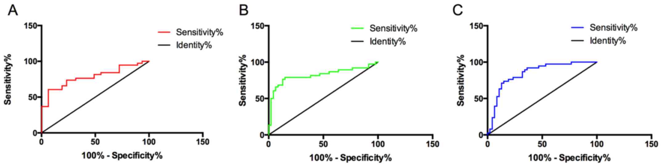 | Figure 1.Diagnostic value of NSE, RI, and PI in
cervical lymph node metastasis of lung cancer. (A) The sensitivity
of NSE in the diagnosis of cervical lymph node metastasis of lung
cancer was 73.68%, the specificity was 72.34%, the diagnostic AUC
was 0.783, and the diagnostic threshold was 38.58 µg/l. (B) The
sensitivity of RI in the diagnosis of cervical lymph node
metastasis of lung cancer was 78.95%, the specificity was 80.85%,
the diagnostic AUC was 0.820, and the diagnostic threshold was
0.745. (C) The sensitivity of PI in the diagnosis of cervical lymph
node metastasis was 81.58%, the specificity was 68.09%, the
diagnostic AUC was 0.844, and the diagnostic threshold was 1.564.
NSE, neuron-specific enolase; RI, resistance index; PI, pulsatility
index. |
| Table III.Diagnostic value of NSE, RI, and PI
alone, and of their combination in cervical lymph node metastasis
of lung cancer. |
Table III.
Diagnostic value of NSE, RI, and PI
alone, and of their combination in cervical lymph node metastasis
of lung cancer.
| Diagnostic
method | Sensitivity (%) | Specificity (%) | AUC |
|---|
| NSE | 73.68 | 72.34 | 0.783 |
| RI | 78.95 | 80.85 | 0.820 |
| PI | 81.58 | 68.09 | 0.844 |
Diagnostic value of NSE combined with RI or PI, and
their combination in cervical lymph node metastasis of lung cancer.
The sensitivity and specificity of NSE combined with RI were 89.47
and 61.70%, respectively, and the diagnostic AUC was 0.881. The
sensitivity and specificity of NSE combined with PI were 92.11 and
74.47%, respectively, and the diagnostic AUC was 0.905. The
sensitivity and specificity of the combination of NSE, RI, and PI
were 97.37 and 57.45%, respectively, and the diagnostic AUC was
0.939. The combination of NSE with RI or PI had good diagnostic
value, however, the combination of the three had the highest
diagnostic value (Table IV and
Fig. 2).
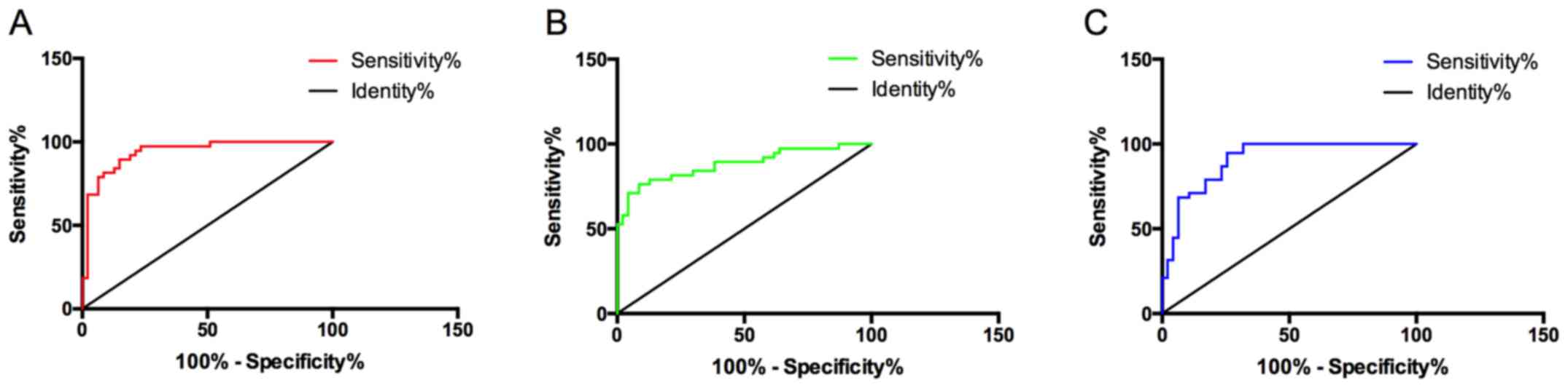 | Figure 2.Diagnostic value of NSE combined with
RI or PI, and their combination in lymph node metastasis of lung
cancer. (A) The sensitivity of NSE combined with RI was 89.47%, the
specificity was 61.70%, and the diagnostic AUC was 0.881. (B) The
sensitivity of NSE combined with PI was 92.11%, the specificity was
74.47%, and the diagnostic AUC was 0.905. (C) The sensitivity of
the combination of NSE, RI, and PI in the diagnosis of cervical
lymph node metastasis was 97.37%, the specificity was 57.45%, and
the diagnostic AUC was 0.939. NSE, neuron-specific enolase; RI,
resistance index; PI, pulsatility index. |
| Table IV.Diagnostic value of NSE combined with
RI or PI, and of their combination in cervical lymph node
metastasis of lung cancer. |
Table IV.
Diagnostic value of NSE combined with
RI or PI, and of their combination in cervical lymph node
metastasis of lung cancer.
| Diagnosis
method | Sensitivity
(%) | Specificity
(%) | AUC |
|---|
| NSE combined with
RI | 89.47 | 61.70 | 0.881 |
| NSE combined with
PI | 92.11 | 74.47 | 0.905 |
| NSE, RI, and PI
combined diagnosis | 97.37 | 57.45 | 0.939 |
Discussion
As a malignant tumor with the highest mortality in
clinical practice, lung cancer has no obvious early symptoms and is
easy to be ignored by patients. Most patients have advanced lung
cancer or metastasis at the time of lung cancer detection (11,12).
Cervical lymph node metastasis is common in lung cancer patients
and one of the main causes of death in lung cancer patients
(13). Therefore, timely diagnosis
and detection of lymph node metastasis in patients with lung cancer
have important clinical significance for effective treatment and
improvement of survival time of patients with lung cancer. However,
the current diagnosis of lymph node metastasis in patients with
lung cancer is mainly pathological examination, which is traumatic
and may cause pain to the patient (14). The main non-invasive diagnostic
method is enhanced CT and PDG-PET, however, they both involve
radiation exposure that some patients are unwilling to accept
(15). In recent years, with the
development of molecular biology, the application of serum tumor
markers in the auxiliary diagnosis of lung cancer and the
monitoring of recurrence and metastasis has achieved good results
(16,17). NSE is recognized as a highly
sensitive tumor marker in lung cancer. It is not only expressed in
nerve tissue, but also a specific marker of neuroendocrine tumors
(18). However, the application of
NSE in the diagnosis of cervical lymph node metastasis of lung
cancer is relatively rare. Studies (19) have explored the mechanism of lymph
node metastasis in lung cancer, suggesting that the condition for
lung cancer metastasis is the continuous survival of
neovascularization. In the present study, the value of ultrasound
hemodynamics in the diagnosis of cervical lymph node metastasis of
lung cancer is investigated. Ultrasound hemodynamics is a
non-invasive method, which has achieved good results as assistant
in the diagnosis of cervical lymph node metastasis of thyroid
cancer in recent years. PI and RI, as important indexes of
hemodynamics, can effectively reflect the status of blood flow
(20).
Therefore, the value of serum tumor marker NSE and
hemodynamic indexes RI and PI individually and jointly in the
diagnosis of cervical lymph node metastasis of lung cancer were
analyzed. NSE in serum of metastatic and non-metastatic patients
was detected by pathology diagnosis. Then, the hemodynamic
parameters of the cervical lymph nodes in the two groups were
measured by ultrasound. The results revealed that NSE, RI and PI
indexes in the metastatic group were significantly higher than
those in the non-metastatic group (P<0.05). These results
suggest that a large amount of NSE is secreted by tumor cells when
cervical lymph node metastasis occurs in patients with lung cancer.
A previous study (21) has explored
the relationship between NSE and prognosis of patients with lung
cancer, indicating that the expression of NSE in serum of patients
with lymph node metastasis is significantly higher than that of
patients without metastasis, which is consistent with our
conclusion. A previous study (22)
has also found that the RI and PI indexes of patients with lymph
node metastasis are significantly higher than those of patients
without lymph node metastasis when examining the hemodynamics of
cervical lymph node metastasis of thyroid cancer. Although this is
not a diagnostic study for cervical lymph node metastasis of lung
cancer, it also confirms our conclusion. At present, there are
numerous clinical methods for the diagnosis of lymph node
metastasis, such as CT (23), cell
biopsy (24) and pathological
diagnosis (25). However, there is
still radiation in the detection by CT. Both cellular biopsy and
pathology are traumatic and can cause more pain to the patient
besides the pain caused by the disease. The value of serum tumor
marker NSE and the hemodynamic indexes RI and PI individually and
jointly were analyzed in the diagnosis of cervical lymph node
metastasis of lung cancer. The results showed that there was no
significant difference between the sensitivity and AUC of NSE, RI
and PI indexes in detecting cervical lymph node metastasis of lung
cancer (P>0.05). However, AUC and the sensitivity of the
combination of NSE, RI, and PI were significantly higher than those
of NSE, RI, and PI alone (P<0.05). The specificity of the
combined diagnosis was shown to be lower than that of each single
diagnosis, which may be a defect of joint diagnosis. These results
suggest that NSE, RI, and PI are valuable for the diagnosis of
cervical lymph node metastasis of lung cancer, however, their
combined diagnosis is more valuable. Up to our knowledge, there are
no relevant studies on the diagnostic value of NSE and hemodynamic
indexes RI and PI in the diagnosis of cervical lymph node
metastasis of lung cancer, and therefore further research it
needed.
In conclusion, the serum NSE and hemodynamic indexes
RI and PI in patients with cervical lymph node metastasis of lung
cancer are higher than those without metastasis, which has certain
diagnostic value for cervical lymph node metastasis of lung cancer.
However, the diagnostic value of their combination is higher. Thus,
it can be used as an auxiliary diagnostic method for cervical lymph
node metastasis of lung cancer. In the present study, the combined
diagnostic value of more serum tumor markers and hemodynamics was
not explored, and there are relatively few reports to confirm our
conclusions at present. Therefore, our conclusions still need to be
further verified.
Acknowledgements
Not applicable.
Funding
No funding was received.
Availability of data and materials
The datasets used and/or analyzed during the present
study are available from the corresponding author on reasonable
request.
Authors' contributions
YK wrote the manuscript. YK and YJ analyzed and
interpreted the patient data. YL performed the experiments and
designed the study. SB was responsible for the analysis and
discussion of the data. All authors read and approved the final
manuscript.
Ethics approval and consent to
participate
The study was approved by the Ethics Committee of
Qingdao Municipal Hospital (Group) (Qingdao, China). Patients who
participated in this research had complete clinical data. Signed
informed consents were obtained from the patients or their
guardians.
Patient consent for publication
Not applicable.
Competing interests
The authors declare that they have no competing
interests.
References
|
1
|
Jones JL, Oien KA, Lee JL and Salto-Tellez
M: Morphomolecular pathology: Setting the framework for a new
generation of pathologists. Br J Cancer. 117:1581–1582. 2017.
View Article : Google Scholar : PubMed/NCBI
|
|
2
|
Braunstein S, Wang L, Newhauser W,
Tenenholz T, Rong Y, van der Kogel A, Dominello M, Joiner MC and
Burmeister J: Three discipline collaborative radiation therapy
(3DCRT) special debate: The United States should build additional
proton therapy facilities. J Appl Clin Med Phys. 20:7–12. 2019.
View Article : Google Scholar : PubMed/NCBI
|
|
3
|
Sun W, Yang X, Liu Y, Yuan Y and Lin D:
Primary tumor location is a useful predictor for lymph node
metastasis and prognosis in lung adenocarcinoma. Clin Lung Cancer.
18:e49–e55. 2017. View Article : Google Scholar : PubMed/NCBI
|
|
4
|
Li N, Tan F, Li J, Shao K, Zhao J, Mu J,
Gao S and He J: Blind spot in lung cancer lymph node metastasis:
Cross-lobe peripheral lymph node metastasis in early stage
patients. Thorac Cancer. 9:480–485. 2018. View Article : Google Scholar : PubMed/NCBI
|
|
5
|
Korkmaz ET, Koksal D, Aksu F, Dikmen ZG,
Icen D, Maden E, Onder S, Akbiyik F and Emri S: Triple test with
tumor markers CYFRA 21.1, HE4, and ProGRP might contribute to
diagnosis and subtyping of lung cancer. Clin Biochem. 58:15–19.
2018. View Article : Google Scholar : PubMed/NCBI
|
|
6
|
Jiang ZF, Wang M and Xu JL: Thymidine
kinase 1 combined with CEA, CYFRA21-1 and NSE improved its
diagnostic value for lung cancer. Life Sci. 194:1–6. 2018.
View Article : Google Scholar : PubMed/NCBI
|
|
7
|
Liu Y, Ren W, Bai Y, Wan L, Sun X, Liu Y,
Xiong W, Zhang YY and Zhou L: Oxyresveratrol prevents murine H22
hepatocellular carcinoma growth and lymph node metastasis via
inhibiting tumor angiogenesis and lymphangiogenesis. J Nat Med.
72:481–492. 2018. View Article : Google Scholar : PubMed/NCBI
|
|
8
|
Pana ZD, Roilides E, Warris A, Groll AH
and Zaoutis T: Epidemiology of invasive fungal disease in children.
J Pediatric Infect Dis Soc. 6 (Suppl 1):S3–S11. 2017. View Article : Google Scholar : PubMed/NCBI
|
|
9
|
Pastushenko I, Van den Eynden GG,
Vicente-Arregui S, Prieto-Torres L, Alvarez-Alegret R, Querol I,
Dirix LY, Carapeto FJ, Vermeulen PB and Van Laere SJ: Increased
angiogenesis and lymphangiogenesis in metastatic sentinel lymph
nodes is associated with nonsentinel lymph node involvement and
distant metastasis in patients with melanoma. Am J Dermatopathol.
38:338–346. 2016. View Article : Google Scholar : PubMed/NCBI
|
|
10
|
Wu X, Takekoshi T, Sullivan A and Hwang
ST: Inflammation and tumor microenvironment in lymph node
metastasis. Cancers (Basel). 3:927–944. 2011. View Article : Google Scholar : PubMed/NCBI
|
|
11
|
Fitzmaurice C, Dicker D, Pain A, Hamavid
H, Moradi-Lakeh M, MacIntyre MF, Allen C, Hansen G, Woodbrook R,
Wolfe C, et al Global burden of disease cancer collaboration, : The
global burden of cancer 2013. JAMA Oncol. 1:505–527. 2015.
View Article : Google Scholar : PubMed/NCBI
|
|
12
|
Vlachogeorgos GS, Manali ED, Blana E,
Legaki S, Karagiannidis N, Polychronopoulos VS and Roussos C:
Placental isoform glutathione S-transferase and P-glycoprotein
expression in advanced nonsmall cell lung cancer: Association with
response to treatment and survival. Cancer. 114:519–526. 2008.
View Article : Google Scholar : PubMed/NCBI
|
|
13
|
Padera TP, Meijer EF and Munn LL: The
lymphatic system in disease processes and cancer progression. Annu
Rev Biomed Eng. 18:125–158. 2016. View Article : Google Scholar : PubMed/NCBI
|
|
14
|
Lee DH, Yoon TM, Lee JK and Lim SC:
Supraclavicular lymph node excision biopsy in patients with
suspected supraclavicular lymph node metastasis of lung cancer:
Experience in a Tertiary Hospital. Chonnam Med J. 53:69–72. 2017.
View Article : Google Scholar : PubMed/NCBI
|
|
15
|
Pak K, Kim K, Kim MH, Eom JS, Lee MK, Cho
JS, Kim YS, Kim BS, Kim SJ and Kim IJ: A decision tree model for
predicting mediastinal lymph node metastasis in non-small cell lung
cancer with F-18 FDG PET/CT. PLoS One. 13:e01934032018. View Article : Google Scholar : PubMed/NCBI
|
|
16
|
Zhang Q, Wang L, Huang D, Xu M, Weng W, Ni
S, Tan C and Sheng W: Pathological risk factors for lymph node
metastasis in patients with submucosal invasive colorectal
carcinoma. Cancer Manag Res. 11:1107–1114. 2019. View Article : Google Scholar : PubMed/NCBI
|
|
17
|
Chen C, Wang Y, Fu S, Pan X, Yang J and
Wang R: The impact on mediastinal recurrence based on the number of
harvested mediastinal lymph nodes and assessed N2 Stations in
patients with stage I invasive lung adenocarcinoma. J Thorac Dis.
10:6803–6810. 2018. View Article : Google Scholar : PubMed/NCBI
|
|
18
|
Shirasawa M, Fukui T, Kusuhara S, Hiyoshi
Y, Ishihara M, Kasajima M, Nakahara Y, Otani S, Igawa S, et al:
Prognostic significance of the 8th edition of the TNM
classification for patients with extensive disease small cell lung
cancer. Cancer Manag Res. 10:6039–6047. 2018. View Article : Google Scholar : PubMed/NCBI
|
|
19
|
Takahashi Y, Suzuki S, Matsutani N and
Kawamura M: 18F-fluorodeoxyglucose positron emission
tomography/computed tomography in the evaluation of clinically
node-negative non-small cell lung cancer. Thorac Cancer.
10:413–420. 2019. View Article : Google Scholar : PubMed/NCBI
|
|
20
|
Hwang HS and Orloff LA: Efficacy of
preoperative neck ultrasound in the detection of cervical lymph
node metastasis from thyroid cancer. Laryngoscope. 121:487–491.
2011. View Article : Google Scholar : PubMed/NCBI
|
|
21
|
Schneider J, Velcovsky HG, Morr H, Katz N,
Neu K and Eigenbrodt E: Comparison of the tumor markers tumor
M2-PK, CEA, CYFRA 21-1, NSE and SCC in the diagnosis of lung
cancer. Anticancer Res. 20:5053–5058. 2000.PubMed/NCBI
|
|
22
|
Shin SH, Kim HS, Jung SH, Xu HD, Jeong YB
and Chung YJ: Implication of leucyl-tRNA synthetase 1 (LARS1)
over-expression in growth and migration of lung cancer cells
detected by siRNA targeted knock-down analysis. Exp Mol Med.
40:229–236. 2008. View Article : Google Scholar : PubMed/NCBI
|
|
23
|
Shao T, Yu L, Li Y and Chen M: Density and
SUV ratios from PET/CT in the detection of mediastinal lymph node
metastasis in non-small cell lung cancer. Zhongguo Fei Ai Za Zhi.
18:155–160. 2015.(In Chinese). PubMed/NCBI
|
|
24
|
Jiang C, Chen Y, Zhu Y and Xu Y:
Systematic review and meta-analysis of the accuracy of 18F-FDG
PET/CT for detection of regional lymph node metastasis in
esophageal squamous cell carcinoma. J Thorac Dis. 10:6066–6076.
2018. View Article : Google Scholar : PubMed/NCBI
|
|
25
|
Kendirlinan R, Ozkan G, Bayram M, Bakan
ND, Tutar M, Gür A and Camsari G: Ultrasound guided fine-needle
aspiration biopsy of metastases in nonpalpable supraclavicular
lymph nodes in lung cancer patients. Multidiscip Respir Med.
6:220–225. 2011. View Article : Google Scholar : PubMed/NCBI
|


