Introduction
Esophageal cancer is a malignant disease associated
with poor prognosis. Furthermore, esophageal squamous cell
carcinoma (ESCC) is a type of esophageal carcinoma that is usually
located in the upper or middle third part of the esophagus
(1), and had a high-incidence rate
between 90–120 per 105 population in the 5-year period
(1995–1999) in the Henan region in China (2).
Current therapeutic regimens for patients with
esophageal cancer include surgery followed by conventional
chemotherapy or radiation; however, the 5-year survival percentage
remains very low at <10% (2,3). Thus,
effective therapeutic agents or regimens are urgently required for
improving the survival rate of patients with esophageal cancer.
Metal chelators are a class of potential
therapeutics that have potent and selective anti-cancer efficacy
(4,5). Most cancer cells have an increasing
demand for metal ions, such as iron or copper, to maintain an
appropriate proliferation rate; therefore, chelators may be a
potential treatment regimen for treating cancer types (6,7).
Previous studies have reported that metal chelators, such as
di-2-pyridyl ketone-4,4-dimethyl-3-thiosemicarbazone (Dp44mT),
possess an inhibitory effect against colon cancer and prostate
cancer (8,9).
Dithiocarbamates are sulfur-containing compounds
with an excellent chelating property toward metal ions that can
regulate crucial molecules involved in reactive oxygen species
accumulation, cell cycle arrest, apoptosis or autophagy (6,10,11).
However, the molecular targets of numerous dithiocarbamates, such
as di-2-pyridylhydrazone dithiocarbamate and pyrrolidine
dithiocarbamate remain unknown. Dixon et al (8) revealed that Dp44mT may exert its
anti-growth activity by inhibiting the oncogenic ERK1/2 signaling
pathway. Moreover, Chen et al (9) reported that the iron chelator
desferrioxamine (DFO) can inhibit epithelial-mesenchymal transition
(EMT) induced by the transforming growth factor-β and by elevating
the protein expression of N-myc downstream-regulated gene 1.
Our previous study synthesized a class of
dithiocarbamate derivative, dipyridylhydrazone dithiocarbamate
(DpdtC), and assessed its anti-cancer activity on hepatocellular
carcinoma cells (6). It was revealed
that DpdtC downregulates erb-b2 receptor tyrosine kinase 2 (ERBB2)
expression and disrupts the formation of a heterodimer between
ERRB2 and epidermal growth factor receptor (EGFR), which further
resulted in the inactivation of ERBB2/ERK 1/2 signaling in
ERBB2-overexpressed ovarian cancer cells (12).
The EGFR/AKT signaling pathway has an important role
in the growth and proliferation of esophageal cancer cells
(13). In the present study, the
antitumor effects of DpdtC on esophageal cancer cells were
evaluated and its potential mechanism of action was investigated,
which may be associated with EGFR/AKT signaling pathway inhibition.
The present study aimed to identify the potential of DpdtC as a
drug candidate for treatment of EGFR-positive esophageal cancer
types, which will aid in the clinical development of esophageal
cancer treatment.
Materials and methods
Cell lines and animals
The human esophageal cancer cell lines KYSE-150 and
KYSE-450 were purchased from the American Type Culture Collection.
Cell were cultured with RPMI-1640 medium (Gibco; Thermo Fisher
Scientific, Inc.) supplemented with 10% Fetal Bovine Serum (Gibco;
Thermo Fisher Scientific, Inc.), 2 mmol/l glutamine, 100 IU/ml
penicillin and 100 mg/ml streptomycin (Invitrogen; Thermo Fisher
Scientific, Inc.) in an incubator at 37°C with 5% CO2
for 48 h. Female BALB/c nude mice (age, 5 weeks; weight, 16–19 g;
n=15) were obtained from the Beijing Vital River Laboratory Animal
Technology Co., Ltd. All animals were treated in accordance with
guidelines of the Committee on Animals of the Xinxiang Medical
University and was approved by Biomedical Ethics Committee of
Xinxiang Medical University.
In vitro cytotoxicity assays
Firstly, the effects of different treatment times
(24, 48 or 72 h) on the cytotoxicity of DpdtC (Henan International
Joint Lab of Recombinant Protein) in esophageal cancer cells was
assessed for determining the appropriate treatment time. Esophageal
cancer KYSE-150 and KYSE-450 cell lines were treated with 10 µM
DpdtC for the aforementioned 3 time points (24, 48 or 72 h) at 37°C
with 5% CO2. Cell viability was then tested using the
Cell Counting 8 (CCK-8) Kit (Dojindo Molecular Technologies, Inc.)
according to the manufacturer's instructions. Next, cells were
treated with DpdtC (Henan International Joint Lab of Recombinant
Protein) at a series of concentrations, which was diluted from 50
µM in a 2Χ dilution manner (50, 25, 12.5, 6.25, 3.125, 1.5625 and
0.78125 µM) at 37°C with 5% CO2 for 48 h. After 2 days,
cell viability was determined using CCK-8 Κit (Dojindo Molecular
Technologies, Inc.). The percentage of surviving cells was
calculated using the following formula: [(A450 of experiment-A450
of background)/(A450 of untreated control-A450 of background)]
×100. The well treated with only medium without DpdtC was the
untreated control. IC50 was calculated using non-linear
regression analyses utilizing GraphPad Prism 5 software (GraphPad
Software, Inc.).
In vivo therapy study
All animal experimentation followed internationally
recognized Animal Research: Reporting of in vivo Experiments
guidelines (14). Female BALB/c nude
(age, 5 weeks) mice were maintained at 22±2°C and 50–60% humidity
in a 12 h dark/light cycle, with continuous free access to food and
water.
KYSE-450 cells (5×10⁶ per mouse) were
inoculated subcutaneously into the right flank of the female BALB/c
nude mice. When tumor volumes reached an average of ~150
mm3, the mice were randomly divided into 3 groups (n=5
in each group): i) A PBS-treated group as control; ii) a
DpdtC-treated group at a low dose (1 mg/kg); and iii) a
DpdtC-treated group at a high dose (3 mg/kg). Mice were
intraperitoneally injected with 200 µl 10 mM PBS or DpdtC (1 or 3
mg/kg) four times (day 0, 2, 4 and 6) as indicated in the period of
14 days. The dosages of DpdtC were chosen according to our previous
study (12). On day 13 post-first
injection, the mice were euthanized using carbon dioxide gas
(20%/min gradual displacement) and monitored for 5 min to confirm
cardiac arrest, and the tumors were removed for subsequent western
blot examination. The tumors were measured with digital calipers,
and tumor volumes were calculated using the formula: Volume=Length
× (Width)2/2. Unspecific toxicity evaluation was
determined in tumor-bearing nude mice injected with PBS control or
DpdtC by monitoring the body weight of these mice at regular
intervals during the whole therapeutic period.
Immunoblotting
Western blot analysis was performed according to
previously described procedures (15). The sample was the protein extracted
from cell pellets or tumor tissues. Cells were treated with DpdtC
(0, 10, 20 or 30 µM) for 24 h before being lysed. Tumor tissues
were removed on day 13 post the first administration of DpdtC.
Then, cells or tumor tissues were lysed in lysis buffer [50 mmol/l
Tris-HCl pH 7.4, 150 mmol/l NaCl, 0.1% SDS, 1% Triton x-100 and
0.5% deoxycholic acid sodium salt (w/v)] supplemented with 2 µl/ml
protease inhibitor cocktail (Sigma-Aldrich; Merck KGaA) on ice for
30 min and centrifuged at 12,000 × g, 4°C for 20 min to remove cell
debris. Subsequently, the protein (20 µg/well) from the cell
lysates were separated using a 10% SDS-PAGE gel and transferred to
PVDF membrane for blocking in 5% skimmed milk for 1 h 30 min at
room temperature. Next, the membrane was immunoblotted with
respective antibodies against EGFR (1:1,000; cat. no. 54359; Cell
Signaling Technology, Inc.), phosphorylated (p)-EGFR (1:1,000; cat.
no. 2234; Cell Signaling Technology, Inc.), AKT (1:1,000; cat. no.
10176-2-AP; ProteinTech Group, Inc.), p-AKT (1:2,000; cat. no.
4060; Cell Signaling Technology, Inc.), Poly (ADP-ribose)
polymerase (PARP; 1:500; cat. no. BM5118; Boster Biological
Technology), BAX (1:1,000; cat. no. 50599; ProteinTech Group,
Inc.), Bcl-2 (1:1,000; cat. no. 4223; Cell Signaling Technology,
Inc.), Bcl-2-binding component 3, isoformed 1/2 (BBC3; 1:1,000;
cat. no. 55120; ProteinTech Group, Inc.) or β-actin (1:5,000; cat.
no. ab179467; Abcam), and subsequently with horseradish
peroxidase-conjugated goat anti-mouse/rabbit secondary antibodies
(1:5,000; cat. no. SA00001-1 or SA00001-2; ProteinTech Group, Inc.)
at room temperature for 45 min. After washing the membrane with
wash buffer (Tris buffered saline with Tween-20, pH 8.0), the bands
were detected using the sensitive ECL reagent (GE Healthcare Life
Sciences), and visualized using a ChemiDoc imaging system (Bio-Rad
Laboratories, Inc.). Relative densitometry analysis of protein
expression level was normalized to control β-actin antibody using
Image J software v.1.46 (National Institute of Health).
Apoptosis analysis
Apoptosis analysis was performed as previously
described (16). For flow cytometry
analysis, KYSE-450 or KYSE-150 cells (1×10⁶/well) were
plated in 6-well plate and treated with DpdtC (0, 10 or 20 µM) for
20 h at 37°C. The cells were then labeled with Annexin V (20 µg/ml)
and propidium iodide (PI) (50 µg/ml) (Dojindo Molecular
Technologies, Inc.) at room temperature for 15 min. Apoptotic rates
were analyzed using a FACSCalibur flow cytometer (BD Biosciences)
and calculated using FlowJo software v.7.6.1 (Treestar, Inc.). The
rate of early apoptosis was calculated by Annexin V (+) and PI (−),
while the rate of late apoptosis was calculated by Annexin V (+)
and PI (+).
Statistical analysis
Statistical analysis was performed with GraphPad
Prism software v.5.0.1 (GraphPad Software, Inc.). All experiments
were repeated 3 times. Numerical values were expressed as the mean
± standard deviation. For the in vitro and in vivo
studies, the differences between the groups were analyzed using
one-way ANOVA analysis with a Dunnett's Multiple Comparison
post-hoc Test. One-way ANOVA analysis with a Tukey's Multiple
Comparison Test was used for analysis of data in Fig. S1. P<0.05 was considered to
indicate a statistically significant difference.
Results
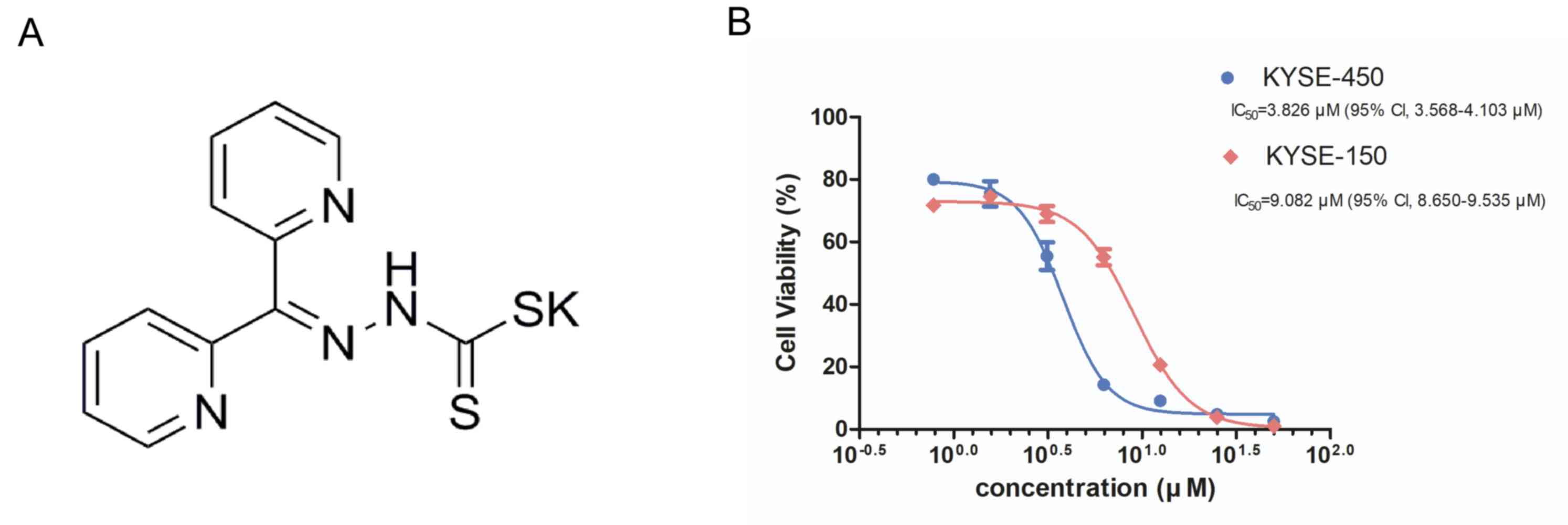
DpdtC exhibits growth inhibitory
effects against esophageal cancer cells in vitro
The cytotoxicity of DpdtC against EGFR-overexpressed
KYSE-150 and KYSE-450 cells was examined (2,13). The
chemical structure of DpdtC is presented in Fig. 1A. Based on the preliminary
experiments, 48 h treatment time with DpdtC was selected for the
following CCK-8 assay (Fig. S1). It
was demonstrated that DpdtC had a concentration-dependent
inhibitory effect in both KYSE-150 and KYSE-450 cells treatment for
48 h. IC50 for KYSE-450 and KYSE-150 cells was 3.826 µM
(95% CI, 3.568–4.103 µM) and 9.082 µM (95% CI, 8.650–9.535 µM),
respectively (Fig. 1B).
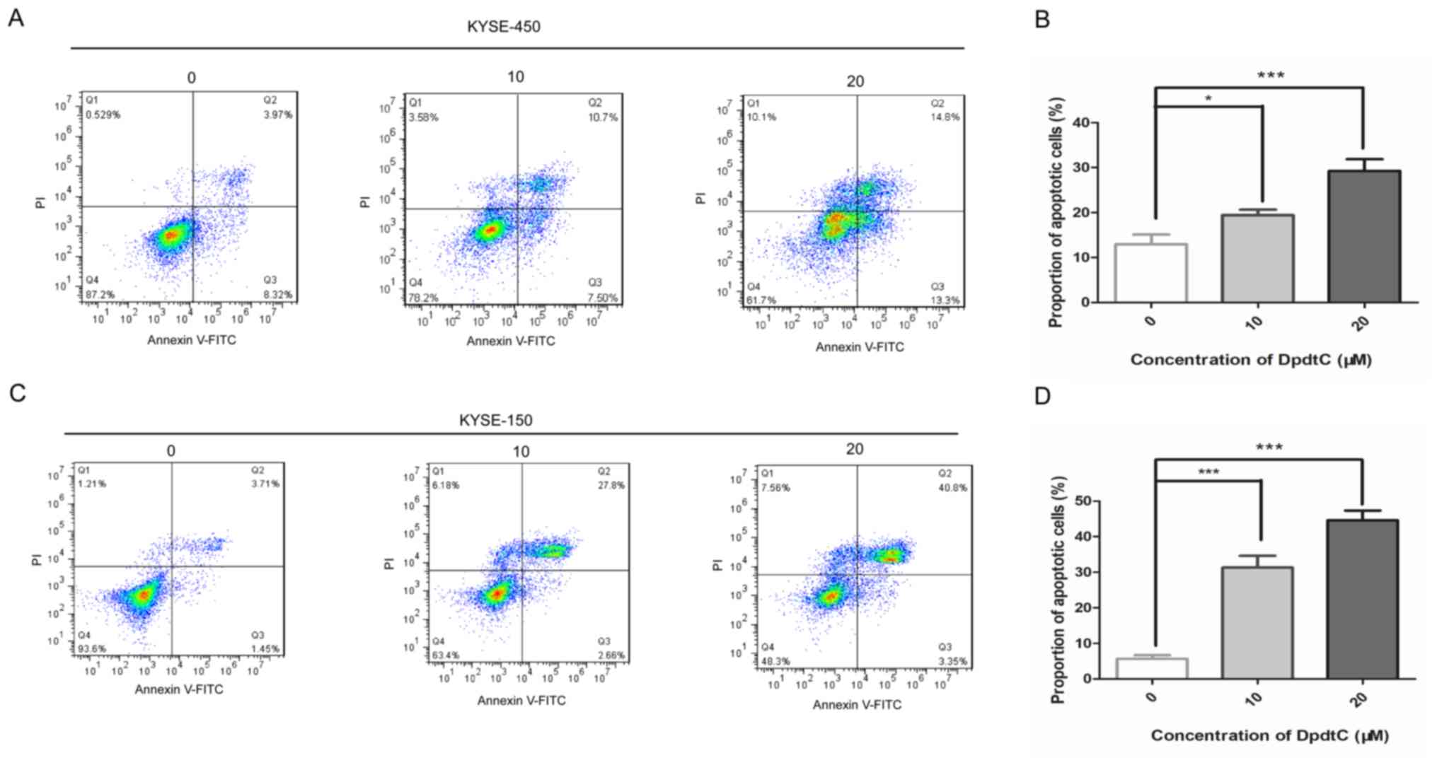
DpdtC induces apoptosis in both
KYSE-150 and KYSE-450 cells
Subsequently, whether DpdtC induces apoptosis was
investigated in KYSE-150 and KYSE-450 cells. The percentage of
apoptotic cells was determined using flow cytometry following
Annexin V and PI staining. The results suggested that the apoptotic
percentage was significantly increased in both KYSE-150 and
KYSE-450 cells treated with an increasing concentration of DpdtC
(Fig. 2). Moreover, a significant
cleavage of PARP, a classical apoptotic initiator, was observed in
both cancer cells treated with DpdtC (Fig. 3). Interestingly, KYSE-450 cells
appeared to be more sensitive to DpdtC treatment in the
cytotoxicity assay, while the apoptosis percentage of KYSE-450
cells was lower compared with that in the KYSE-150 cells at the
same concentration of DpdtC (Figs. 1
and 2).
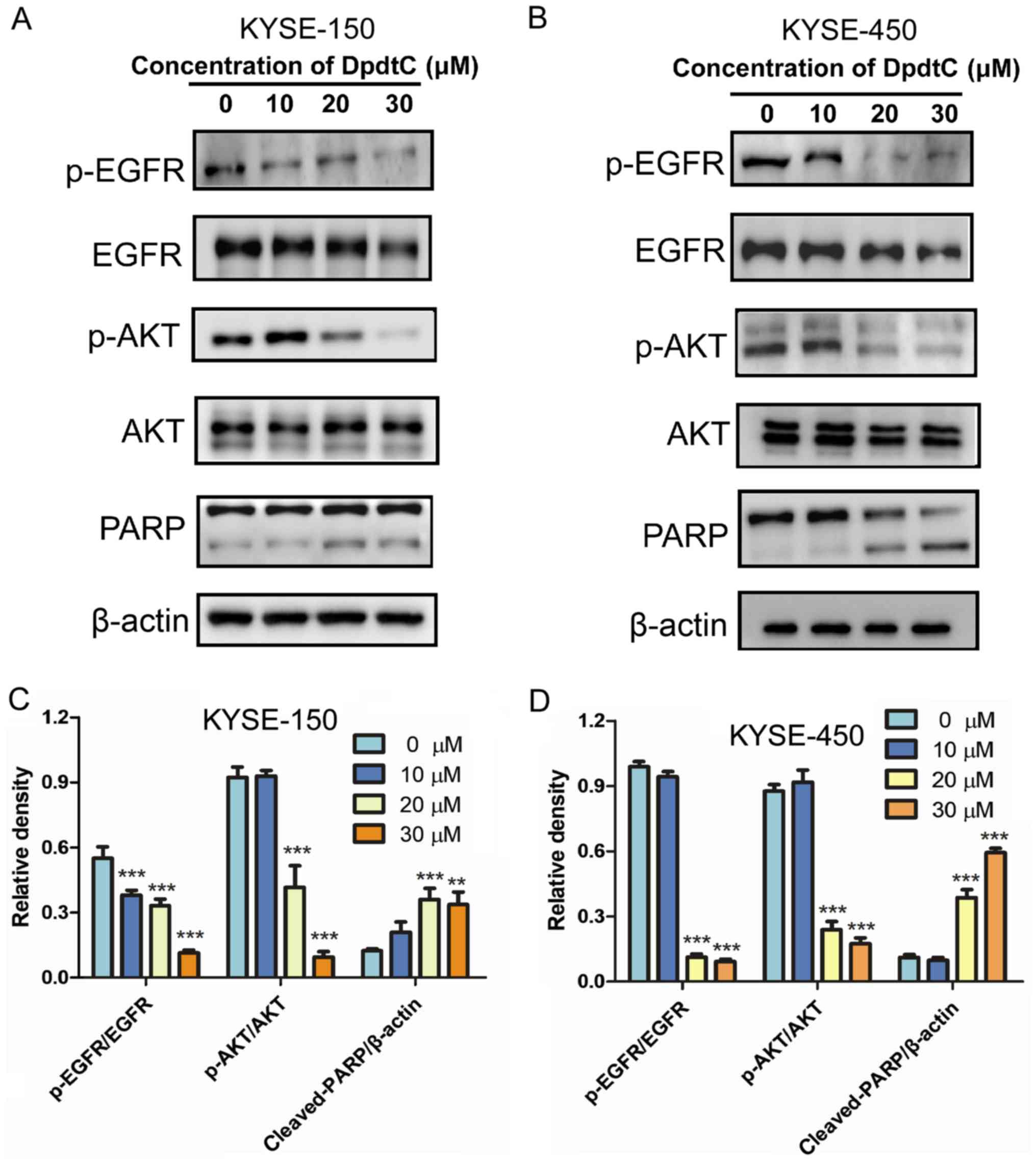 | Figure 3.DpdtC inhibits the EGFR/AKT signaling
pathway in KYSE-150 and KYSE-450 cells. (A) KYSE-150 and (B)
KYSE-450 cells were treated with DpdtC (0, 10, 20 or 30 µM) for 24
h before being evaluated. EGFR, p-EGFR, AKT, p-AKT and PARP were
analyzed by immunoblotting assay. Quantification of protein signal
intensity in (C) KYSE-150 and (D) KYSE-450 cells and expressed
relative to the β-actin or total EGFR/AKT protein using ImageJ
software. Data are presented as the mean ± SD of three independent
experiments. **P<0.01, ***P<0.001 vs. untreated control
cells. DpdtC, Dipyridylhydrazone dithiocarbamate; p-,
phosphorylated; PARP, Poly (ADP-ribose) polymerase; EGFR, epidermal
growth factor receptor. |
DpdtC inhibits the EGFR/AKT signaling
pathway in KYSE-150 and KYSE-450 cells
The expression of EGFR is increased in esophageal
cancer cells KYSE-150 and KYSE-450 (2,13). In
the present study, a significant decrease in the phosphorylation
levels of EGFR and AKT was identified in both KYSE-150 and KYSE-450
cells treated with DpdtC. Moreover, this decrease was
dose-dependent (Fig. 3).
DpdtC inhibits the growth of KYSE-450
tumor xenografts
To evaluate the inhibitory activity of DpdtC in
vivo, the therapeutic effect of DpdtC was examined in nude mice
with KYSE-450 ×enograft tumors. It was found that DpdtC
significantly decreased tumor growth compared with that in the
control-PBS treatment group (Fig. 4A and
B). Moreover, while both concentrations of DpdtC reduced the
growth of tumors, the reduction was higher with 3 mg/kg DpdtC. In
addition, DpdtC treatment did not result in loss in body weight
(Fig. S2). Collectively, the
results suggested that DpdtC had a dose-dependent inhibitory effect
against KYSE-450 esophageal cancer in vivo.
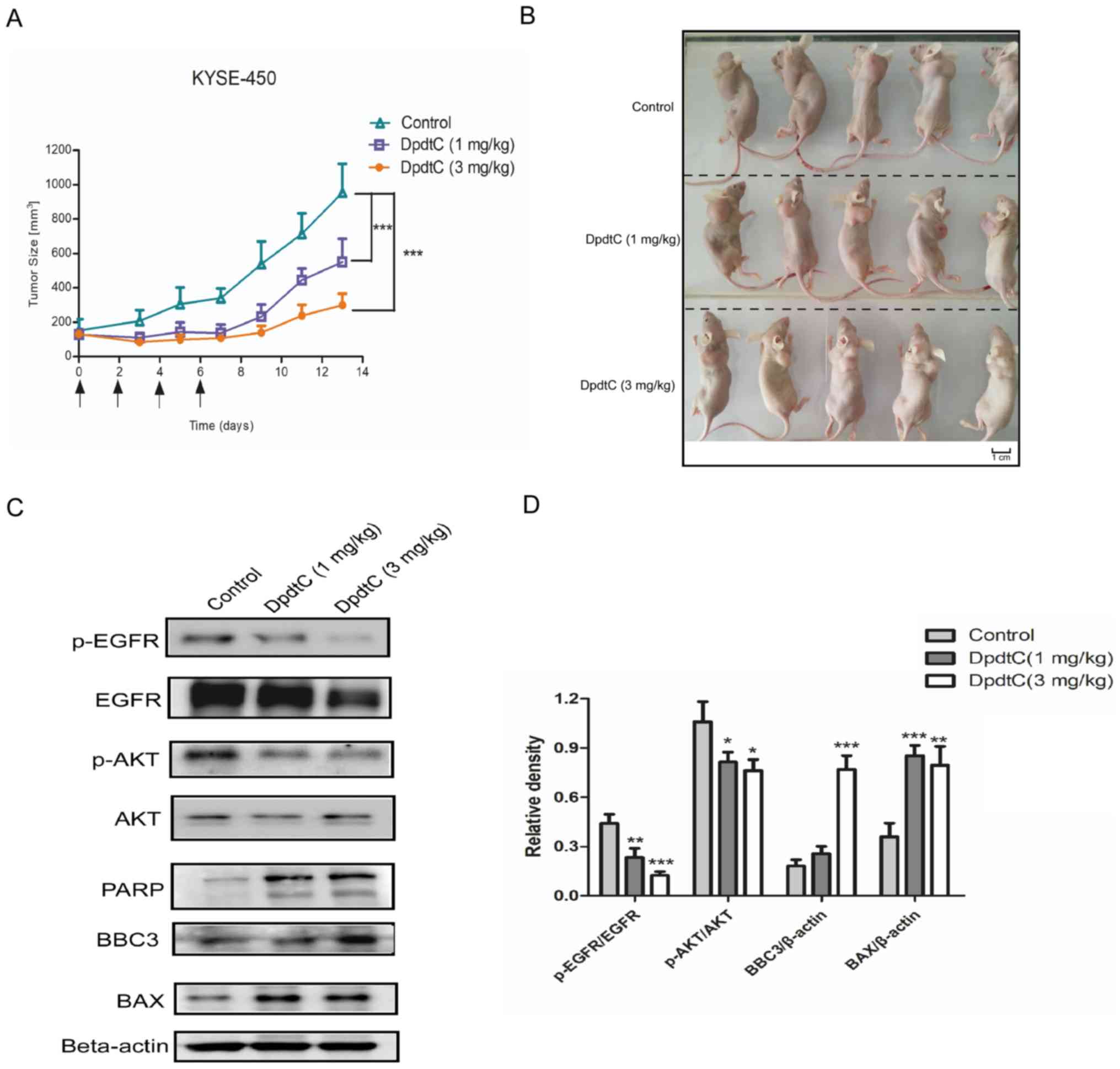 | Figure 4.In vivo effects of DpdtC in the
KYSE-450 tumor-bearing nude mice. (A) Mean tumor volumes of mice
xenografted with KYSE-450 cells and treated with different doses of
DpdtC (1 or 3 mg/kg). n=5. The black arrows indicate the DpdtC
treatment times. (B) KYSE-450 tumor-bearing mice were imaged and
tumors were removed on day 13 post-administration. Tumor tissues
were isolated from KYSE-450 ×enografts following treatment with
control (PBS) or DpdtC (1 or 3 mg/kg), then the expression levels
of EGFR, p-EGFR, AKT, p-AKT, PARP, BBC3, BAX and β-actin were
determined using (C) western blot analysis and the results were (D)
quantified and the protein levels expressed relative to the β-actin
or total EGFR/AKT protein using ImageJ software. Data are presented
as the mean ± SD of three independent experiments. *P<0.05,
**P<0.01, ***P<0.001 vs. control group. DpdtC,
Dipyridylhydrazone dithiocarbamate; p-, phosphorylated; PARP, Poly
(ADP-ribose) polymerase; EGFR, epidermal growth factor receptor;
BBC3, Bcl-2-binding component 3. |
DpdtC induces apoptosis and inhibits
the EGFR/AKT signaling pathway in vivo
To further assess the mechanism of action of DpdtC
in vivo, tumor samples from treated mice were collected and
analyzed using an immunoblotting assay. Related apoptotic markers,
including PARP, BBC3 and BAX were also examined to evaluate the
apoptosis level in vivo. It was found that the expression
levels of p-EGFR and p-AKT in tumors were significantly decreased
compared with that in the control-PBS treatment group. Furthermore,
PARP was significantly cleaved, while BBC3 and BAX expression
levels were significantly elevated in the DpdtC-treated groups
(Fig. 4C and D).
Discussion
Metal chelators possess a potent and targeted
antitumor activity against a variety of types of cancer including
myeloid leukemia, hepatocellular carcinoma and breast cancers etc.
(6,17–19).
Previous studies have reported that Dp44mT or DFO may inhibit
cancer cell proliferation by regressing multiple signaling pathways
including transforming growth factor-β, AKT, ERK and c-Abelson
murine leukemia viral oncogene homolog 1-adaptor molecule CrkII
pathways related to tumor progression and metastasis (4,20,21).
Dithiocarbamates are sulfur-containing compounds with a strong
chelating ability toward metal ions (11,22).
Moreover, their derivatives, such as the synthetic gold(III)
dithiocarbamate and pyrrolidine derivative of dithiocarbamate may
be inhibitors targeting NF-κB (23),
inhibitors against proteasome (24),
DNA intercalators (25) and
inactivators of metal-containing enzymes (26); however, the underlying mechanism
remains to be elucidated.
Our previous study characterized and assessed the
chemical properties of DpdtC (6). To
the best of our knowledge, the present study was the first to
examine the antitumor efficacy of DpdtC against esophageal cancer
cells and to investigate its mechanism of action. The present
results suggested that DpdtC exhibited effective antitumor effects
by inhibiting the EGFR/AKT signaling pathway and inducing apoptosis
of esophageal cancer cells in vitro and in vivo.
Furthermore, it was found that DpdtC induced the downregulation of
EGFR at a relatively high concentration (30 µM) in vitro or
at a high dose (3 mg/kg) in vivo, which suggested that the
growth inhibition caused by DpdtC may be associated with EGFR
downregulation. In addition, the results suggested that treatment
with DpdtC was well tolerated by the mice, without affecting their
body weights. Therefore, it was hypothesized that DpdtC exerted its
antitumor effects primarily by suppressing the EGFR/AKT signaling
pathway. DpdtC has a relative weaker potency on inducing apoptosis
in KYSE-450 cells compared with that in the KYSE-150 cells;
however, it may more effectively inhibit the phosphorylation of
EGFR and AKT in KYSE-450 cells compared with that in KYSE-150
cells. Collectively, the results suggested that DpdtC may exert its
effects by inhibiting the EGFR/AKT signaling pathway and inducing
apoptosis, thus suggesting the potential of DpdtC as a drug
candidate for the treatment of EGFR-positive esophageal cancer
types.
Currently, esophageal cancer lacks potent treatment
regimens, and the 5-year survival rate is <10%, thus the
development of more effective therapeutic agents is required
(3). Our previous study developed a
novel EGFR-targeted antibody-drug, denoted as PT that exerts
antitumor effects on esophageal cancer types by inhibiting the
EGFR/ERK1/2 pathway and inducing apoptosis via blockade of the
nuclear factor erythroid 2-related factor 2/Kelch-like
ECH-associated protein 1 pathway (13). The present study evaluated in
vivo the novel drug, DpdtC which has the potential to be used
in clinical treatment. Moreover, we hypothesized that the
combination of EGFR-targeted antibody drugs with DpdtC could
achieve greater antitumor effects by synergistically inhibiting the
EGFR downstream signaling pathway and inducing apoptosis in
esophageal cancer types, which will be further investigated in
future studies.
In conclusion, the present study identified a
promising anti-cancer agent, DpdtC, which targets the EGFR/AKT
pathway and induces apoptosis, and thus has potential to be a novel
drug candidate in treating esophageal cancer types.
Supplementary Material
Supporting Data
Acknowledgements
The authors would like to thank Professor Changzheng
Li (School of Basic Medical Sciences, Xinxiang Medical University)
for his technical assistance on the design of the study.
Funding
This study was supported by grants from the Natural
Science Foundation of China (grant nos. 81703054 and 81803076),
Henan Provincial Medical Science and Technology Research Project
(grant no. SB201901064), Science and Technology Project for Young
Talents of Henan Province (grant no. 2020HYTP048), Key Project of
School of Basic Medical Sciences in Xinxiang Medical University
(grant nos. JCYXYKY201901 and JCYXYKY201907), Key Science and
Technology Program of Henan Province (grant nos. 192102310414,
182102310259 and 182102310436) and Innovation Project of Graduate
in Xinxiang Medical University (grant no. YJSCX201933Y).
Availability of data and materials
The datasets used and/or analyzed during the current
study are available from the corresponding author on reasonable
request.
Authors' contributions
YY and SD designed the study. YY, ZT, XZ and YL
collected the data and performed the experiments. YY and SD
performed the statistical analyses. YY and SD wrote and revised the
manuscript. All authors read and approved the final manuscript.
Ethics approval and consent to
participate
All animal experiments were approved by the
Biomedical Ethics Committee of Xinxiang Medical University.
Patient consent for publication
Not applicable.
Competing interests
The authors declare that they have no competing
interests.
References
|
1
|
Wu C, Li D, Jia W, Hu Z, Zhou Y, Yu D,
Tong T, Wang M, Lin D, Qiao Y, et al: Genome-wide association study
identifies common variants in SLC39A6 associated with length of
survival in esophageal squamous-cell carcinoma. Nat Genet.
45:632–638. 2013. View
Article : Google Scholar : PubMed/NCBI
|
|
2
|
Guo XF, Zhu XF, Yang WC, Zhang SH and Zhen
YS: An EGFR/HER2-Bispecific and enediyne-energized fusion protein
shows high efficacy against esophageal cancer. PLoS One.
9:e929862014. View Article : Google Scholar : PubMed/NCBI
|
|
3
|
Campbell NP and Villaflor VM: Neoadjuvant
treatment of esophageal cancer. World J Gastroenterol.
16:3793–3803. 2010. View Article : Google Scholar : PubMed/NCBI
|
|
4
|
Kovacevic Z, Chikhani S, Lovejoy DB and
Richardson DR: Novel thiosemicarbazone iron chelators induce
up-regulation and phosphorylation of the metastasis suppressor
N-myc down-stream regulated gene 1: A new strategy for the
treatment of pancreatic cancer. Mol Pharmacol. 80:598–609. 2011.
View Article : Google Scholar : PubMed/NCBI
|
|
5
|
Wang J, Yin D, Xie C, Zheng T, Liang Y,
Hong X, Lu Z, Song X, Song R, Yang H, et al: The iron chelator
Dp44mT inhibits hepatocellular carcinoma metastasis via N-Myc
downstream-regulated gene 2 (NDRG2)/gp130/STAT3 pathway.
Oncotarget. 5:8478–8491. 2014. View Article : Google Scholar : PubMed/NCBI
|
|
6
|
Wang T, Fu Y, Huang T, Liu Y, Wu M, Yuan
Y, Li S and Li C: Copper ion attenuated the antiproliferative
activity of Di-2-pyridylhydrazone dithiocarbamate derivative;
however, there was a lack of correlation between ros generation and
antiproliferative activity. Molecules. 21(pii): E10882016.
View Article : Google Scholar : PubMed/NCBI
|
|
7
|
Fu Y, Liu Y, Wang J and Li C, Zhou S, Yang
Y, Zhou P, Lu C and Li C: Calcium release induced by
2-pyridinecarboxaldehyde thiosemicarbazone and its copper complex
contributes to tumor cell death. Oncol Rep. 37:1662–1670. 2017.
View Article : Google Scholar : PubMed/NCBI
|
|
8
|
Dixon KM, Lui GY, Kovacevic Z, Zhang D,
Yao M, Chen Z, Dong Q, Assinder SJ and Richardson DR: Dp44mT
targets the AKT, TGF-β and ERK pathways via the metastasis
suppressor NDRG1 in normal prostate epithelial cells and prostate
cancer cells. Br J Cancer. 108:409–419. 2013. View Article : Google Scholar : PubMed/NCBI
|
|
9
|
Chen Z, Zhang D, Yue F, Zheng M, Kovacevic
Z and Richardson DR: The iron chelators Dp44mT and DFO inhibit
TGF-β-induced epithelial-mesenchymal transition via up-regulation
of N-Myc downstream-regulated gene 1 (NDRG1). J Biol Chem.
287:17016–17028. 2012. View Article : Google Scholar : PubMed/NCBI
|
|
10
|
Orrenius S, Nobel CS, van den Dobbelsteen
DJ, Burkitt MJ and Slater AF: Dithiocarbamates and the redox
regulation of cell death. Biochem Soc Trans. 24:1032–1038. 1996.
View Article : Google Scholar : PubMed/NCBI
|
|
11
|
Buac D, Schmitt S, Ventro G, Kona FR and
Dou QP: Dithiocarbamate-based coordination compounds as potent
proteasome inhibitors in human cancer cells. Mini Rev Med Chem.
12:1193–1201. 2012. View Article : Google Scholar : PubMed/NCBI
|
|
12
|
Yang Y, Liu Y, Guo R, Fu Y, Zhang Z, Zhang
P, Zhou P, Wang T, Huang T, Li X and Li C: The novel
dithiocarbamate, DpdtC suppresses HER2-overexpressed cancer cells
by up-regulating NDRG1 via inactivation of HER2-ERK 1/2 signaling.
Sci Rep. 8:33982018. View Article : Google Scholar : PubMed/NCBI
|
|
13
|
Yang Y, Tian Z, Ding Y, Li X, Zhang Z,
Yang L, Zhao F, Ren F and Guo R: EGFR-targeted immunotoxin exerts
antitumor effects on esophageal cancers by increasing ROS
accumulation and inducing apoptosis via inhibition of the
Nrf2-keap1 pathway. J Immunol Res. 2018:10902872018. View Article : Google Scholar : PubMed/NCBI
|
|
14
|
Jones-Bolin S: Guidelines for the care and
use of laboratory animals in biomedical research. Curr Protoc
Pharmacol Appendix 4: Appendix 4B. 2012. View Article : Google Scholar
|
|
15
|
Yang Y, Guo R, Tian X, Zhang Z, Zhang P,
Li C and Feng Z: Synergistic anti-tumor activity of Nimotuzumab in
combination with Trastuzumab in HER2-positive breast cancer.
Biochem Biophys Res Commun. 489:523–527. 2017. View Article : Google Scholar : PubMed/NCBI
|
|
16
|
Zhang L, Jiang G, Yao F, He Y, Liang G,
Zhang Y, Hu B, Wu Y, Li Y and Liu H: Growth inhibition and
apoptosis induced by osthole, a natural coumarin, in hepatocellular
carcinoma. PLoS One. 7:e378652012. View Article : Google Scholar : PubMed/NCBI
|
|
17
|
Yuan J, Lovejoy DB and Richardson DR:
Novel di-2-pyridyl-derived iron chelators with marked and selective
antitumor activity: In vitro and in vivo assessment. Blood.
104:1450–1458. 2004. View Article : Google Scholar : PubMed/NCBI
|
|
18
|
Whitnall M, Howard J, Ponka P and
Richardson DR: A class of iron chelators with a wide spectrum of
potent antitumor activity that overcomes resistance to
chemotherapeutics. Proc Natl Acad Sci USA. 103:14901–14906. 2006.
View Article : Google Scholar : PubMed/NCBI
|
|
19
|
Ohyashiki JH, Kobayashi C, Hamamura R,
Okabe S, Tauchi T and Ohyashiki K: The oral iron chelator
deferasirox represses signaling through the mTOR in myeloid
leukemia cells by enhancing expression of REDD1. Cancer Sci.
100:970–977. 2009. View Article : Google Scholar : PubMed/NCBI
|
|
20
|
Le NT and Richardson DR: Iron chelators
with high antiproliferative activity up-regulate the expression of
a growth inhibitory and metastasis suppressor gene: A link between
iron metabolism and proliferation. Blood. 104:2967–2975. 2004.
View Article : Google Scholar : PubMed/NCBI
|
|
21
|
Liu W, Yue F, Zheng M, Merlot A, Bae DH,
Huang M, Lane D, Jansson P, Lui GY, Richardson V, et al: The
proto-oncogene c-Src and its downstream signaling pathways are
inhibited by the metastasis suppressor, NDRG1. Oncotarget.
6:8851–8874. 2015. View Article : Google Scholar : PubMed/NCBI
|
|
22
|
Li C, Liu Y, Fu Y, Huang T and Kang L: The
antiproliferative activity of di-2-pyridylketone dithiocarbamate is
partly attributed to catalase inhibition: Detailing the interaction
by spectroscopic methods. Mol Biosyst. 13:1817–1826. 2017.
View Article : Google Scholar : PubMed/NCBI
|
|
23
|
Schreck R, Meier B, Mannel DN, Droge W and
Baeuerle PA: Dithiocarbamates as potent inhibitors of nuclear
factor kappa B activation in intact cells. J Exp Med.
175:1181–1194. 1992. View Article : Google Scholar : PubMed/NCBI
|
|
24
|
Milacic V, Chen D, Ronconi L,
Landis-Piwowar KR, Fregona D and Dou QP: A novel anticancer
gold(III) dithiocarbamate compound inhibits the activity of a
purified 20S proteasome and 26S proteasome in human breast cancer
cell cultures and xenografts. Cancer Res. 66:10478–10486. 2006.
View Article : Google Scholar : PubMed/NCBI
|
|
25
|
Ronconi L, Marzano C, Zanello P, Corsini
M, Miolo G, Maccà C, Trevisan A and Fregona D: Gold(III)
dithiocarbamate derivatives for the treatment of cancer: Solution
chemistry, DNA binding, and hemolytic properties. J Med Chem.
49:1648–1657. 2006. View Article : Google Scholar : PubMed/NCBI
|
|
26
|
Nobel CS, Burgess DH, Zhivotovsky B,
Burkitt MJ, Orrenius S and Slater AF: Mechanism of dithiocarbamate
inhibition of apoptosis: Thiol oxidation by dithiocarbamate
disulfides directly inhibits processing of the caspase-3 proenzyme.
Chem Res Toxicol. 10:636–643. 1997. View Article : Google Scholar : PubMed/NCBI
|