Introduction
Colorectal cancer (CRC) represents a major threat to
global health, with an estimated 1,096,601 new cases and 551,269
CRC-associated mortalities predicted worldwide in 2018 (1). The prognosis of CRC remains poor,
particularly in the advanced stages; this is due to the symptoms
frequently appearing in the later stages of disease, resulting in
delayed diagnosis and treatment (2).
Chemotherapy is still the preferred adjuvant therapy for patients
with CRC undergoing radical resection (3). In addition, an increased range of
targeted therapies, such as EGFR or KRAS targeted therapies are
being applied in clinical practice (4), of which the suppression of immune
checkpoint pathways may represent the most promising approach
(5). However, the discovery of
reliable biomarkers for screening the target population remains an
essential factor for successful targeted therapy.
Programmed death 1 (PD-1) and programmed death
ligand 1 (PD-L1) are important regulators of immune checkpoints
that induce tumor cell immune escape (6). The expression levels of PD-L1 were
discovered to be increased in various types of tumor, including
non-small cell lung cancer, gastric cancer, breast cancer and
colorectal adenocarcinoma (7–10). The
PD-1/PD-L1 blockade has been used as a novel oncotherapy for
multiple types of cancer, including CRC (11). However, studies assessing the
prognostic significance of PD-L1 expression in CRC remain
controversial (10).
Additionally, various pathways are involved in the
upregulation of PD-L1 in several cancers of the digestive system,
such as the epidermal growth factor receptor/ERK signaling pathway
in esophageal squamous cell carcinoma (12), the janus kinase (JAK)/STAT signaling
pathway in gastric cancer (13) and
the ERK/mitogen-activated protein kinase pathway in hepatocellular
carcinoma (14). Nonetheless, the
mechanisms by which PD-L1 expression is regulated in CRC are yet to
be fully elucidated. Notably, the interferon-γ (IFN-γ)/JAK/STAT
signaling pathway has been confirmed to induce PD-L1 expression in
myeloid leukemia cells, and pancreatic and gastric cancer (13,15,16).
However, the roles of the IFN-γ/JAK/STAT signaling pathway in
regulating PD-L1 expression in CRC remain to be determined.
Therefore, the present study aimed to determine the predictive
value of PD-L1 in the prognosis of CRC and the mechanisms of action
of PD-L1 regulation, with a focus on the IFN-γ/JAK/STAT signaling
pathway in vitro and in patients with CRC.
Materials and methods
Patient studies
The present study was approved by the Institutional
Review Board of China-Japan Union Hospital of Jilin University
(Changchun, China) and written informed consent was provided by all
patients. Patients with colorectal adenocarcinoma were randomly
recruited from the Department of Gastric and Colorectal Surgery in
the China-Japan Union Hospital of Jilin University between January
2010 and December 2015. Patients enrolled in the present study
adhered to the following inclusion criteria: i) Initially diagnosed
with colorectal adenocarcinoma; ii) had undergone tumorectomy; and
iii) had not received chemotherapy or radiotherapy before surgery.
The exclusion criteria were as follows: i) Patients with distant
metastases and positive surgical margins; and ii) patients who had
succumbed to postoperative complications within 30 days following
surgery. Patient diagnosis was independently confirmed by two
pathologists. Finally, 183 patients were randomly selected from the
patients that meet the inclusion and exclusion criteria above.
Clinicopathological data
The following principal clinicopathological
parameters were obtained from the patients: Sex, age, World Health
Organization classification (17),
the primary tumor, tumor size, vascular lymphatic infiltration,
perineurium invasion, tumor location, tumor differentiation and
tumor-node-metastasis (TNM) stage according to the American Joint
Committee on Cancer/Leading the global fight against cancer 2010
classifications (18). All patients
underwent follow-up after surgery in the first, third and sixth
month in the first year, and every year by phone until death or the
last scheduled follow-up. Survival time was defined as the duration
between the date of surgery to the date of death or the final
successful follow-up date. Patients who succumbed to surgical
complications during the perioperative period or who were lost to
follow-up at the time of the first interview were excluded from the
survival analysis. A total of 181 patients were included in the
survival analysis.
Gene set enrichment analysis
(GSEA)
RNA-sequencing data (level 3 with RPKM files) were
downloaded from The Cancer Genome Atlas (TCGA; http://gdc-portal.nci.nih.gov). This data set
comprised the gene expression data from cancerous and healthy
normal tissue of 276 patients with colorectal adenocarcinoma
(19). These data were preprocessed
using TCGA biolinks and annotated with Entrez ID v.17.0 (https://cancergenome.nih.gov/). The co-expression of
PD-L1 with other genes whose sequences were present in this
database was determined using the cBioPortal for Cancer Genomics
v.3.2.13 (20,21). Signaling pathway enrichment was
performed using the Kyoto Encyclopedia of Genes and Genomes (KEGG)
database (http://www.genome.jp/kegg) (22).
Cell culture and treatment
The HCT 116 human CRC cell line (cat. no. CBP60028,
Cobioer) was cultured in DMEM (HyClone; GE Healthcare Life
Sciences), supplemented with 10% FBS (Gibco; Thermo Fisher
Scientific, Inc.) and 100 U/ml penicillin/streptomycin. The cells
were maintained at 37°C (5% CO2) in a humidified
incubator. Recombinant human IFN-γ (R&D Systems, Inc.) was
diluted with PBS to a concentration of 0.2 mg/ml and stored at
70°C. Cells were seeded into 6-well plates at 2×10⁵
cells/well, incubated overnight and then treated with 10 or 20
ng/ml IFN-γ for 24 h at 37°C.
Immunohistochemistry (IHC)
Cancer tissue and paired normal tissue were obtained
from the all of the 181 patients included in the survival analysis
following surgery. Tissue microarray slides of embedded tumor
specimens from patients with colorectal adenocarcinoma were used
for IHC staining. Briefly, tissues were fixed in 10% formalin for
24 h and embedded in paraffin at 65°C. Paraffin-embedded tissues
were subsequently cut to a thickness of 5 µm. After washing with
xylene for 20 min twice at room temperature and rehydration in
descending alcohol series for 5 min in different concentrations
(100, 90 and 80%), the slides were boiled for 20 min in
ethylenediamine tetraacetic acid without high pressure for antigen
retrieval. Endogenous peroxidase activity was blocked using 3%
H2O2. The sections were blocked with 10%
normal goat serum (cat. no. SP-B5; Fuzhou Maixin Biotech Co., Ltd)
for 10 min at room temperature prior to incubation with primary
antibody. The sections were incubated with a primary monoclonal
antibody against PD-L1 (1:200; cat. no. 13684; Cell Signaling
Technology, Inc.) at room temperature for 90 min. Following the
primary antibody incubation, the sections were incubated with a
horseradish peroxidase (HRP)-conjugated secondary antibody (cat.
no. 5020; Fuzhou Maixin Biotech Co., Ltd) for 15 min at room
temperature. The level of protein expression was determined using
the 3,3-diaminobenzidine (DAB) (cat. no. DAB-0031; Maixin Biotech,
Co., Ltd) and a light microscope (magnification ×100) was used to
visualize the slides. PD-L1 expression was observed in the cell
membrane and cytoplasm. The staining intensity was defined as: i)
0, no immunostaining (<5% expression); ii) 1 weak staining
(5–19% expression); iii) 2, moderate staining (20–49% expression);
or iv) 3, strong staining (≥50% expression). The sum of the
intensity and percentage scores resulted in an immunoreactive score
value ranging from 0–6 and a total score of >2 was defined as
positive PD-L1 expression.
Western blotting
Total protein from HTC116 cell lines was extracted
using a mammalian protein extraction kit (cat. no. CW0891M; CoWin
Biosciences) and used according to the manufacturer's instructions.
Total protein was quantified using a bicinchoninic acid assay kit
(cat. no. CW0014S; CoWin Biosciences). The mass of protein loaded
per lane were 10–40 µg on a 12.5% SDS-PAGE gel. The proteins were
transferred to PVDF membranes. The membranes were blocked with 5%
no-fat milk at room temperature for 1 h. The membranes were
incubated with the following primary antibodies overnight at 4°C:
Anti-PD-L1 (1:2,000; cat. no. 13684; Cell Signaling Technology,
Inc.), anti-JAK2 (1:2,000; cat. no. ab39636; Abcam),
anti-phosphorylated (p)-JAK2 (1:2,000; cat. no. ab32101; Abcam),
anti-STAT1 (1:2,000; cat. no. ab31369; Abcam), anti-p-STAT1
(1:1,000; cat. no. ab109461; Abcam) and anti-GAPDH (1:1,000; cat.
no. ab181602; Abcam). The membranes were incubated with an
HRP-conjugated goat anti-rabbit IgG secondary antibody at room
temperature for 25 min (1:10,000; cat. no. ab6721; Abcam). Protein
bands were visualized using ECL reagents (iBright cat. no. CL750;
Thermo Fisher Scientific, Inc.) and a ChemiDoc XRS + imaging system
(Bio-Rad Laboratories, Inc.).
Reverse transcription-quantitative PCR
(RT-qPCR)
Total RNA was extracted from the tumor tissues of 45
patients from whom fresh tissues were available using RNA
extraction buffer from the Promega SV Total RNA Isolation System
kit, (cat. no. Z3100; Promega Corporation) according to the
manufacturer's instructions. Total RNA was reverse transcribed into
cDNA using the cDNA synthesis kit Roche Transcriptor cDNA Synth.
Kit 2 (cat. no. 4897030001; Roche Diagnostics) according to the
manufacturer's instructions (first step 65°C for 10 min, then 65°C
for 30 min and finally 85°C for 5 min). qPCR was subsequently
performed using SYBR Master Mix (Roche Diagnostics) on a
LightCycler 480 Real Time PCR system (Roche Diagnostics). The
thermocycling conditions were as follows: 94°C for 3 min, followed
by 30 cycles of 94°C for 30 sec, 55°C for 30 sec, 72°C for 1 min
and 72°C for 1 min with a final extension step at 72°C for 10 min.
The primers used for RT-qPCR were as follows: PD-L1, forward:
5′-GTACCGCTGCATGATCAGCTAT-3′ and reverse:
5′-GGCATTGACTTTCACAGTAATTCG-3′; IFN-γ, forward:
5′-TCTGGATCCATGAACGCTACACACTGC-3′ and reverse:
5′-ACTAAGCTTTCAGCAGCGACTCCTTTTCC-3′; JAK2, forward:
5′-CTGCAGGAAGGAGAGAGGAAGAGGA-3′ and reverse:
5′-GAATGTTATTGGCAGTCAG-3′; STAT1, forward:
5′-CCACTGAGACATCCTGCCACC-3′ and reverse:
5′-CCACTGAGACATCCTGCCACC-3′ and GAPDH, forward:
5′-ACCACAGTCCATGCCATCACT-3′ and reverse:
5′-ACTGTGCCGTTGAATTTGCC-3′. GAPDH was used as the endogenous
reference gene. Expression levels of PD-L1, IFNG, JAK2 and STAT1
were quantified using the 2−ΔΔCq method (23).
Statistical analysis
Statistical analysis was performed using SPSS
version 17.0 software (SPSS, Inc.) or GraphPad Prism 5.0 software
(GraphPad Software, Inc.). Experiments were repeated 3 times and
the data are presented as the mean ± SD. Statistical differences
between groups were determined using one-way ANOVA and a Fisher's
Least Significant Difference post-hoc test. Categorical variables
are presented as frequencies (percentages) and were compared using
a χ2 test. The log-rank test was used to determine the
significance between the Kaplan-Meier survival curves generated by
SPSS v.17.0 (SPSS Inc.). The variables with P<0.1 in log-rank
test were included in the subsequent multivariate analysis.
Multivariate Cox regression analysis was performed to assess the
hazard ratio (HR) and 95% CI of possible prognostic factors. The
correlations between mRNA expression levels in the tumor tissues
were calculated using Spearman's rank correlation analysis.
P<0.05 was considered to indicate a statistically significant
difference.
Results
Association between PD-L1 expression
levels and the clinicopathological features of patients with
CRC
Among the 181 patients with CRC, 17.1% exhibited
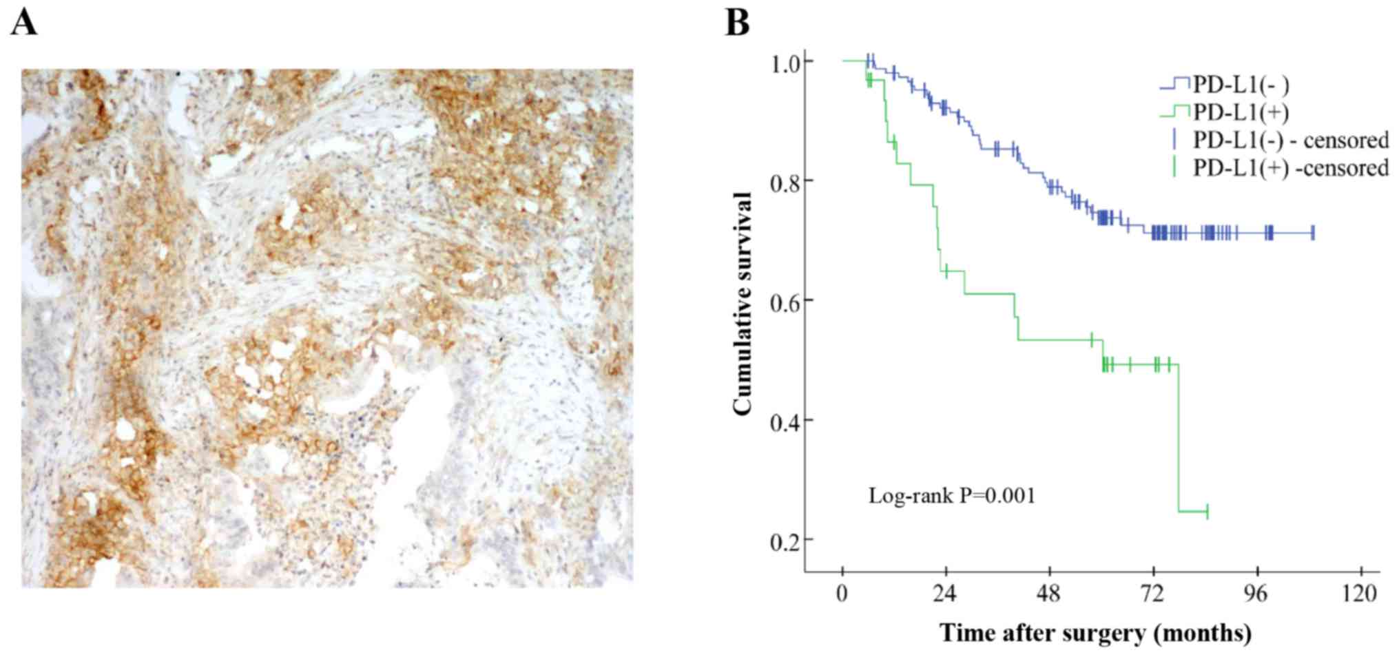
PD-L1 expression in the tumor tissues (Fig. 1A). The association between PD-L1
expression levels and clinicopathological features are presented in
Table I. The expression levels of
PD-L1 were found to be significantly associated with a tumor size
of >5 cm, positive vascular or lymphatic infiltration and a
poorly differentiated stage in the patients (all P<0.01;
Table I).
 | Table I.Association between PD-L1 expression
levels and clinicopathological variables of patients with
colorectal cancer. |
Table I.
Association between PD-L1 expression
levels and clinicopathological variables of patients with
colorectal cancer.
| Variable | PD-L1 (+), n
(%) | PD-L1 (−), n
(%) |
χ2-value | P-value |
|---|
| Age, years |
|
|
|
|
|
>65 | 13 (41.9) | 59 (39.3) | 0.073 | 0.788 |
|
≤65 | 18 (58.1) | 91 (60.7) |
|
|
| Sex |
|
|
|
|
|
Male | 13 (41.9) | 86 (57.3) | 2.458 | 0.117 |
|
Female | 18 (58.1) | 64 (42.7) |
|
|
| Tumor size |
|
|
|
|
| >5
cm | 10 (32.3) | 92 (61.3) | 8.830 | 0.003 |
| ≤5
cm | 21 (67.7) | 58 (38.7) |
|
|
| Tumor
classification |
|
|
|
|
| Tubular
adenocarcinoma | 22 (71.0) | 121 (80.7) | 4.002 | 0.135 |
|
Mucinous adenocarcinoma | 5 (16.1) | 23 (15.3) |
|
|
|
Other | 4 (12.9) | 6 (4.0) |
|
|
| Vascular and
lymphatic infiltration |
|
|
|
|
| No | 11 (35.5) | 95 (63.3) | 8.221 | 0.004 |
|
Yes | 20 (64.5) | 55 (36.7) |
|
|
| Perineurium
invasion |
|
|
|
|
| No | 21 (67.7) | 119 (79.3) | 1.970 | 0.160 |
|
Yes | 10 (32.3) | 31 (20.7) |
|
|
| Tumor location |
|
|
|
|
|
Rectum | 12 (38.7) | 74 (49.3) | 1.163 | 0.281 |
|
Colon | 19 (61.3) | 76 (50.7) |
|
|
| Tumor
differentiation |
|
|
|
|
| Medium
and high differentiation | 13 (41.9) | 134 (89.3) | 37.831 | <0.001 |
| Poorly
differentiated | 18 (58.1) | 16 (10.7) |
|
|
|
Tumor-node-metastasis stage |
|
|
|
|
|
I–II | 11 (35.5) | 85 (56.7) | 4.628 | 0.031 |
|
III | 20 (64.5) | 65 (43.3) |
|
|
PD-L1 expression levels are associated
with a poor prognosis in patients with CRC
The median follow-up time of the patients was 72.9
months (range, 5.4–109.0 months). At the end of the follow-up
period, 105 (58.0%) patients remained alive, 49 (27.1%) patients
had died and 27 (14.9%) were lost to follow-up. A log-rank test
demonstrated that PD-L1 expression levels were significantly
associated with a poor prognosis in patients with CRC (Fig. 1B and Table SI). In addition, the tumor
classification as a mucinous adenocarcinoma, positive vascular and
lymphatic infiltration, positive perineurium invasion and higher
TNM stages were also associated with a poor prognosis of patients
with CRC (P<0.05; Table SI).
Variables with a P-value of <0.1 in the log-rank test were
subsequently included in the multivariate analysis. Multivariate
Cox regression with a stepwise method analysis revealed that the
PD-L1 expression levels in tumor cells was an independent risk
factor for the poor survival of the patients with CRC (HR=1.937;
95% CI=1.038–3.616; P=0.038; Table
II). In addition, positive perineural invasion (P=0.004) and
higher TNM stages (P=0.013) were also discovered to be
independently associated with a poor survival of patients with CRC.
The variables, tumor classification (P=0.391) and vascular &
lymphatic infiltration (P=0.368), were excluded from the final
multivariate analysis in Table
II.
| Table II.Multivariate Cox's regression
analysis for PD-L1 expression levels and the prognosis of patients
with colorectal cancer. |
Table II.
Multivariate Cox's regression
analysis for PD-L1 expression levels and the prognosis of patients
with colorectal cancer.
| Variable | Hazard ratio | 95% CI | P-value |
|---|
| PD-L1 |
|
Positive vs. Negative | 1.937 | 1.038–3.616 | 0.038 |
| Perineural
invasion |
|
Positive vs. Negative | 2.416 | 1.330–4.389 | 0.004 |
| TNM stage |
| III vs.
I–II | 2.206 | 1.181–4.123 | 0.013 |
PD-L1 expression levels are correlated
with IFNG, JAK2 and STAT1 expression levels at the mRNA level
Based on the dataset from TCGA, the genes that were
co-expressed with PD-L1 were sorted using Spearman's rank
correlation analysis (Table SII).
Based on r >0.6, 164 genes were selected for pathway enrichment
analysis using the KEGG pathway database. The first 13 pathways
sorted by the number of hits from the 164 aforementioned genes are
presented in Table SIII. Following
pathway enrichment analysis, 9 of the 164 genes were discovered to
be enriched in the JAK/STAT signaling pathway (Fig. 2A and Table III).
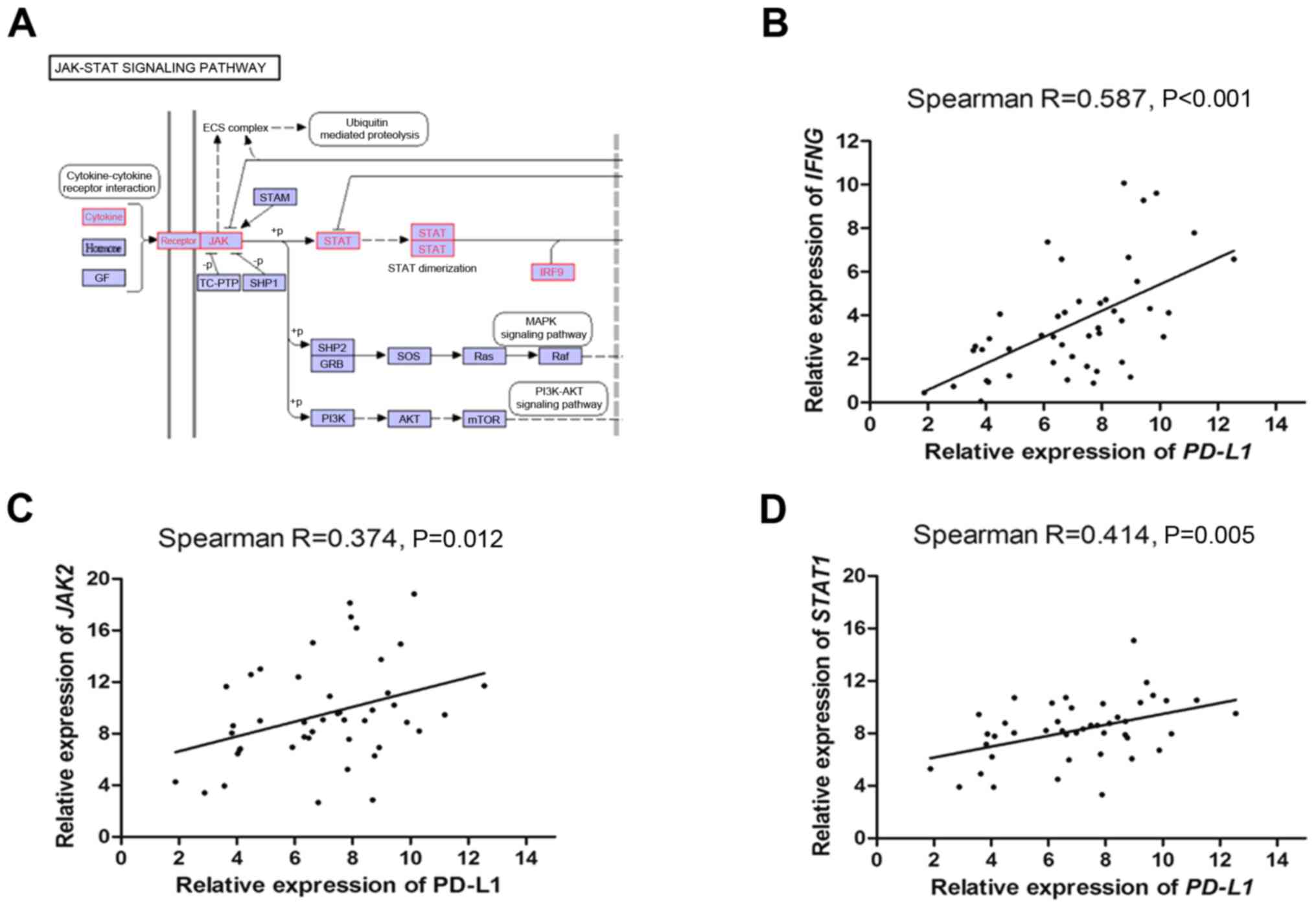 | Figure 2.PD-L1 mRNA expression levels
are correlated the expression levels of IFNG, JAK2 and
STAT1. (A) Kyoto Encyclopedia of Genes and Genomes analysis
revealed nine genes that were enriched in the JAK/STAT signaling
pathway. Spearman's rank correlation analysis between the mRNA
expression levels of (B) PD-L1 and IFNG, (C) PD-L1 and JAK2 and (D)
PD-L1 and STAT1. PD-L1, programmed death ligand 1; JAK, janus
kinase; GF, galphaf; STAM, signal transducing adaptor molecule;
TC-PTP, protein tyrosine phosphatase non-receptor; SHP1, the
protein-tyrosine phosphatase Shp1; SHP2, the protein-tyrosine
phosphatase SHP2; GRB, growth factor receptor; IRF9, interferon
regulatory factor 9; GRB, growth factor receptor bound protein;
SOS, SOS Ras/Rac guanine nucleotide exchange factor. |
| Table III.Gene enrichment in the JAK/STAT
signaling pathway. |
Table III.
Gene enrichment in the JAK/STAT
signaling pathway.
| Correlated
gene | Cytoband | Spearman's
r-value | P-value |
|---|
| STAT1 | 2q32.2 | 0.7929 |
5.00×10−54 |
| Interferon γ | 12q15 | 0.7021 |
1.50×10−37 |
| IL2RA | 22q12.3 | 0.6837 |
5.71×10⁻³⁵ |
| IL2RB | 22q12.3 | 0.6837 |
5.71×10⁻³⁵ |
| STAT2 | 12q13.3 | 0.6530 |
4.84×10⁻³¹ |
| JAK2 | 9p24.1 | 0.6516 |
7.08×10⁻³¹ |
| STAT4 | 2q32.2-q32.3 | 0.6445 |
4.94×10⁻³⁰ |
| IL10RA | 11q23.3 | 0.6060 |
7.56×10⁻²⁶ |
| IRF9 | 14q12 | 0.6039 |
1.22×10⁻²⁵ |
Tumors from 45 randomly selected patients with
colorectal adenocarcinoma (among the 181 recruited patients) were
investigated to confirm the correlation between PD-L1 and IFNG,
JAK2 and STAT1 mRNA expression levels. The results revealed that
PD-L1 expression levels were significantly positively correlated
with the expression levels of IFNG, JAK2 and STAT1 (Fig. 2B-D).
PD-L1 expression levels are increased
by treatment with IFN-γ via the JAK2/STAT1 signaling pathway using
the HCT 116 cell line
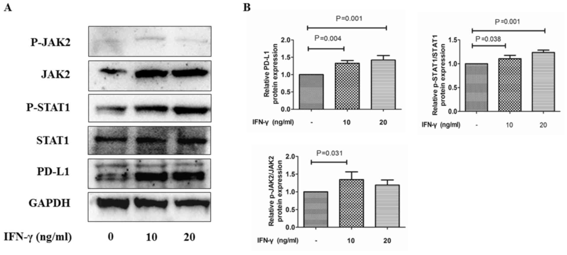
Following treatment with 10 or 20 ng/ml IFN-γ for 24
h, HCT 116 cells exhibited significantly increased expression
levels of PD-L1 expression compared with the untreated group
(Fig. 3A and B). Consistent with
these results, the expression levels of JAK2 and p-STAT1 were also
significantly increased in the IFN-γ treatment groups compared with
the untreated group (Fig. 3A and B).
The p-JAK2/JAK2 and p-STAT1/STAT1 ratios were also increased
following the treatment with IFN-γ compared with the untreated
group. These findings indicated that the JAK2/STAT1 signaling
pathway may influence IFN-γ-mediated upregulation of PD-L1
(Fig. 3).
Discussion
In the present study, increased expression levels of
PD-L1 were discovered to be associated with a poor prognosis in
patients with CRC. This result was consistent with the studies of
Zhu et al (24) and Enkhbat
et al (25), but paradoxical
to the findings reported by Liu et al (26). However, in the latter study, the
subjects were patients with metastatic CRC, whereas in the present
study, patients with distant metastases were excluded. PD-L1
expression was also observed in a large proportion of patients with
tumors of >5 cm, the presence of positive vascular or lymphatic
infiltration and poorly differentiated tumors. These three
clinicopathological variables were all associated with a poor
prognosis in patients with CRC, which suggests that a poor patient
prognosis may be associated with the expression levels of
PD-L1.
Anti-PD-L1 therapy works by blocking the binding of
PD-1 to PD-L1 to inhibit negative signaling transmission,
eliminating the T cell immune inhibitory effects and activating the
immune microenvironment, and thus, antitumor cells (27). Immunotherapy targeting the PD-1/PD-L1
checkpoint pathway is effective in a variety of tumors, such as
non-small cell lung cancer, Hodgkin's lymphoma and malignant
melanoma; in fact, the FDA has reported an objective remission rate
of >65% following treatment with approved PD-L1 inhibitors in
advanced or metastatic hepatocellular carcinoma (28). Notably, CRC treatment with PD-L1
inhibitors was reported to be ineffective (29). Furthermore, a proportion of patients
with CRC are PD-L1-negative and even those with PD-L1-positive CRC
do not always respond to checkpoint inhibitors (30). Therefore, continued investigations
into the molecular mechanisms underlying the action of PD-L1 in CRC
are essential.
The JAK/STAT signaling pathway regulates the
upregulation of PD-L1 in pancreatic, gastric, and head and neck
cancers (13,16,31).
However, the role of the JAK/STAT signaling pathway in CRC remains
unclear. In the present study, PD-L1 expression in a cohort of 181
patients was discovered to be significantly positively correlated
with the expression levels of IFNG, JAK2 and STAT1, which was
consistent with the data extracted from TCGA database.
Additionally, IFN-γ was demonstrated to increase the expression
levels of PD-L1 at the protein level, which was suggested to be
mediated via the increased expression levels of p-JAK2 and p-STAT1.
The current findings indicate that the activation of the JAK2/STAT1
signaling pathway may regulate PD-L1 expression in CRC.
Previous studies have reported that PD-L1 expression
on tumor-infiltrating immune cells is correlated with the survival
of patients with CRC (32–34). Moreover, the tumor microenvironment
in CRC becomes infiltrated with T lymphocytes, which have the
potential to activate the JAK/STAT signaling pathway following
IFN-γ stimulation (35). Thus, it
was hypothesized that in CRC, IFN-γ released from
tumor-infiltrating T lymphocytes may activate the JAK2/STAT1
signaling pathway and promote the expression of PD-L1. Other
studies have indicated that dysregulated JAK/STAT signaling
represents a promising therapeutic target for modulating immune
responses (36). Furthermore,
JAK1/JAK2 mutations were revealed to block PD-L1 induction,
protecting cancer cells from immune attack (37). In concordance with the findings of
the present study, these findings suggested that blocking the
JAK/STAT signaling pathway may affect the efficacy of PD-L1
inhibitors in tumor immunotherapy, though further research is
required to confirm this hypothesis.
There are several limitations to the current study.
Firstly, previous studies have reported that PD-L1 expression in
tumor-infiltrating immune lymphocytes has a prognostic value in CRC
(32,38). However, it was difficult to determine
PD-L1 expression levels in the tumor-infiltrating immune cells
using the tissue microarray slides in the present IHC staining
experiments. Furthermore, comparing the current IHC data with
paired, normal healthy tissues for PD-L1 staining is required to
validate our findings in further studies. Secondly, previous
studies have suggested that PD-L1 inhibitors demonstrate good
efficacy in patients with microsatellite instability (MSI) and
cancers of the digestive system (39,40);
however, information on the patient MSI was not collected in the
present study and a relative analysis could therefore not be
conducted. Thirdly, experiments using pharmacological inhibitors of
JAK/STAT in the presence of IFN-γ are required to be performed in
future research to validate the IFN-γ/JAK/STAT/PD-L1 hypothesis.
Finally, according to the GSEA, STAT2 and STAT4 are also enriched
in the PD-L1-related pathway; however, whether they regulate PD-L1
expression in CRC requires further clarification.
In conclusion, the findings of the present study
suggest that increased expression levels of PD-L1 may be associated
with a poor prognosis in patients with CRC. Furthermore, the
IFN-γ-mediated upregulation of PD-L1 expression may be regulated
via the JAK2/STAT1 signaling pathway in CRC cells.
Supplementary Material
Supporting Data
Acknowledgements
Not applicable.
Funding
The present study was supported by the Jilin
Province Department of Finance (grant nos. 2018sc2006 and
sczsyz01506), the Health Commission of Jilin Province (grant no.
2018Q021) and The Education Department of Jilin Province (grant no.
JJKH20190077KJ).
Availability of data and materials
The datasets used and/or analyzed during the present
study are available from the corresponding author on reasonable
request.
Authors' contributions
BZ conceived and designed the study and guaranteed
its integrity; TCZ contributed to the design of the experiment,
performed the literature research and conducted the statistical
analysis; and YZL and JYZ performed the clinical and experimental
studies. All authors read and approved the final manuscript.
Ethics approval and consent to
participate
The present study was approved by the Institutional
Review Board of China-Japan Union Hospital of Jilin University
(approval number, 2018-NFSC-046, Changchun, China) and written
informed consent provided by from all patients.
Patient consent for publication
Not applicable.
Competing interests
The authors declare that they have no competing
interests.
Glossary
Abbreviations
Abbreviations:
|
PD-L1
|
programmed death ligand 1
|
|
CRC
|
colorectal cancer
|
|
GSEA
|
gene set enrichment analysis
|
|
TCGA
|
The Cancer Genome Atlas
|
|
KEGG
|
Kyoto Encyclopedia of Genes and
Genomes
|
References
|
1
|
Bray F, Ferlay J, Soerjomataram I, Siegel
RL, Torre LA and Jemal A: Global cancer statistics 2018: GLOBOCAN
estimates of incidence and mortality worldwide for 36 cancers in
185 countries. CA Cancer J Clin. 68:394–424. 2018. View Article : Google Scholar : PubMed/NCBI
|
|
2
|
Maida M, Macaluso FS, Ianiro G, Mangiola
F, Sinagra E, Hold G, Maida C, Cammarota G, Gasbarrini A and
Scarpulla G: Screening of colorectal cancer: Present and future.
Expert Rev Anticancer Ther. 17:1131–1146. 2017. View Article : Google Scholar : PubMed/NCBI
|
|
3
|
Binefa G, Rodriguez-Moranta F, Teule A and
Medina-Hayas M: Colorectal cancer: From prevention to personalized
medicine. World J Gastroenterol. 20:6786–6808. 2014. View Article : Google Scholar : PubMed/NCBI
|
|
4
|
Tapia Rico G, Townsend AR, Broadbridge V
and Price TJ: Targeted therapies in elderly patients with
metastatic colorectal cancer: A review of the evidence. Drugs
Aging. 34:173–189. 2017. View Article : Google Scholar : PubMed/NCBI
|
|
5
|
Passardi A, Canale M, Valgiusti M and
Ulivi P: Immune checkpoints as a target for colorectal cancer
treatment. Int J Mol Sci. 18:E13242017. View Article : Google Scholar : PubMed/NCBI
|
|
6
|
Iwai Y, Ishida M, Tanaka Y, Okazaki T,
Honjo T and Minato N: Involvement of PD-L1 on tumor cells in the
escape from host immune system and tumor immunotherapy by PD-L1
blockade. Proc Natl Acad Sci USA. 99:12293–12297. 2002. View Article : Google Scholar : PubMed/NCBI
|
|
7
|
Wu Y, Cao D, Qu L, Cao X, Jia Z, Zhao T,
Wang Q and Jiang J: PD-1 and PD-L1 co-expression predicts favorable
prognosis in gastric cancer. Oncotarget. 8:64066–64082. 2017.
View Article : Google Scholar : PubMed/NCBI
|
|
8
|
Song P, Cui X, Bai L, Zhou X, Zhu X, Zhang
J, Jin F, Zhao J, Zhou C, Zhou Y, et al: Molecular characterization
of clinical responses to PD-1/PD-L1 inhibitors in non-small cell
lung cancer: Predictive value of multidimensional immunomarker
detection for the efficacy of PD-1 inhibitors in Chinese patients.
Thorac Cancer. 10:1303–1309. 2019. View Article : Google Scholar : PubMed/NCBI
|
|
9
|
Li S, Chen L and Jiang J: Role of
programmed cell death ligand-1 expression on prognostic and overall
survival of breast cancer: A systematic review and meta-analysis.
Medicine (Baltimore). 98:e152012019. View Article : Google Scholar : PubMed/NCBI
|
|
10
|
Li Y, He M, Zhou Y, Yang C, Wei S, Bian X,
Christopher O and Xie L: The prognostic and clinicopathological
roles of PD-L1 expression in colorectal cancer: A systematic review
and meta-analysis. Front Pharmacol. 10:1392019. View Article : Google Scholar : PubMed/NCBI
|
|
11
|
Yaghoubi N, Soltani A, Ghazvini K,
Hassanian SM and Hashemy SI: PD-1/PD-L1 blockade as a novel
treatment for colorectal cancer. Biomed Pharmacother. 110:312–318.
2019. View Article : Google Scholar : PubMed/NCBI
|
|
12
|
Ng HY, Li J, Tao L, Lam AK, Chan KW, Ko
JMY, Yu VZ, Wong M, Li B and Lung ML: Chemotherapeutic treatments
increase PD-L1 expression in esophageal squamous cell carcinoma
through EGFR/ERK activation. Transl Oncol. 11:1323–1333. 2018.
View Article : Google Scholar : PubMed/NCBI
|
|
13
|
Mimura K, Teh JL, Okayama H, Shiraishi K,
Kua LF, Koh V, Smoot DT, Ashktorab H, Oike T, Suzuki Y, et al:
PD-L1 expression is mainly regulated by interferon gamma associated
with JAK-STAT pathway in gastric cancer. Cancer Sci. 109:43–53.
2018. View Article : Google Scholar : PubMed/NCBI
|
|
14
|
Qin X, Liu C, Zhou Y and Wang G: Cisplatin
induces programmed death-1-ligand 1(PD-L1) over-expression in
hepatoma H22 cells via Erk/MAPK signaling pathway. Cell Mol Biol
(Noisy-le-grand). 11:OL1366–OL1372. 2010.
|
|
15
|
Bellucci R, Martin A, Bommarito D, Wang K,
Hansen SH, Freeman GJ and Ritz J: Interferon-γ-induced activation
of JAK1 and JAK2 suppresses tumor cell susceptibility to NK cells
through upregulation of PD-L1 expression. Oncoimmunology.
4:e10088242015. View Article : Google Scholar : PubMed/NCBI
|
|
16
|
Imai D, Yoshizumi T, Okano S, Itoh S,
Ikegami T, Harada N, Aishima S, Oda Y and Maehara Y: IFN-γ promotes
epithelial-mesenchymal transition and the expression of PD-L1 in
pancreatic cancer. J Surg Res. 240:115–123. 2019. View Article : Google Scholar : PubMed/NCBI
|
|
17
|
Nagtegaal ID, Odze RD, Klimstra D, Paradis
V, Rugge M, Schirmacher P, Washington KM, Carneiro F and Cree IA;
WHO Classification of Tumours Editorial Board, : The 2019 WHO
classification of tumours of the digestive system. Histopathology.
76:182–188. 2019. View Article : Google Scholar : PubMed/NCBI
|
|
18
|
Cuccurullo V and Mansi L: AJCC Cancer
staging Handbook: From the AJCC cancer staging manual (7th
edition). Eur J Nucl Med Mol Imaging. 38:408. 2011. View Article : Google Scholar
|
|
19
|
Cancer Genome Atlas Network: Comprehensive
molecular characterization of human colon and rectal cancer.
Nature. 487:330–337. 2012. View Article : Google Scholar : PubMed/NCBI
|
|
20
|
Gao J, Aksoy BA, Dogrusoz U, Dresdner G,
Gross B, Sumer SO, Sun Y, Jacobsen A, Sinha R, Larsson E, et al:
Integrative analysis of complex cancer genomics and clinical
profiles using the cBioPortal. Sci Signal. 6:pl12013. View Article : Google Scholar : PubMed/NCBI
|
|
21
|
Cerami E, Gao J, Dogrusoz U, Gross BE,
Sumer SO, Aksoy BA, Jacobsen A, Byrne CJ, Heuer ML, Larsson E, et
al: The cBio cancer genomics portal: An open platform for exploring
multidimensional cancer genomics data. Cancer Discov. 2:401–404.
2012. View Article : Google Scholar : PubMed/NCBI
|
|
22
|
Kanehisa M, Furumichi M, Tanabe M, Sato Y
and Morishima K: KEGG: New perspectives on genomes, pathways,
diseases and drugs. Nucleic Acids Res. 45:D353–D361. 2017.
View Article : Google Scholar : PubMed/NCBI
|
|
23
|
Livak KJ and Schmittgen TD: Analysis of
relative gene expression data using real-time quantitative PCR and
the 2(-Delta Delta C(T)) method. Methods. 25:402–408. 2001.
View Article : Google Scholar : PubMed/NCBI
|
|
24
|
Zhu H, Qin H, Huang Z, Li S, Zhu X, He J,
Yang J, Yu X and Yi X: Clinical significance of programmed death
ligand-1 (PD-L1) in colorectal serrated adenocarcinoma. Int J Clin
Exp Pathol. 8:9351–9359. 2015.PubMed/NCBI
|
|
25
|
Enkhbat T, Nishi M, Takasu C, Yoshikawa K,
Jun H, Tokunaga T, Kashihara H, Ishikawa D and Shimada M:
Programmed cell death ligand 1 expression is an independent
prognostic factor in colorectal cancer. Anticancer Res.
38:3367–3373. 2018. View Article : Google Scholar : PubMed/NCBI
|
|
26
|
Liu R, Peng K, Yu Y, Liang L, Xu X, Li W,
Yu S and Liu T: Prognostic value of immunoscore and PD-L1
expression in metastatic colorectal cancer patients with different
RAS status after palliative operation. Biomed Res Int.
2018:59206082018.PubMed/NCBI
|
|
27
|
Francisco LM, Salinas VH, Brown KE,
Vanguri VK, Freeman GJ, Kuchroo VK and Sharpe AH: PD-L1 regulates
the development, maintenance, and function of induced regulatory T
cells. J Exp Med. 206:3015–3029. 2009. View Article : Google Scholar : PubMed/NCBI
|
|
28
|
Carretero-Gonzalez A, Lora D, Ghanem I,
Zugazagoitia J, Castellano D, Sepúlveda JM, López-Martin JA,
Paz-Ares L and de Velasco G: Analysis of response rate with ANTI
PD1/PD-L1 monoclonal antibodies in advanced solid tumors: A
meta-analysis of randomized clinical trials. Oncotarget.
9:8706–8715. 2018. View Article : Google Scholar : PubMed/NCBI
|
|
29
|
Asaoka Y, Ijichi H and Koike K: PD-1
Blockade in tumors with mismatch-repair deficiency. N Engl J Med.
373:19792015. View Article : Google Scholar : PubMed/NCBI
|
|
30
|
O'Neil BH, Wallmark J, Lorente D, Elez E,
Raimbourg J, Gomez-Roca C, Ejadi S, Piha-Paul SA, Moss RA, Siu LL,
et al: Pembrolizumab (MK-3475) for patients (pts) with advanced
colorectal carcinoma (CRC): Preliminary results from KEYNOTE-028.
Eur J Cancer. 51:4332015. View Article : Google Scholar
|
|
31
|
Concha-Benavente F, Srivastava RM, Trivedi
S, Lei Y, Chandran U, Seethala RR, Freeman GJ and Ferris RL:
Identification of the cell-intrinsic and -extrinsic pathways
downstream of EGFR and IFNγ that induce PD-L1 expression in head
and neck cancer. Cancer Res. 76:1031–1043. 2016. View Article : Google Scholar : PubMed/NCBI
|
|
32
|
Li Y, Liang L, Dai W, Cai G, Xu Y, Li X,
Li Q and Cai S: Prognostic impact of programed cell death-1 (PD-1)
and PD-ligand 1 (PD-L1) expression in cancer cells and tumor
infiltrating lymphocytes in colorectal cancer. Mol Cancer.
15:552016. View Article : Google Scholar : PubMed/NCBI
|
|
33
|
Kong P, Wang J, Song Z, Liu S, He W, Jiang
C, Xie Q, Yang L, Xia X and Xia L: Circulating lymphocytes, PD-L1
expression on tumor-infiltrating lymphocytes, and survival of
colorectal cancer patients with different mismatch repair gene
status. J Cancer. 10:1745–1754. 2019. View Article : Google Scholar : PubMed/NCBI
|
|
34
|
Calik I, Calik M, Turken G, Ozercan IH,
Dagli AF, Artas G and Sarikaya B: Intratumoral cytotoxic
T-lymphocyte density and PD-L1 expression are prognostic biomarkers
for patients with colorectal cancer. Medicina (Kaunas).
55:E7232019. View Article : Google Scholar : PubMed/NCBI
|
|
35
|
Sudoyo AW, Kurniawan AN, Kusumo GD, Putra
TP, Rexana FA, Yunus M, Budiyati AD, Kurniawan D, Utama A and Utomo
AR: Increased CD8 tumor infiltrating lymphocytes in colorectal
cancer microenvironment supports an adaptive immune resistance
mechanism of PD-L1 expression. Asian Pac J Cancer Prev.
20:3421–3427. 2019. View Article : Google Scholar : PubMed/NCBI
|
|
36
|
Meyer SC: Mechanisms of resistance to JAK2
inhibitors in myeloproliferative neoplasms. Hematol Oncol Clin
North Am. 31:627–642. 2017. View Article : Google Scholar : PubMed/NCBI
|
|
37
|
Gotthardt D, Putz EM, Grundschober E,
Prchal-Murphy M, Straka E, Kudweis P, Heller G, Bago-Horvath Z,
Witalisz-Siepracka A, Cumaraswamy AA, et al: STAT5 is a key
regulator in NK cells and acts as a molecular switch from tumor
surveillance to tumor promotion. Cancer Discov. 6:414–429. 2016.
View Article : Google Scholar : PubMed/NCBI
|
|
38
|
Valentini AM, Di Pinto F, Cariola F,
Guerra V, Giannelli G, Caruso ML and Pirrelli M: PD-L1 expression
in colorectal cancer defines three subsets of tumor immune
microenvironments. Oncotarget. 9:8584–8596. 2018. View Article : Google Scholar : PubMed/NCBI
|
|
39
|
Oliveira AF, Bretes L and Furtado I:
Review of PD-1/PD-L1 inhibitors in metastatic dMMR/MSI-H colorectal
cancer. Front Oncol. 9:3962019. View Article : Google Scholar : PubMed/NCBI
|
|
40
|
Angell HK, Lee J, Kim KM, Kim K, Kim ST,
Park SH, Kang WK, Sharpe A, Ogden J, Davenport A, et al: PD-L1 and
immune infiltrates are differentially expressed in distinct
subgroups of gastric cancer. Oncoimmunology. 8:e15444422019.
View Article : Google Scholar : PubMed/NCBI
|