Introduction
Lung cancer remains the most prevalent malignancy
and a leading cause of global cancer-associated mortalities
(1). According to the statistics
from 2012, there were 1.8 million new lung cancer cases and 1.59
million deaths worldwide (2).
Non-small cell lung cancer (NSCLC) is the most common subtype of
lung cancer, accounting for ~80% of all cases (3). Metastasis and invasion are considered
two major biological characteristics of NSCLC, which pose treatment
challenges and continue to increase mortality in patients with
NSCLC (4). A lack of typical
clinical manifestations in patients with lung cancer further
contributes to NSCLC mortality, as it is difficult to effectively
diagnose NSCLC at the early stages (5). Despite major advancements in
therapeutic strategies, such as surgery, chemotherapy and
radiotherapy, the 5-year overall survival rate of patients with
NSCLC remains <15% (6). Thus, it
is critical to identify and develop novel biomarkers involved in
tumor progression for effective NSCLC diagnosis, prognosis and
treatment.
Currently, a number of tumor-associated molecules
have been confirmed to be involved in NSCLC progression, such as
coiled-coil domain containing 106, long non-coding RNA XIST and
microRNA-16 (7,8). microRNAs (miRNAs/miR) are a group of
non-coding small RNA molecules that function in regulating tumor
initiation and progression (9).
miRNAs regulate gene expression by directly binding to the 3′-
untranslated region of target mRNAs (10), and influence several cellular
processes, including cell proliferation, migration and invasion
(11). Previous studies have
reported the pivotal roles of miRNAs in different types of human
cancer, such as glioma, breast cancer and NSCLC (12–15).
Furthermore, the clinical significance of miRNAs has been
highlighted through their effective diagnostic and prognostic
values in different types of cancer (16,17). For
example, serum elevated miR-191 and miR-425 levels are biomarkers
for gastric cancer diagnosis and prognosis (18). The increased expression of miR-665 in
patients with lung cancer has been reported to predict poor
prognosis (19).
miR-518b is a member of the functional miRNAs
(20), which has been investigated
in hepatocellular carcinoma (21,22),
chondrosarcoma (23) and esophageal
squamous cell carcinoma (24).
Regarding hepatocellular carcinoma, Zheng et al (21) and Wang et al (22) demonstrated that miR-518b expression
is elevated in tumor samples compared with the normal controls,
while miR-518b expression is downregulated in chondrosarcoma
(23) and esophageal squamous cell
carcinoma (24). These results
suggest that miR-518b expression varies in different types of human
cancer. In NSCLC, an in silico study reported that miR-518b
expression was higher in tumor samples compared with the normal
controls (25). However, the precise
expression patterns of miR-518b in NSCLC clinical samples, as well
as its role in tumor progression remain unclear.
The present study aimed to determine the biological
role and clinical significance of miR-518b in patients with NSCLC,
and investigate the regulatory effects of miR-518b on NSCLC cell
proliferation, migration and invasion. Taken together, the results
of the present study suggest that miR-518b may serve as a novel
potential diagnostic and prognostic biomarker, and a candidate
therapeutic target for NSCLC treatment.
Materials and methods
Patients and serum and tissue sample
collection
The present study was approved by the Ethics
Committee of Qilu Hospital Huantai Branch (Zibo, China), and
written informed consent was provided by all participants prior to
the study start. A total of 118 patients, including 48 females and
70 males with a mean age of 58.37±12.37 years (age range, 34–85
years), who were pathologically diagnosed with NSCLC at the Qilu
Hospital Huantai Branch, were enrolled in the present study between
January 2011 and December 2013. The inclusion criteria were as
follows: i) All cases received their first surgical resection at
the Qilu Hospital Huantai Branch and were pathologically diagnosed
with NSCLC; ii) Patients who had not received any previous
preoperative antitumor therapy; iii) Patients who had no history of
exposure to asbestos; and iv) Patients with complete
clinicopathological data and follow-up information. A total of 60
healthy volunteers, including 25 females and 35 males with a mean
age of 57.62±12.06 years (age range, 35–83 years), who had no
history of malignancy, were also enrolled in the present study as
the controls. Blood samples were collected from the patients and
healthy individuals, and immediately centrifuged at 1500 × g at 4°C
for 10 min for serum extraction. Tumor tissues and adjacent normal
tissues (controls; at least 3 cm from the edge of tumor) were
collected from patients during surgical resection. The
Tumor-Node-Metastasis (TNM) stage of the tumor tissues was
determined using the criteria from the American Joint Committee on
Cancer classification (26). All
serum and tissue samples were stored at −80ºC until
subsequent experimentation. The demographic and clinical
characteristics of the patients and the survival information
obtained from a 5-year follow-up survey were recorded for
subsequent analyses. During the 5-year follow-up, the patients were
followed up every 3 months in the first 2 years, then after every 6
months for the subsequent 2 years and annually for the last
year.
Cell culture and transfection
A normal human lung epithelial cell line (BEAS-2B)
and four NSCLC cell lines (A549, H1299, H1975 and PC9) were
purchased from the Shanghai Institutes for Biological Sciences of
the Chinese Academy of Sciences. Cells were cultured in DMEM
supplemented with 10% FBS (Invitrogen; Thermo Fisher Scientific,
Inc.), 100 U/ml penicillin and 100 µg/ml streptomycin at 37
ºC in 5% CO2.
A549 and PC9 cells were seeded into 6-well plates at
a density of 5×10⁴ cells/well and transfected with 50 nM
of miR-518b mimic, miR-518b inhibitor or non-targeting miRNA
negative control (miR-NC) using Lipofectamine® 3000
reagent (Invitrogen; Thermo Fisher Scientific, Inc.), according to
the manufacturer's protocol. Following were the sequences of the
vectors: miR-518b mimic, 5′-CAAAGCGCUCCCCUUUAGAGGU-3′; miR-518b
inhibitor, 5′-ACCUCUAAAGGGGAGCGCUUUG-3′; miR-NC,
5′-UUCUCCGAACGUGUCACGU-3′. All vectors were synthesized by Shanghai
GenePharma Co., Ltd., and all the experiments were performed in
triplicate. Subsequent experiments were performed 48 h
post-transfection.
Reverse transcription-quantitative
(RT-q)PCR
Total RNA was extracted from serum of patients and
healthy controls, tissues of patients and NSCLC cell lines using
TRIzol® reagent (Invitrogen; Thermo Fisher Scientific,
Inc.). Total RNA was reverse transcribed into cDNA using the
PrimeScript™ RT reagent kit (Takara Bio, Inc.). All the experiments
were performed following manufacturer's protocols. qPCR was
subsequently performed using the SYBR Green I Master mix kit
(Invitrogen; Thermo Fisher Scientific, Inc.) and a 7500 Real-Time
PCR System (Applied Biosystems; Thermo Fisher Scientific, Inc.).
The thermocycling conditions were as follows: Initial denaturation
at 95°C for 10 min; 40 cycles of denaturation at 95°C for 30 sec,
annealing at 60°C for 20 sec and elongation at 72°C for 30 sec; and
final extension at 72°C for 10 min. The following primer sequences
were used for qPCR: miR-518b forward, 5′-GCCGAGCAAAGCGCTCCCCT-3′,
and reverse, 5′-CTCAACTGGTGTCGTGGA-3′; and U6 forward,
5′-CTCGCTTCGGCAGCACA-3′ and reverse, 5′-AACGCTTCACGAATTTGCGT-3′.
Relative miR-518b expression levels were measured using the
2−ΔΔCq method (27) and
normalized to the internal reference gene U6.
Cell Counting Kit-8 (CCK-8) assay
The CCK-8 assay was performed to assess NSCLC cell
proliferation. A549 and PC9 were seeded into 96-well plates at
density of 5×10³ cells/well (100 µl/well) and cultured
at 37ºC for 72 h. A volume of 10 µl CCK-8 reagent
(Sigma-Aldrich; Merck KGaA) was added to the plates at 0, 24, 48
and 72 h, and incubated at 37ºC for 2 h at each time
point according to the manufacturer's instructions. Cell
proliferation was subsequently analyzed at a wavelength of 450 nm,
using a microplate reader (BioTek).
Migration and invasion assays
A549 and PC9 cells were plated in the upper chambers
(cell density of 3×10⁵ cells/well) of Transwell plates
in serum-free DMEM medium (Invitrogen; Thermo Fisher Scientific,
Inc.) and incubated at 37ºC for 24 h. Transwell
membranes were pre-coated with Matrigel (Corning, Inc.) at 37°C for
1 h for the invasion assay. The DMEM medium supplemented with 10%
FBS was plated in the lower chambers. After 24 h of incubation at
37°C, the migratory and invasive cells in the lower chambers were
stained with 0.1% crystal violet at room temperature for 10 min and
counted in five randomly-selected fields using a light microscope
(magnification, ×200).
Statistical analysis
Statistical analysis was performed using SPSS
(version 21.0; IBM Corp.) and GraphPad Prism (version 7.0; GraphPad
Software, Inc.) software. Data are presented as the mean ± standard
deviation and all experiments were performed in triplicate. Paired
Student's t-test was used to compare differences of miR-518b
expression between tumor tissues and non-tumor tissues, and
unpaired Student's t-test was used to compared the serum expression
of miR-518b between patients with NSCLC and healthy controls, while
one-way ANOVA, followed by Tukey's post-hoc test was used to
compare differences between multiple groups. The expression of
miR-518b was divided into low and high expression group based on
the mean expression value (1.20 for serum miR-518b expression; 3.45
for tissue miR-518b expression), then the association between
miR-518b expression and clinicopathological characteristics of
patients with NSCLC was determined using the χ2 test. A
receiver operating characteristic (ROC) curve was plotted to
determine the diagnostic value of miR-518b, while a Kaplan-Meier
survival curve was generated to assess the prognostic value of
miR-518b in patients with NSCLC and the log-rank test was used to
compare the differences between the survival curves. A multivariate
Cox regression analysis was performed to verify miR-518b as a
prognostic indicator. P<0.05 was considered to indicate a
statistically significant difference.
Results
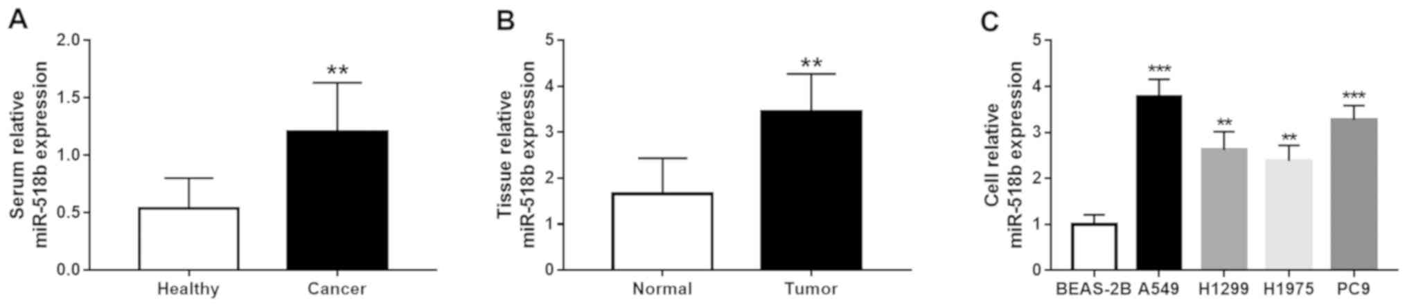
miR-518b expression is upregulated in
NSCLC serum, tissues and cell lines
The results demonstrated that miR-518b expression
levels in the serum and tissue samples significantly increased in
patients with NSCLC compared with the healthy controls and adjacent
normal tissues, respectively (both P<0.01; Fig. 1A and B). Similarly, miR-518b
expression significantly increased in all four NSCLC cell lines
compared with BEAS-2B cells (all P<0.01; Fig. 1C), and a greater significance was
observed for the upregulation of miR-518b in A549 and PC9 cell
lines (both P<0.001).
Association between miR-518b
expression and clinicopathological characteristics of patients with
NSCLC
The clinicopathological characteristics of patients
with NSCLC are presented in Table I.
Patients were divided into low (n=58) and high (n=60) miR-518b
expression groups based on serum mean expression value of miR-518b.
Meanwhile, according to the mean value of miR-518b in tumor
tissues, patients were grouped into low miR-518b group (n=54) and
high miR-518b group (n=64). The results demonstrated that serum
miR-518b expression was significantly associated with tumor size
(P=0.042), TNM stage (P=0.006) and lymph node metastasis (P=0.039).
Similarly, tissue miR-518b expression was significantly associated
with tumor size (P=0.014), TNM stage (P=0.006) and lymph node
metastasis (P=0.031) in patients with NSCLC. However, no
significant associations were observed between miR-518b expression
and age, sex, smoking status, histological type and degree of
differentiation (all P>0.05).
 | Table I.Association between miR-518b
expression and clinicopathological characteristics of patients with
non-small cell lung cancer (n=118). |
Table I.
Association between miR-518b
expression and clinicopathological characteristics of patients with
non-small cell lung cancer (n=118).
|
|
| Serum miR-518b
expression |
| Tissue miR-518b
expression |
|
|---|
|
|
|
|
|
|
|
|---|
| Characteristic | Patients, n | Low (n=58) | High (n=60) | P-value | Low (n=54) | High (n=64) | P-value |
|---|
| Age, years |
|
|
| 0.969 |
|
| 0.569 |
|
≤60 | 47 | 23 | 24 |
| 20 | 27 |
|
|
>60 | 71 | 35 | 36 |
| 34 | 37 |
|
| Sex |
|
|
| 0.879 |
|
| 0.265 |
|
Female | 48 | 24 | 24 |
| 19 | 29 |
|
|
Male | 70 | 34 | 36 |
| 35 | 35 |
|
| Smoking status |
|
|
| 0.732 |
|
| 0.554 |
|
Never | 49 | 25 | 24 |
| 24 | 25 |
|
|
Previous/Current | 69 | 33 | 36 |
| 30 | 39 |
|
| Histological
type |
|
|
| 0.791 |
|
| 0.946 |
|
Adenocarcinoma | 70 | 34 | 36 |
| 32 | 38 |
|
|
Squamous cell carcinoma | 36 | 19 | 17 |
| 16 | 20 |
|
|
Othersa | 12 | 5 | 7 |
| 6 | 6 |
|
| Tumor size, cm |
|
|
| 0.042 |
|
| 0.014 |
| ≤3 | 62 | 36 | 26 |
| 35 | 27 |
|
|
>3 | 56 | 22 | 34 |
| 19 | 37 |
|
|
Differentiation |
|
|
| 0.127 |
|
| 0.097 |
|
Well/Moderate | 69 | 38 | 31 |
| 36 | 33 |
|
|
Poor | 49 | 20 | 29 |
| 18 | 31 |
|
| TNM stage |
|
|
| 0.006 |
|
| 0.006 |
|
I–II | 58 | 36 | 22 |
| 34 | 24 |
|
|
III–IV | 60 | 22 | 38 |
| 20 | 40 |
|
| Lymph node
metastasis |
|
|
| 0.039 |
|
| 0.031 |
|
Negative | 66 | 38 | 28 |
| 36 | 30 |
|
|
Positive | 52 | 20 | 32 |
| 18 | 34 |
|
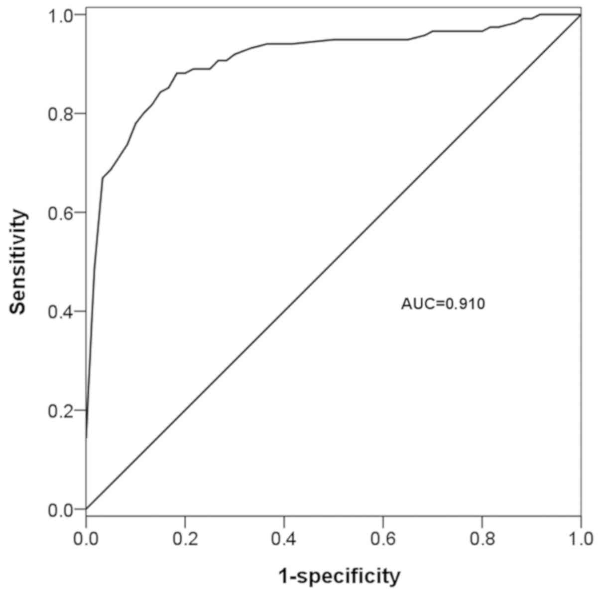
Diagnostic value of miR-518b in
patients with NSCLC
Molecules aberrantly expressed in the serum of
patients with cancer are considered effective diagnostic tools
(28). In the present study, the
diagnostic value of serum miR-518b was determined by analyzing its
deregulated expression in patients with NSCLC. The ROC curve based
on serum miR-518b expression exhibited an area under the curve
value of 0.910, with 88.1% sensitivity and 81.7% specificity, under
the cut-off value of 0.745 (Fig. 2),
which indicated the diagnostic accuracy of serum miR-518b in
patients with NSCLC.
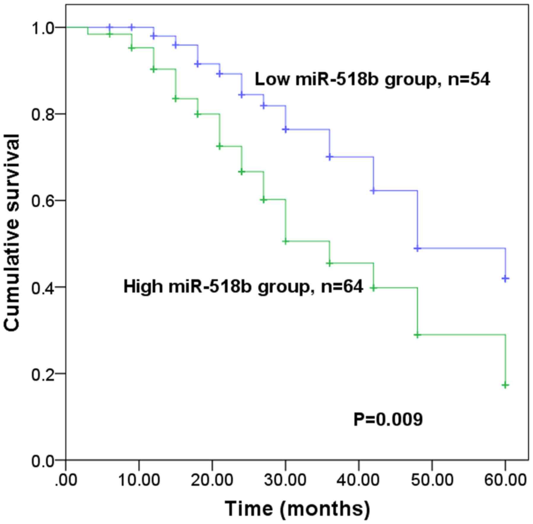
Prognostic value of miR-518b in
patients with NSCLC
The present study further investigated the
prognostic value of miR-518b in patients with NSCLC. The results
demonstrated that patients with high miR-518b expression levels
experienced a shorter survival time than those with low miR-518b
expression levels (P=0.009; Fig. 3).
Furthermore, Cox regression analysis indicated that miR-518b
expression may serve as an independent prognostic indicator in
patients with NSCLC (P=0.012; hazard ratio=2.270; 95% confidence
interval=1.197–4.305; Table
II).
| Table II.Multivariate Cox regression analysis
for miR-518b in patients with non-small cell lung cancer. |
Table II.
Multivariate Cox regression analysis
for miR-518b in patients with non-small cell lung cancer.
|
| Cox regression
analysis |
|---|
|
|
|
|---|
| Characteristic | HR | 95% CI | P-value |
|---|
| miR-518b | 2.270 | 1.197–4.305 | 0.012 |
| Age | 1.064 | 0.601–1.884 | 0.832 |
| Sex | 1.060 | 0.583–1.927 | 0.850 |
| Smoking status | 1.425 | 0.802–2.531 | 0.227 |
| Histological
type | 2.408 | 0.562–10.326 | 0.495 |
| Tumor size | 1.123 | 0.610–2.068 | 0.710 |
|
Differentiation | 1.198 | 0.652–2.198 | 0.560 |
| TNM | 5.359 | 1.177–24.389 | 0.030 |
| Lymph node
metastasis | 5.957 | 1.374–25.829 | 0.017 |
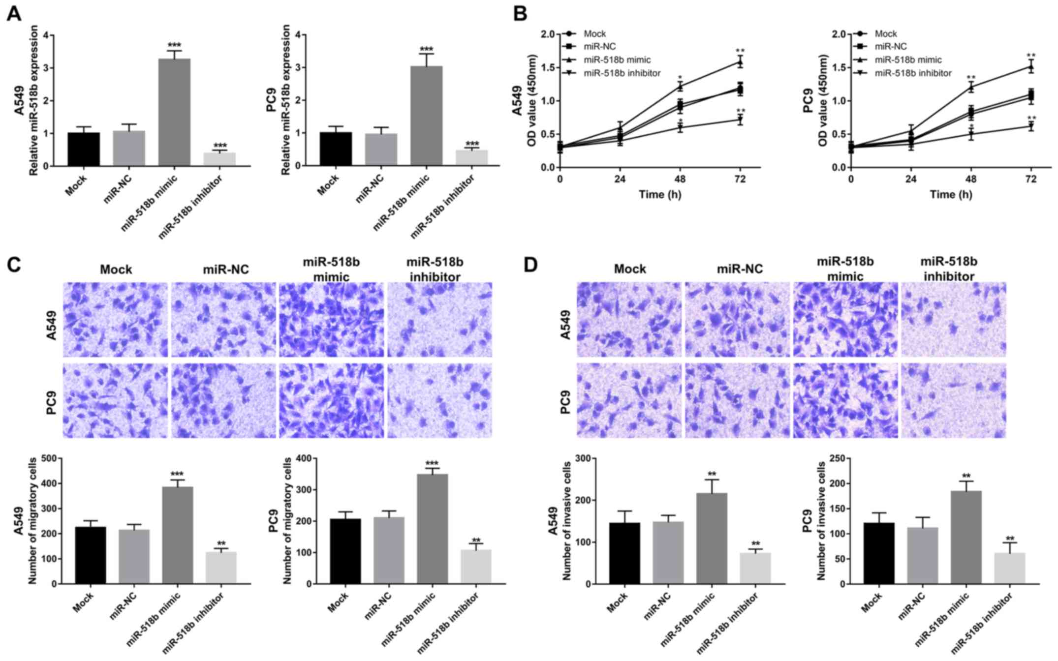
Overexpression of miR-518b facilitates
NSCLC cell proliferation, migration and invasion
The biological function of miR-518b in NSCLC
progression was further investigated in A549 and PC9 cells as the
expression of miR-518b was significantly elevated compared with
normal cells. miR-518b expression was successfully regulated in
vitro via cell transfection, evidenced by increased miR-518b
expression induced by miR-518 mimic, and decreased miR-518b
expression induced by miR-518b inhibitor (all P<0.001; Fig. 4A). Results from the CCK-8 assay, and
cell migration and invasion assays demonstrated that overexpression
of miR-518b in NSCLC cells enhanced cell proliferation, migration
and invasion, while miR-518b knockdown inhibited NSCLC cell
proliferation, migration and invasion, respectively (all P<0.05;
Fig. 4B-D).
Discussion
Several aberrantly expressed miRNAs have been
reported to serve crucial roles in tumor pathology of different
types of human cancer, such as gastric (29), breast (30) and lung cancer (31). The present study aimed to determine
the clinical significance and biological function of miR-518b in
NSCLC. The results of the present study demonstrated significantly
increased miR-518b expression in NSCLC serum, tissues and cell
lines compared with the corresponding normal controls. Furthermore,
elevated miR-518b expression levels in serum and tissues were
associated with tumor size, lymph node metastasis and TNM stage of
patients with NSCLC. Serum miR-518b expression had potential
diagnostic value to distinguish patients with NSCLC from healthy
individuals, and miR-518b expression in tumor tissues was
identified as an independent prognostic indicator in patients with
NSCLC. The gain- and loss-of-function experiments demonstrated that
the cell proliferation, migration and invasion abilities of NSCLC
cells were enhanced following overexpression of miR-518b, while
miR-518b knockdown reversed these effects. Taken together, the
results of the present study suggest that miR-518b may represent a
novel molecule that can be used improve NSCLC diagnosis and
prognosis. Furthermore, determining the biological function of
miR-518b may help to better understand its underlying molecular
mechanisms in the pathogenesis of NSCLC.
The significant roles of miRNAs have been
highlighted in human malignancies in recent decades. For example,
aberrantly expressed miRNAs are associated with tumorigenesis and
have attracted considerable attention in their role as diagnostic
and prognostic biomarkers in several types of cancer, such as
bladder cancer and hepatocellular carcinoma (32,33).
Thus, the expression profiles of miRNAs remain an important focus
in the research field regarding the treatment of human
malignancies. In patients with NSCLC, several aberrantly expressed
miRNAs have been identified. For example, Du et al (34) demonstrated that miR-335-3p expression
is downregulated in NSCLC tissues compared with normal tissues.
Furthermore, downregulated miR-7-5p expression has been reported in
NSCLC tissues and cell lines, which exerts regulatory effects on
tumor cell biological processes (35). Overexpression of miR-100 in NSCLC
tissues has been demonstrated to predict the poor prognosis of this
malignancy (36). miR-518b
expression has been demonstrated to be downregulated in
chondrosarcoma (23) and esophageal
squamous cell carcinoma (24), and
upregulated in hepatocellular carcinoma (21,22).
RT-qPCR analysis in the present study indicated that miR-518b
expression levels were elevated in NSCLC serum and tissue samples
compared with healthy control and normal tissues, respectively,
which was consistent with a previous in silico study that
reported increased miR-518b expression in NSCLC (25). Furthermore, miR-518b expression was
demonstrated to be significantly associated with tumor size, lymph
node metastasis and TNM stage in patients with NSCLC. Taken
together, the results of the present study suggest that miR-518b
may influence the progression of NSCLC.
A lack of typical clinical symptoms and the
complexity of tumor pathogenesis means that diagnosis and
prediction of prognosis are problematic, both of which are
important for effective cancer management and treatment (37). miRNAs are a group of well-established
biomarkers for cancer diagnosis and prognosis (38). Increased serum miR-484 expression has
been identified as a potential diagnostic and prognostic biomarker
for patients with NSCLC (39).
Furthermore, upregulated miR-25 expression has been associated with
poor overall survival of patients with NSCLC, and is considered to
serve as an independent prognostic indicator (40). Li et al (41) reported that patients with NSCLC, with
high miR-421 expression had a shorter overall survival time
compared with low miR-421 expression levels. These previous
findings indicate the significant clinical significance of miRNAs
in the diagnosis and prognosis of NSCLC. In the present study, a
ROC curve was plotted according to serum miR-518b expression, which
demonstrated the diagnostic accuracy of miR-518b in differentiating
between patients with NSCLC and healthy individuals. Furthermore,
the sensitivity and specificity of serum miR-518b were 88.1 and
81.7%, respectively, indicating the potential of miR-518b as a
novel candidate diagnostic biomarker of NSCLC. The survival
analysis implied that miR-518b was associated with overall survival
time, thus may function as an independent prognostic biomarker in
patients with NSCLC. However, the present study is not without
limitations. A small sample size was implemented, thus prospective
studies will aim to use larger cohorts to validate the clinical
significance of miR-518b, in order to determine whether it can be
used as an early biomarker in NSCLC.
It has been reported that miRNAs serve critical
regulatory functions in several biological processes, such as cell
proliferation, migration and invasion (42). Previous studies have investigated the
functional roles of miRNAs in tumorigenesis in different types of
human cancer, including NSCLC (43–45). For
example, miR-650 has been reported to be highly expressed in NSCLC
tissues and cells, which promotes tumor cell proliferation and
invasion (46). Furthermore, Tian
et al (47) reported that
overexpression of miR-16 in NSCLC cells suppresses cell
proliferation, migration and invasion abilities, indicating the
potential of miR-16 as a therapeutic target of NSCLC. In the
present study, cell experiments were also performed, which provided
evidence supporting the role of miR-518b as an oncogenic miRNA. The
results demonstrated that overexpression miR-518b enhanced NSCLC
cell proliferation, migration and invasion, while miR-518b
knockdown resulted in the opposite effects. Although the present
study provided novel insight into the functional role of miR-518b,
the underlying molecular mechanisms remain unclear.
Rap1b has been identified as a target gene for
miR-518b during its inhibiting effects on the cell proliferation
and invasion of esophageal squamous cell carcinoma (24). Furthermore, Kushwaha et al
(48) reported that miR-518b
regulates epithelial lineage development by targeting forkhead box
N1 (FOXN1). Notably, FOXN1 has been identified as a tumor
suppressor in NSCLC cells and exerts its effects via inhibiting
tumor cell proliferation and invasion (49). Thus, it is speculated that miR-518b
may regulate tumor progression in NSCLC cells by targeting FOXN1.
However, further studies are required to determine whether FOXN1
has the ability to mediate the biological function of miR-518b in
NSCLC progression.
In conclusion, the results of the present study
demonstrated that miR-518b expression was upregulated in serum,
tissues and cell lines in NSCLC, thus miR-518b may serve as a
candidate non-invasive biomarker for the diagnosis and prognosis of
NSCLC. Furthermore, miR-518b may function as a potential oncogene
in NSCLC tumorigenesis as its knockdown resulted in the inhibition
of tumor cell proliferation, migration and invasion abilities,
indicating that downregulated miR-518b expression may improve the
treatment of NSCLC.
Acknowledgements
Not applicable.
Funding
No funding was received.
Availability of data and materials
The datasets used and/or analyzed during the current
study are available from the corresponding author on reasonable
request.
Authors' contributions
XZ and CZ designed the study, collected and analyzed
the clinical data, drafted and revised the manuscript. YH and CG
conducted the cell experiments and analyzed the data. All authors
have read and approved the manuscript.
Ethics approval and consent to
participate
The present study was approved by the Ethics
Committee of Qilu Hospital Huantai Branch (Zibo, China; approval
no. ZQH-001086), and written informed consent was provided by all
participants prior to the study start.
Patients consent for publication
Not applicable.
Competing interests
The authors declare that they have no competing
interests.
References
|
1
|
Torre LA, Siegel RL and Jemal A: Lung
cancer statistics. Adv Exp Med Biol. 893:1–19. 2016. View Article : Google Scholar : PubMed/NCBI
|
|
2
|
Torre LA, Bray F, Siegel RL, Ferlay J,
Lortet-Tieulent J and Jemal A: Global cancer statistics, 2012. CA
Cancer J Clin. 65:87–108. 2015. View Article : Google Scholar : PubMed/NCBI
|
|
3
|
Akhurst T: Staging of Non-Small-Cell Lung
Cancer. PET Clin. 13:1–10. 2018. View Article : Google Scholar : PubMed/NCBI
|
|
4
|
Valentino F, Borra G, Allione P and Rossi
L: Emerging targets in advanced non-small-cell lung cancer. Future
Oncol. 14:61–72. 2018. View Article : Google Scholar : PubMed/NCBI
|
|
5
|
Hirsch FR, Scagliotti GV, Mulshine JL,
Kwon R, Curran WJ Jr..Wu YL and Paz-Ares L: Lung cancer: Current
therapies and new targeted treatments. Lancet. 389:299–311. 2017.
View Article : Google Scholar : PubMed/NCBI
|
|
6
|
Heist RS and Engelman JA: SnapShot:
Non-small cell lung cancer. Cancer Cell. 21:448.e22012. View Article : Google Scholar : PubMed/NCBI
|
|
7
|
Zhang X, Zheng Q, Wang C, Zhou H, Jiang G,
Miao Y, Zhang Y, Liu Y, Li Q, Qiu X and Wang E: CCDC106 promotes
non-small cell lung cancer cell proliferation. Oncotarget.
8:26662–26670. 2017. View Article : Google Scholar : PubMed/NCBI
|
|
8
|
Zhou X, Xu X, Gao C and Cui Y: XIST
promote the proliferation and migration of non-small cell lung
cancer cells via sponging miR-16 and regulating CDK8 expression. Am
J Transl Res. 11:6196–6206. 2019.PubMed/NCBI
|
|
9
|
Peng H, Pan X, Su Q, Zhu LS and Ma GD:
MiR-372-3p promotes tumor progression by targeting LATS2 in
colorectal cancer. Eur Rev Med Pharmacol Sci. 23:8332–8344.
2019.PubMed/NCBI
|
|
10
|
Shi C, Yang Y, Zhang L, Yu J, Qin S, Xu H
and Gao Y: MiR-200a-3p promoted the malignant behaviors of ovarian
cancer cells through regulating PCDH9. Onco Targets Ther.
12:8329–8338. 2019. View Article : Google Scholar : PubMed/NCBI
|
|
11
|
Zhang Z, Zhang L, Wang B, Wei R, Wang Y,
Wan J, Zhang C, Zhao L, Zhu X, Zhang Y, et al: MiR-337-3p
suppresses proliferation of epithelial ovarian cancer by targeting
PIK3CA and PIK3CB. Cancer Lett. 469:54–67. 2019. View Article : Google Scholar : PubMed/NCBI
|
|
12
|
Chen Y, Gao DY and Huang L: In vivo
delivery of miRNAs for cancer therapy: Challenges and strategies.
Adv Drug Deliv Rev. 81:128–141. 2015. View Article : Google Scholar : PubMed/NCBI
|
|
13
|
Xu X, Ban Y, Zhao Z, Pan Q and Zou J:
MicroRNA-1298-3p inhibits proliferation and invasion of glioma
cells by downregulating Nidogen-1. Aging (Albany NY). 12:2020.(Epub
ahead of print).
|
|
14
|
Wu X: Expressions of miR-21 and miR-210 in
breast cancer and their predictive values for prognosis. Iran J
Public Health. 49:21–29. 2020.PubMed/NCBI
|
|
15
|
Wang J, Shu H and Guo S: MiR-646
suppresses proliferation and metastasis of non-small cell lung
cancer by repressing FGF2 and CCND2. Cancer Med. Apr 29–2020.(Epub
ahead of print). View Article : Google Scholar
|
|
16
|
Qiu Z, Li H, Wang J and Sun C: miR-146a
and miR-146b in the diagnosis and prognosis of papillary thyroid
carcinoma. Oncol Rep. 38:2735–2740. 2017. View Article : Google Scholar : PubMed/NCBI
|
|
17
|
Yuan Z, Baker K, Redman MW, Wang L, Adams
SV, Yu M, Dickinson B, Makar K, Ulrich N, Bohm J, et al: Dynamic
plasma microRNAs are biomarkers for prognosis and early detection
of recurrence in colorectal cancer. Br J Cancer. 117:1202–1210.
2017. View Article : Google Scholar : PubMed/NCBI
|
|
18
|
Bie LY, Li N, Deng WY, Lu XY, Guo P and
Luo SX: Serum miR-191 and miR-425 as diagnostic and prognostic
markers of advanced gastric cancer can predict the sensitivity of
FOLFOX chemotherapy regimen. Onco Targets Ther. 13:1705–1715. 2020.
View Article : Google Scholar : PubMed/NCBI
|
|
19
|
Xia J, Li D, Zhu X, Xia W, Qi Z, Li G and
Xu Q: Upregulated miR-665 expression independently predicts poor
prognosis of lung cancer and facilitates tumor cell proliferation,
migration and invasion. Oncol Lett. 19:3578–3586. 2020.PubMed/NCBI
|
|
20
|
Xing Z, Li S, Liu Z, Zhang C and Bai Z:
CTCF-induced upregulation of HOXA11-AS facilitates cell
proliferation and migration by targeting miR-518b/ACTN4 axis in
prostate cancer. Prostate. 80:388–398. 2020. View Article : Google Scholar : PubMed/NCBI
|
|
21
|
Zheng J, Sadot E, Vigidal JA, Klimstra DS,
Balachandran VP, Kingham TP, Allen PJ, D'Angelica MI, DeMatteo RP,
Jarnagin WR and Ventura A: Characterization of hepatocellular
adenoma and carcinoma using microRNA profiling and targeted gene
sequencing. PLoS One. 13:e02007762018. View Article : Google Scholar : PubMed/NCBI
|
|
22
|
Wang W, Zhao LJ, Tan YX, Ren H and Qi ZT:
MiR-138 induces cell cycle arrest by targeting cyclin D3 in
hepatocellular carcinoma. Carcinogenesis. 33:1113–1120. 2012.
View Article : Google Scholar : PubMed/NCBI
|
|
23
|
Liang W, Li X, Li Y, Li C, Gao B, Gan H,
Li S, Shen J, Kang J, Ding S, Lin X and Liao L: Gallic acid induces
apoptosis and inhibits cell migration by upregulating miR-518b in
SW1353 human chondrosarcoma cells. Int J Oncol. 44:91–98. 2014.
View Article : Google Scholar : PubMed/NCBI
|
|
24
|
Zhang M, Zhou S, Zhang L, Zhang J, Cai H,
Zhu J, Huang C and Wang J: miR-518b is down-regulated, and involved
in cell proliferation and invasion by targeting Rap1b in esophageal
squamous cell carcinoma. FEBS Lett. 586:3508–3521. 2012. View Article : Google Scholar : PubMed/NCBI
|
|
25
|
Xu C, Zheng Y, Lian D, Ye S, Yang J and
Zeng Z: Analysis of microRNA expression profile identifies novel
biomarkers for non-small cell lung cancer. Tumori. 101:104–110.
2015. View Article : Google Scholar : PubMed/NCBI
|
|
26
|
Singletary SE, Allred C, Ashley P, Bassett
LW, Berry D, Bland KI, Borgen PI, Clark GM, Edge SB, Hayes DF, et
al: Staging system for breast cancer: Revisions for the 6th edition
of the AJCC cancer staging manual. Surg Clin North Am. 83:803–819.
2003. View Article : Google Scholar : PubMed/NCBI
|
|
27
|
Livak KJ and Schmittgen TD: Analysis of
relative gene expression data using real-time quantitative PCR and
the 2(-Delta Delta C(T)) Method. Methods. 25:402–408. 2001.
View Article : Google Scholar : PubMed/NCBI
|
|
28
|
Chu GCW, Lazare K and Sullivan F: Serum
and blood based biomarkers for lung cancer screening: A systematic
review. BMC Cancer. 18:1812018. View Article : Google Scholar : PubMed/NCBI
|
|
29
|
Shin VY and Chu KM: MiRNA as potential
biomarkers and therapeutic targets for gastric cancer. World J
Gastroenterol. 20:10432–10439. 2014. View Article : Google Scholar : PubMed/NCBI
|
|
30
|
Adhami M, Haghdoost AA, Sadeghi B and
Malekpour Afshar R: Candidate miRNAs in human breast cancer
biomarkers: A systematic review. Breast Cancer. 25:198–205. 2018.
View Article : Google Scholar : PubMed/NCBI
|
|
31
|
Hashemi ZS, Khalili S, Forouzandeh
Moghadam M and Sadroddiny E: Lung cancer and miRNAs: A possible
remedy for anti-metastatic, therapeutic and diagnostic
applications. Expert Rev Respir Med. 11:147–157. 2017. View Article : Google Scholar : PubMed/NCBI
|
|
32
|
Yang X and Wang P: MiR-188-5p and
MiR-141-3p influence prognosis of bladder cancer and promote
bladder cancer synergistically. Pathol Res Pract. 215:1525982019.
View Article : Google Scholar : PubMed/NCBI
|
|
33
|
Ning S, Liu H, Gao B, Wei W, Yang A, Li J
and Zhang L: miR-155, miR-96 and miR-99a as potential diagnostic
and prognostic tools for the clinical management of hepatocellular
carcinoma. Oncol Lett. 18:3381–3387. 2019.PubMed/NCBI
|
|
34
|
Du W, Tang H, Lei Z, Zhu J, Zeng Y, Liu Z
and Huang JA: miR-335-5p inhibits TGF-beta1-induced
epithelial-mesenchymal transition in non-small cell lung cancer via
ROCK1. Respir Res. 20:2252019. View Article : Google Scholar : PubMed/NCBI
|
|
35
|
Li Q, Wu X, Guo L, Shi J and Li J:
MicroRNA-7-5p induces cell growth inhibition, cell cycle arrest and
apoptosis by targeting PAK2 in non-small cell lung cancer. FEBS
Open Bio. 9:1983–1993. 2019. View Article : Google Scholar : PubMed/NCBI
|
|
36
|
Ma X, Zhou J, Mo H and Ying Y: Association
of miR-100 expression with clinicopathological features and
prognosis of patients with lung cancer. Oncol Lett. 18:1318–1322.
2019.PubMed/NCBI
|
|
37
|
Mott TF: Lung Cancer: Management. FP
Essent. 464:27–30. 2018.PubMed/NCBI
|
|
38
|
Yang X, Zhang Q, Zhang M, Su W, Wang Z, Li
Y, Zhang J, Beer DG, Yang S and Chen G: Serum microRNA signature is
capable of early diagnosis for non-small cell lung cancer. Int J
Biol Sci. 15:1712–1722. 2019. View Article : Google Scholar : PubMed/NCBI
|
|
39
|
Zhuang Z, Sun C and Gong H: High serum
miR-484 expression is associated with the diagnosis and prognosis
of patients with non-small cell lung cancer. Exp Ther Med.
18:4095–4102. 2019.PubMed/NCBI
|
|
40
|
Zhang YL, Zhang ZL, Zhu XB, Xu L, Lu P, Xu
M, Liu WJ, Zhang XY, Yao HM and Ye XW: Low plasma miR-25 expression
is a favorite prognosis factor in non-small cell lung cancer. Eur
Rev Med Pharmacol Sci. 23:5251–5259. 2019.PubMed/NCBI
|
|
41
|
Li Y, Cui X, Li Y, Zhang T and Li S:
Upregulated expression of miR-421 is associated with poor prognosis
in non-small-cell lung cancer. Cancer Manag Res. 10:2627–2633.
2018. View Article : Google Scholar : PubMed/NCBI
|
|
42
|
Hu C, Peng J, Lv L, Wang X, Zhou Y, Huo J
and Liu D: miR-196a regulates the proliferation, invasion and
migration of esophageal squamous carcinoma cells by targeting
ANXA1. Oncol Lett. 17:5201–5209. 2019.PubMed/NCBI
|
|
43
|
Arabsorkhi Z, Gharib E, Yaghmoorian
Khojini J, Farhadieh ME, Nazemalhosseini-Mojarad E and Zali MR:
miR-298 plays a pivotal role in colon cancer invasiveness by
targeting PTEN. J Cell Physiol. 235:4335–4350. 2019. View Article : Google Scholar : PubMed/NCBI
|
|
44
|
Han X, Du C, Chen Y, Zhong X, Wang F, Wang
J, Liu C, Li M, Chen S and Li B: Overexpression of miR-939-3p
predicts poor prognosis and promotes progression in lung cancer.
Cancer Biomark. 25:325–332. 2019. View Article : Google Scholar : PubMed/NCBI
|
|
45
|
Huang T, Wang G, Yang L, Peng B, Wen Y,
Ding G and Wang Z: MiR-186 inhibits proliferation, migration, and
invasion of non-small cell lung cancer cells by downregulating Yin
Yang 1. Cancer Biomark. 21:221–228. 2017. View Article : Google Scholar : PubMed/NCBI
|
|
46
|
Tang X, Ding Y, Wang X, Wang X, Zhao L and
Bi H: miR-650 promotes non-small cell lung cancer cell
proliferation and invasion by targeting ING4 through
Wnt-1/β-catenin pathway. Oncol Lett. 18:4621–4628. 2019.PubMed/NCBI
|
|
47
|
Tian G, Wang SW, Song M, Hu YF, Cao XN and
Ge JW: MicroRNA-16 inhibits the proliferation, migration and
invasion of non-small cell lung carcinoma cells by down-regulating
matrix metalloproteinase-19 expression. Eur Rev Med Pharmacol Sci.
23:5260–5269. 2019.PubMed/NCBI
|
|
48
|
Kushwaha R, Thodima V, Tomishima MJ, Bosl
GJ and Chaganti RS: miR-18b and miR-518b Target FOXN1 during
epithelial lineage differentiation in pluripotent cells. Stem Cells
Dev. 23:1149–1156. 2014. View Article : Google Scholar : PubMed/NCBI
|
|
49
|
Ji X, Ji Y, Wang W and Xu X: Forkhead box
N1 inhibits the progression of non-small cell lung cancer and
serves as a tumor suppressor. Oncol Lett. 15:7221–7230.
2018.PubMed/NCBI
|