Introduction
Breast cancer is a common malignancy in women, which
has a high incidence rate. Currently, it has become the principal
disease affecting women's life and health (1). Moreover, breast cancer is characterized
by high migration, which is mainly caused by lymphatic vessels and
reflux veins in breasts of women. Usually, tumor spread and
migration are detected in most patients at a late stage, leading to
poor surgical treatment efficacy, so the death rate of patients is
relatively high (2). Based on
statistics, it is found that women suffering from breast cancer are
getting younger in China, and the incidence and fatality rates of
breast cancer are increasing year by year (3). With the development of molecular
biology and evidence-based medicine in recent years, the clinical
treatment methods for breast cancer mainly include surgical
resection, radiotherapy, chemotherapy and targeted therapy
(4).
Fibulin-2 is one of the largest isomer proteins in
the Fibulin family and also an important component of the
extracellular matrix basement membrane and elastic basal fibers.
Extracellular matrix is the environment in which cells live.
Therefore, Fibulin-2 is capable of affecting cell metabolism,
migration, proliferation and differentiation by regulating
signaling pathways (5,6). Fibulin-2 is an important component of
cells to keep normal physiological activities, which exerts various
functions such as regulation of cell metabolism and survival by
binding to various extracellular ligands and calcium ions (7). Studies have manifested that knockout of
Fibulin-2 gene in mice results in abnormal development and multiple
pathological symptoms of mice. Therefore, Fibulin-2 is closely
correlated with the normal survival of mice. It was also found that
Fibulin-2 plays a key role in the occurrence and development of
tumors and is expected to be a new target for overcoming tumors
(8–10).
In this study, breast cancer MDA-MB-231, BT483,
MCF-7 and SK-BR-3 cell lines was selected and subjected to in
vitro culture, followed by upregulation and downregulation of
Fibulin-2 expression using ribonucleic acid interference (RNAi)
technology and lentiviral transfection technique, respectively,
expression levels of Fibulin-2 messenger RNA (mRNA) and protein
were measured via quantitative real-time reverse transcription
polymerase chain reaction (qRT-PCR) and western blotting,
respectively, the proliferation ability of breast cancer cells
after transfection was detected by Cell Counting Kit-8 (CCK8)
assay, and wound healing assay and Transwell chamber assay were
performed to detect the impact of Fibulin-2 on the migration and
invasion of breast cancer cells, respectively, to study the effects
of Fibulin-2 expression on the proliferation, migration and
invasion of breast cancer cells, thus providing a basis for
research on Fibulin-2 as a therapeutic target for breast
cancer.
Materials and methods
Materials and reagents
Breast cancer MDA-MB-231, BT483, MCF-7 and SK-BR-3
cell lines (the Cell Bank of the Shanghai Institutes for Biological
Sciences of the Chinese Academy of Sciences, Shanghai, China), CCK8
kit and Giemsa stain (Sigma-Aldrich; Merck KGaA), Fibulin-2 small
interfering RNA (siRNA) (sc-43119, Santa Cruz Biotechnology, Inc.),
negative control (NC) siRNA (sc-37007, Santa Cruz Biotechnology,
Inc.), Fibulin-2 lentiviral activation particles (LAP) and
pLenti-C-Myc-DDK-P2A-Puro Tagged Cloning Vector (cat. nos.
RC218622L3V, PS100092, OriGene Technologies, Inc.), Fibulin-2
primary antibody and horseradish peroxidase (HRP)-labeled secondary
antibody (Proteintech Group, Inc.), TB Green™ Fast qPCR Mix
(Takara); Roswell Park Memorial Institute (RPMI)-1640 medium and
fetal bovine serum (FBS) (Gibco; Thermo Fisher Scientific, Inc.),
and TRIzol kit (Invitrogen; Thermo Fisher Scientific, Inc.).
Sixteen BALB/C-nu female nude mice, 4–5 weeks old and 15–18 g, were
purchased by SPF (Beijing) Biotechnology Co., Ltd. The study was
approved by the Ethics Committee of Navy General Hospital (Beijing,
China)
Cell culture
MDA-MB-231, BT483, MCF-7 and SK-BR-3 cells were
cultured with RPMI-1640 medium containing 10% FBS in an incubator
at 37°C and 5% CO2. The medium was changed every 24 h.
Cells were subjected to digestion and passaged when they were
confluent.
Transfection with Fibulin-2 siRNA and
Fibulin-2 overexpression lentivirus
SK-BR-3 cells (Fibulin-2 overexpression) were
selected as siRNA interference model and MDA-MB-231 cells
(expression deletion) as Fibulin-2 overexpression model. Breast
cancer cells in the logarithmic growth phase were collected and
subjected to digestion process. Then, cells were divided into 4
groups: SK-BR-3 NC siRNA group, SK-BR-3 Fibulin-2 siRNA group,
MDA-MB-231 NC LAP group and MDA-MB-231 Fibulin-2 LAP group.
Interfering and overexpressing were conducted in accordance with
the instructions of Santa Cruz Biotechnology, Inc. siRNA
Transfection and LAP Transduction, respectively, establishing
Fibulin-2 siRNA-transiently-transfected cell line and
Fibulin-2-overexpressed stable cell line for subsequent
experiments.
Detection of the effects of siRNA
interference and lentivirus overexpression on Fibulin-2 mRNA
expression via qRT-PCR
TRIzol assay was applied to extract RNA from cells
in each group. Then, the concentration and purity were measured.
After that, qualified RNA was selected for reverse transcription
that was performed based on the instructions of the reverse
transcription kit (Eppendorf). The system was 10 µl. The specific
reaction conditions: 37°C for 15 min and 95°C for 5 min, followed
by cooling on ice. Thereafter, routine qRT-PCR was conducted, with
primer sequences are shown in Table
I. β-actin was selected as the internal reference. The reaction
conditions: 94°C for 5 min, and then 94°C for 30 sec, 52°C for 30
sec and 72°C for 30 sec, with 40 cycles of amplification in total,
followed by 72°C for 5 min. Cycle threshold (Ct) value was output
from the instrument (Bio-Rad Laboratories, Inc.), and the relative
expression level of Fibulin-2 mRNA was calculated using the
2−ΔCT method.
 | Table I.RT-PCR primer sequences. |
Table I.
RT-PCR primer sequences.
| Gene | Primer name | Primer sequence |
|---|
| β-actin | Forward primer |
5′-GCTTGGAATGAGACTGCTGA-3′ |
|
| Reverse primer |
5′-CTGGCCATATCCACCAGAGT-3′ |
| Fibulin-2 | Forward primer |
5′-GCAGCTCTTCTCCTGCAAGT-3′ |
|
| Reverse primer |
5′-CAGACCCCAACTCTGTCCAT-3′ |
Determination of the influences of
siRNA interference and lentivirus overexpression on Fibulin-2
protein expression through western blotting
Subsequently cells in each group were collected, the
total protein was extracted. Then, bicinchoninic acid (BCA) assay
was adopted to measure protein concentration, and corresponding
loading buffer was added, followed by heating for protein
denaturation. Afterwards, 40 µg protein was taken from each group
for sodium dodecyl sulfate polyacrylamide gel electrophoresis
(SDS-PAGE), followed by wet constant current (300 mA) membrane
transfer and 1 h of blocking with 5% skim milk powder at room
temperature. Thereafter, the membrane was incubated with primary
antibody (diluted at 1:1000) at 4°C overnight and washed with
Tris-buffered saline with Tween-20® (TBST) 3 times (8
min/time). Then, incubation with secondary antibody (diluted at
1:2,000) at room temperature for 1 h and washing with TBST 3 times
(8 min/time) were performed, followed by dark room development with
enhanced chemiluminescence (ECL) and scanning and photographing
using a gel imager. With glyceraldehyde-3-phosphate dehydrogenase
(GAPDH) as the internal reference, gray analysis and comparison
were conducted.
Detection of the effects of siRNA
interference and lentivirus overexpression on cell proliferation
ability using CCK-8 assay
Cells in each group were inoculated into a 96-well
plate at a concentration of 5×10⁴/ml, with 100 µl in
each well. After cells were routinely cultured for 48 h, 20 µl CCK8
was added into each well, followed by 2 h of culture in an
incubator. Then, cells were taken out to measure the absorbance (A)
value at 450 nm using a microplate reader. The white well was used
for zero adjustment. Cell proliferation rate was calculated
according to the following formula: proliferation rate (%) =
optical density (OD) value in experimental group / OD value in
normal control group × 100%.
Determination of the impact of siRNA
interference and lentivirus overexpression on cell metastatic
ability via wound healing assay
Wound healing assay was carried out to detect the
metastatic ability of cells. Before the inoculation of cells, a
marking pen was used to mark a transverse line on the back of a
6-well plate. Then, cells were inoculated into the 6-well plate at
the appropriate density after they were digested. The next day,
cell scratches were made perpendicular to the plate using a 1 ml
tip. Then, cell medium was aspirated, and the plate was washed with
phosphate-buffered saline (PBS) 3 times. Then, serum-free medium
was added, followed by photography, and the plate was placed in an
incubator and taken out for photography every 6 h. The experimental
results were analyzed based on the collected image data.
Detection of the influence of siRNA
interference and lentivirus overexpression on cell invasion
capacity through Transwell assay
The invasive ability of cells was detected using
Transwell assay. First, Transwell chambers were coated with
Matrigel. Then, cells in each group were inoculated into Transwell
chambers at a concentration of 4×10⁴/ml, with 100 µl per
well. Thereafter, 100 µl cell suspension and 100 µl serum-free
medium were evenly added into upper chambers, and lower chambers
were added with 500 µl medium containing 30% FBS. After cells were
incubated in the incubator for 48 h, chambers were taken out, and
then cells were fixed with formaldehyde, stained with crystal
violet, photographed by a microscope and analyzed.
Establishment of tumor transplantation
model in nude mice
MDA-MB-231 NC LAP and MDA-MB-231 Fibulin-2 LAP cells
were cultured in vitro. When the cells were in logarithmic
growth phase, cells was subjected to digestion process and counted.
The cell suspension was prepared with PBS and the concentration was
1×10⁷/ml. Each nude mouse was inoculated with 0.2 ml
cell suspension under the right anterior armpit. The tumor volume
was measured every 3 days after inoculation. On the 21st day, the
nude mice were sacrificed and the tumor was taken out and weighed.
The mice were kept in cages with controlled temperature, light
cycles and humidity (24°C and 12/12-h light cycles, 60±10%) and
free access to water. When the tumor diameter of mice was 10 mm,
the mice were anesthetized with 400 mg/kg chloral hydrate, then
tumor tissue was taken and the mice were sacrificed by cervical
dislocation. No sign of peritonitis was observed.
Statistical processing
Statistical Product and Service Solutions (SPSS)
19.0 software (IBM Corp.) was used for data processing. All data
were expressed as mean ± standard deviation (SD). t-test was
employed for comparisons among groups. P<0.05 indicates that the
difference was statistically significant.
Results
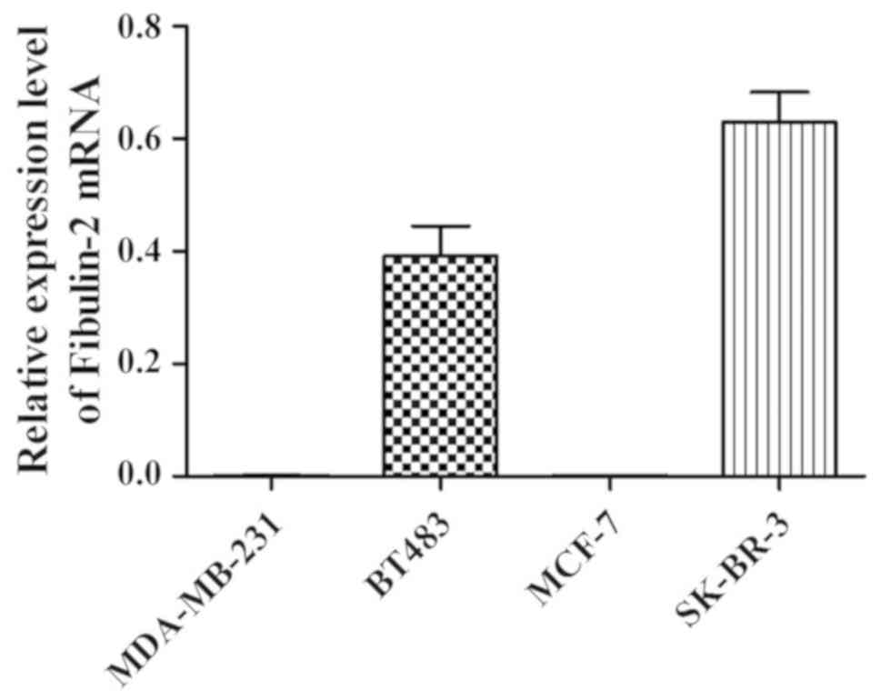
Fibulin-2 mRNA expression in different
breast cancer cells detected via qRT-PCR
Fibulin-2 mRNA expression was not detected in
MDA-MB-231 and MCF-7 cells, but Fibulin-2 expression was detected
in BT483 and SK-BR-3 cells (Fig.
1).
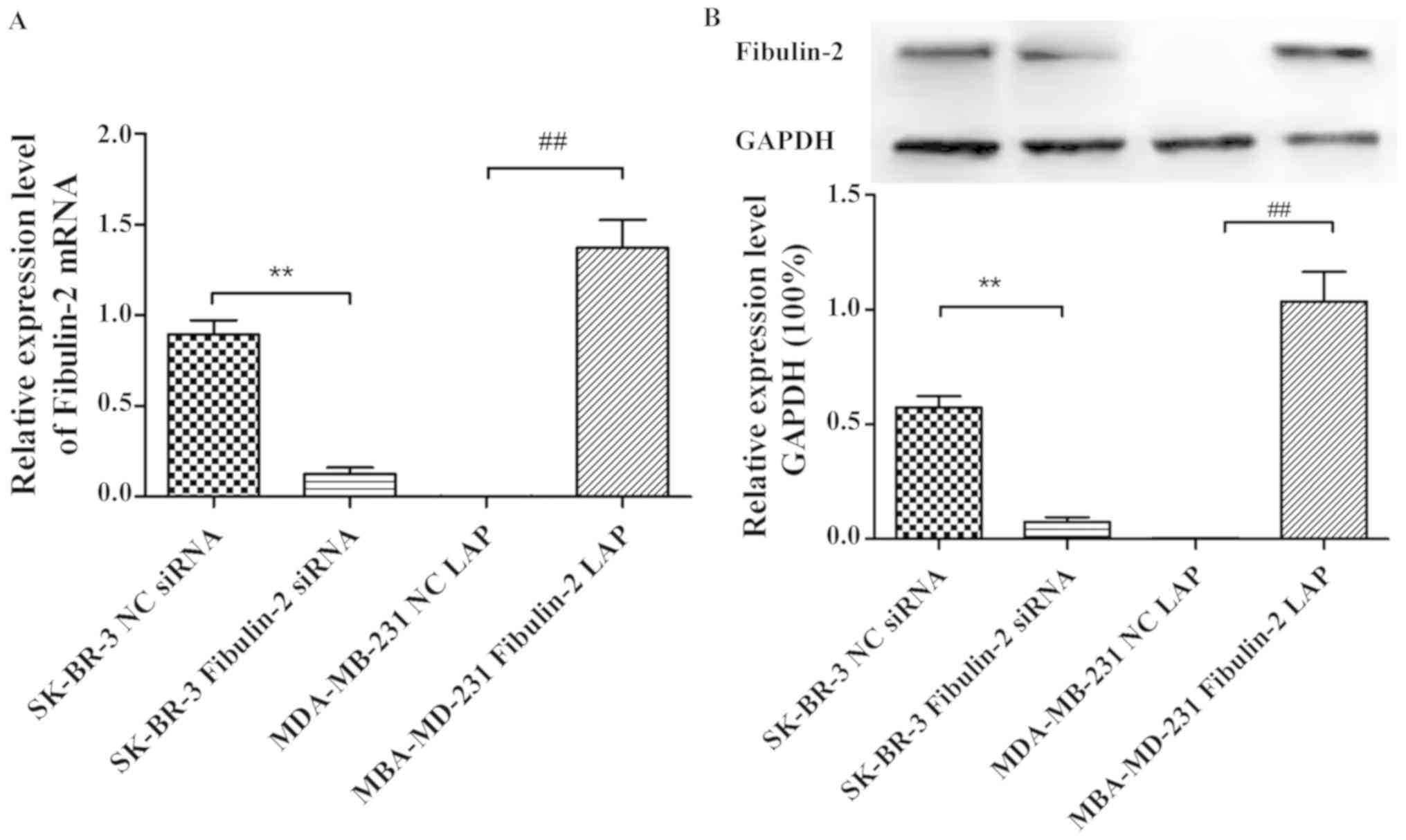
Effects of Fibulin-2 siRNA
interference and lentivirus overexpression on the expression of
Fibulin-2
The expression of Fibulin-2 was detected by qRT-PCR
and western blotting showed that the expression of Fibulin-2 mRNA
and protein in SK-BR-3 Fibulin-2 siRNA group was significantly
lower than that in SK-BR-3 NC siRNA group 48 h after transfection
(P<0.01). The expression of Fibulin-2 mRNA and protein in
MDA-MB-231 Fibulin-2 LAP group was significantly higher than that
in MDA-MB-231 NC LAP group (P<0.01) (Fig. 2).
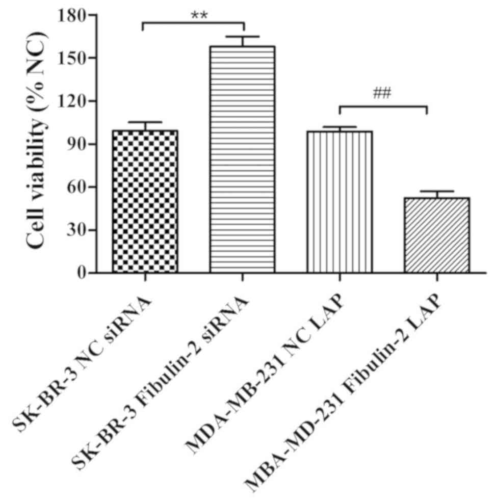
Effect of Fibulin-2 on the
proliferation of breast cancer cells
The changes of cell proliferation ability were
detected by CCK8 assay showing that the cell proliferation ability
of SK-BR-3 Fibulin-2 siRNA group was significantly higher than that
of SK-BR-3 NC siRNA group (P<0.01), while that of MDA-MB-231
Fibulin-2 LAP group was significantly lower than that of MDA-MB-231
NC LAP group (P<0.01) (Fig.
3).
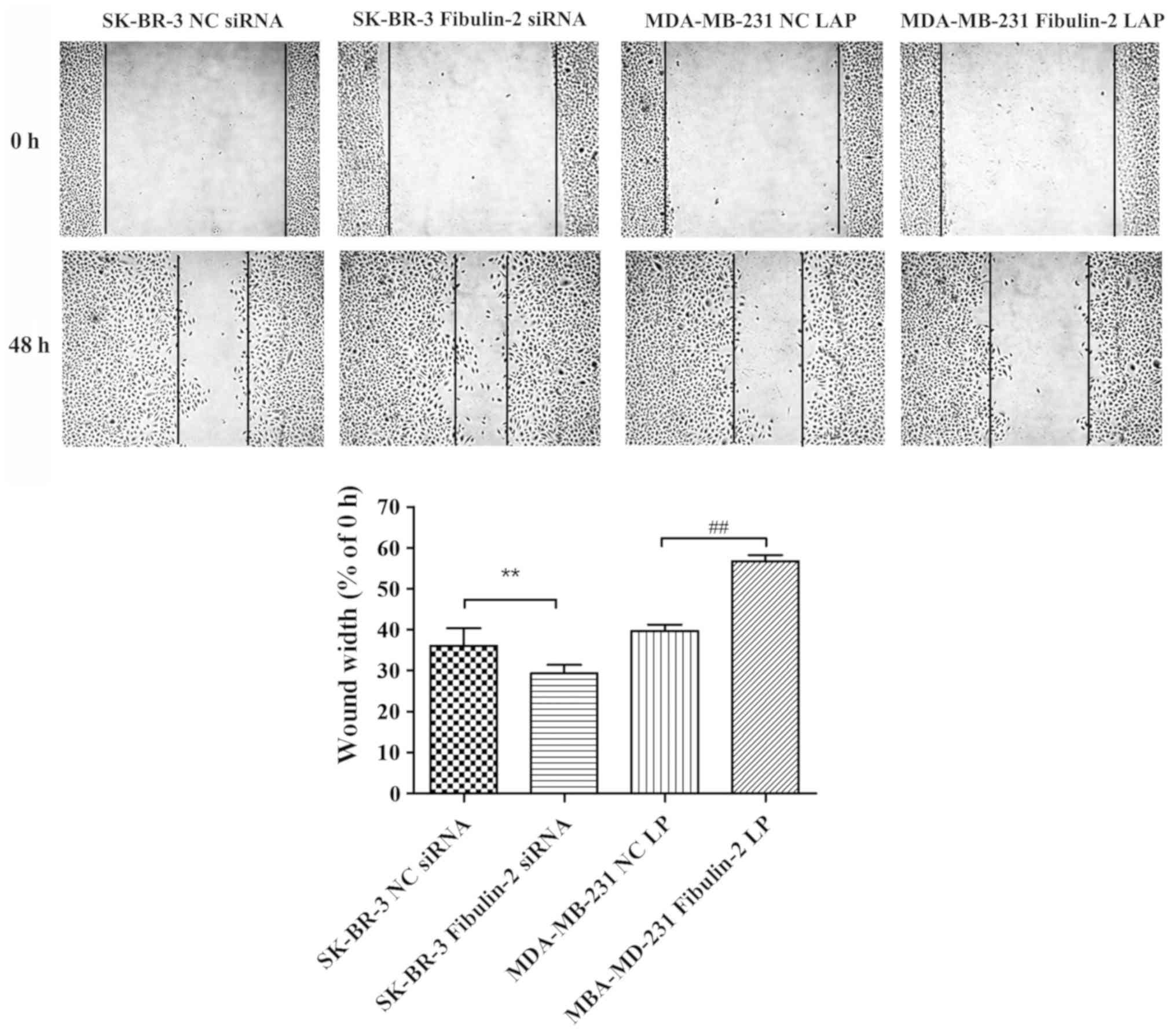
Influence of Fibulin-2 on the
migration of breast cancer cells
The cell migration ability of SK-BR-3 Fibulin-2
siRNA group was significantly higher than that of SK-BR-3 NC siRNA
group (P<0.01, while that of MDA-MB-231 Fibulin-2 LAP group was
significantly lower than that of MDA-MB-231 NC LAP group
(P<0.01) (Fig. 4).
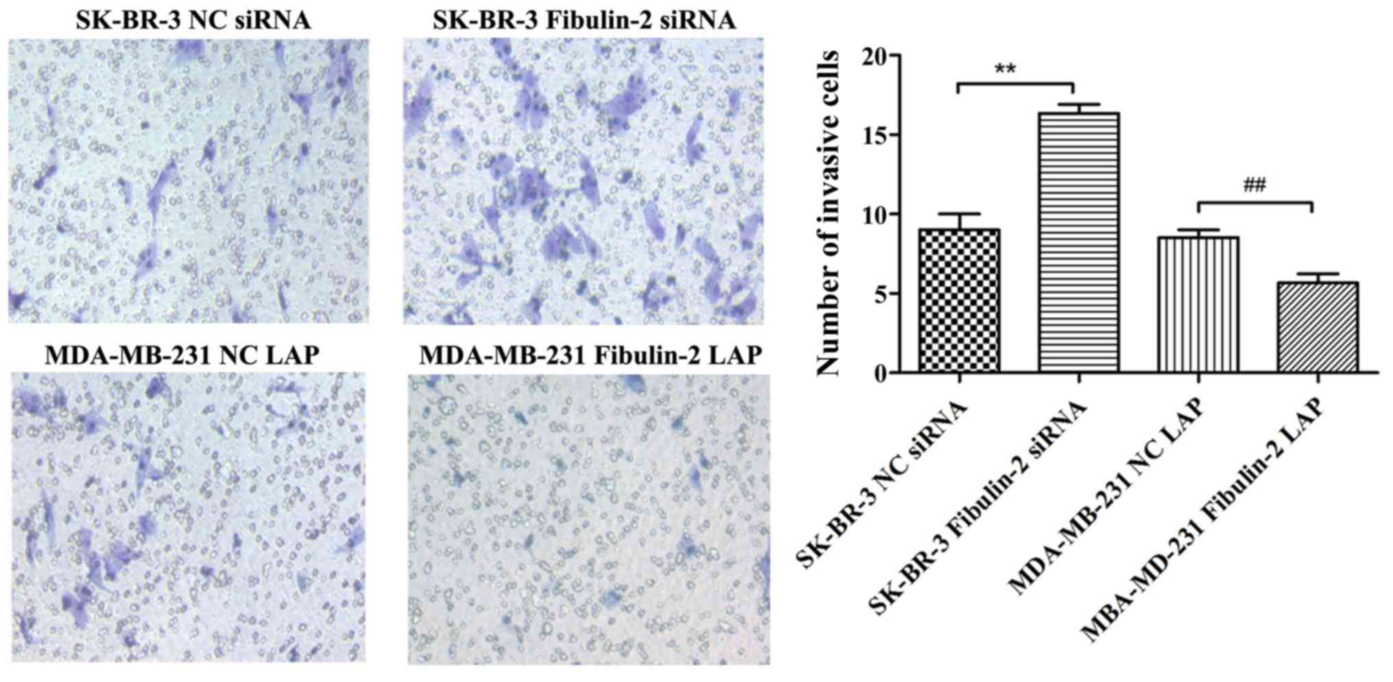
Impact of Fibulin-2 on the invasion of
breast cancer cells
The invasive ability of cells in SK-BR-3 Fibulin-2
siRNA group was significantly higher than that in SK-BR-3 NC siRNA
group (P<0.01), while that in MDA-MB-231 Fibulin-2 LAP group was
significantly lower than that in MDA-MB-231 NC LAP group
(P<0.01) (Fig. 5).
Effect of Fibulin-2 on tumor growth in
nude mice
Compared with MDA-MB-231 NC LAP group, the tumor
weight of nude mice in MDA-MB-231 Fibulin-2 LAP group was
significantly lower than that in MDA-MB-231 NC LAP group
(p<0.01), and the tumor growth and tumor volume in MDA-MB-231
Fibulin-2 LAP group were significantly lower than those in
MDA-MB-231 NC LAP group (P<0.01) (Fig. 6).
Discussion
Breast cancer has become a growing threat to women's
health, and its age of onset is getting younger. Furthermore, its
incidence rate ranks first among urban female malignant tumors.
With the increase in the incidence rate of breast cancer, the side
effects of chemotherapy drugs and the huge demand of clinical
treatment, increasing number of researchers pay attention to the
exploration of new mechanisms and targets of breast cancer
(11).
Remodeling of extracellular matrix can be achieved
by the synthesis and degradation of matrix proteins. A study
manifested that extracellular matrix is closely associated with the
differentiation, migration and migration of most tumors. After
remodeling, there are obvious changes in physical properties and
spatial structure of extracellular matrix proteins. Therefore, the
characteristics and molecules relating to extracellular matrix
remodeling can be used as important indicators for early diagnosis
and clinical staging of tumors (12). Another study revealed that Fibulin-2
protein, as an important component of the extracellular matrix, is
closely correlated with the occurrence of cancer and plays a dual
role in the occurrence and development of tumors, i.e., it inhibits
tumorigenesis under certain conditions and also promotes the growth
of tumor cells under such conditions (13). Studies have revealed that Fibulin-2
suppresses the migration and invasion of tumors such as
nasopharyngeal carcinoma and astrocytoma, but it promotes migration
and invasion of tumor cells in lung adenocarcinoma (14–16).
A recent study found that there is no expression of
Fibulin-2 in breast cancer patients, whereas normal in
paracancerous tissues of breast cancer, which confirmed that
Fibulin-2 can stabilize the basement membrane, and its
downregulation is related to the loss of basement membrane and
early invasion (17). Tan et
al (18) showed that Fibulin-2
was expressed in breast ducts and perivascular tissues in normal
breast tissues. In malignant breast tissues, collagen IV was
integrated around ducts, while Fibulin-2 was not fully expressed
around collagen IV, which showed that Fibulin-2 was involved in
tumor invasion and migration. Moreover, it has been shown that
Fibulin2 is a new matrix of ADAMTS5 and the degradation of Fibulin2
is associated with the enhancement of the invasion potential of
SKBR3 cells, and this proteolytic hydrolysis can alter the cell
microenvironment and affects the balance between tumor and
anti-tumor effects associated with Fibulin2 and ADAMTS
metalloproteinase (19).
Studies have verified that Fibulin-2 is capable of
promoting the development and progression of lung cancer and
pancreatic cancer, with different expression levels in lung cancer,
pancreatic cancer and adjacent normal tissues and a mouse lung
cancer cell line. It was discovered in a previous study that
Fibulin-2 is highly expressed in human lung adenocarcinoma
extracellular matrix and metastatic lung adenocarcinoma mouse tumor
cell lines. A study found that knockout of Fibulin-2 leads to small
tumor growth and low migration rate by constructing stable mouse
lung adenocarcinoma tumor cells without Fibulin-2 gene and
establishing a nude-mouse model of transplantation. Research at the
cellular level discovered that the migration and invasion ability
of tumor cells is weakened after knockdown of Fibulin-2 gene, and
Fibulin-2 promotes cell collagen cross-linking and adhesion in lung
tumor (20–22). Studies have confirmed that Fibulin-2
is overexpressed in different solid tumor tissues, fully proving
that Fibulin-2 plays a vital role in the occurrence and development
of tumors (23).
In this investigation, a variety of breast cancer
cells were cultured in vitro. qRT-PCR results showed that
MDA-MB-231 and MCF-7 cells did not express Fibulin-2, while BT483
and SK-BR-3 cells expressed Fibulin-2. Expression of Fibulin-2 was
upregulated and downregulated using RNAi technology and lentiviral
transfection techniques, respectively, and the expression levels of
Fibulin-2 mRNA and protein were evaluated via qRT-PCR and western
blotting, respectively. The results showed that the expression of
Fibulin-2 mRNA and protein in SK-BR-3 Fibulin-2 siRNA group was
significantly lower than that in SK-BR-3 NC siRNA group, while the
expression of Fibulin-2 mRNA and protein in MDA-MB-231 Fibulin-2
LAP group was significantly higher than that in MDA-MB-231 NC LAP
group 48 h after transfection. The results of cell proliferation,
Scratch and Transwell tests showed that compared with SK-BR-3 NC
siRNA group, the ability of cell proliferation, migration and
invasion in SK-BR-3 Fibulin-2 siRNA group was enhanced. Compared
with MDA-MB-231 NC LAP group, the ability of cell proliferation,
migration and invasion in MDA-MB-231 Fibulin-2 LAP group was
reduced. It was found that the tumor weight and volume of
MDA-MB-231 Fibulin-2 LAP group decreased significantly after
upregulating the expression of Fibulin-2 in nude mice.
In conclusion, this study demonstrates that
Fibulin-2 expression can greatly affect the proliferation,
migration and invasion of human breast cancer cell lines.
Therefore, Fibulin-2 may be a potential therapeutic target and a
prognostic index for breast cancer.
Acknowledgements
Not applicable.
Funding
No funding was received.
Availability of data and materials
The datasets used and/or analyzed during the current
study are available from the corresponding author on reasonable
request.
Authors' contributions
XZ wrote the manuscript. XZ and LD performed PCR and
western blot. YZ and HZ were responsible for cell culture and
transfection. XY and CZ contributed to wound healing assay and
Transwell assay. All authors read and approved the final
manuscript.
Ethics approval and consent to
participate
The study was approved by the Ethics Committee of
Navy General Hospital (Beijing, China).
Patient consent for publication
Not applicable.
Competing interests
The authors declare that they have no competing
interests.
References
|
1
|
Ducros E, Berthaut A, Mirshahi P,
Lemarchand S, Soria J, Legeais JM and Mirshahi M: Expression of
extracellular matrix proteins fibulin-1 and fibulin-2 by human
corneal fibroblasts. Curr Eye Res. 32:481–490. 2007. View Article : Google Scholar : PubMed/NCBI
|
|
2
|
Wight TN: The extracellular matrix and
atherosclerosis. Curr Opin Lipidol. 6:326–334. 1995. View Article : Google Scholar : PubMed/NCBI
|
|
3
|
Serra N, Rosales R, Masana L and Vallvé
JC: Simvastatin increases fibulin-2 expression in human coronary
artery smooth muscle cells via RhoA/Rho-kinase signaling pathway
inhibition. PLoS One. 10:e01338752015. View Article : Google Scholar : PubMed/NCBI
|
|
4
|
Aspberg A, Adam S, Kostka G, Timpl R and
Heinegård D: Fibulin-1 is a ligand for the C-type lectin domains of
aggrecan and versican. J Biol Chem. 274:20444–20449. 1999.
View Article : Google Scholar : PubMed/NCBI
|
|
5
|
Olin AI, Mörgelin M, Sasaki T, Timpl R,
Heinegård D and Aspberg A: The proteoglycans aggrecan and Versican
form networks with fibulin-2 through their lectin domain binding. J
Biol Chem. 276:1253–1261. 2001. View Article : Google Scholar : PubMed/NCBI
|
|
6
|
Kwan ML, Kushi LH, Weltzien E, Maring B,
Kutner SE, Fulton RS, Lee MM, Ambrosone CB and Caan BJ:
Epidemiology of breast cancer subtypes in two prospective cohort
studies of breast cancer survivors. Breast Cancer Res. 11:R312009.
View Article : Google Scholar : PubMed/NCBI
|
|
7
|
Siegel R, Naishadham D and Jemal A: Cancer
statistics, 2013. CA Cancer J Clin. 63:11–30. 2013. View Article : Google Scholar : PubMed/NCBI
|
|
8
|
Chen WQ, Zeng HM, Zheng RS, Zhang SW and
He J: Cancer incidence and mortality in China, 2007. Chin J Cancer
Res. 24:1–8. 2012. View Article : Google Scholar : PubMed/NCBI
|
|
9
|
Werb Z: ECM and cell surface proteolysis:
Regulating cellular ecology. Cell. 91:439–442. 1997. View Article : Google Scholar : PubMed/NCBI
|
|
10
|
Wu TS and Hammond GL: Naturally occurring
mutants inform SHBG structure and function. Mol Endocrinol.
28:1026–1038. 2014. View Article : Google Scholar : PubMed/NCBI
|
|
11
|
Shamay M, Barak O and Shaul Y: HBXAP, a
novel PHD-finger protein, possesses transcription repression
activity. Genomics. 79:523–529. 2002. View Article : Google Scholar : PubMed/NCBI
|
|
12
|
Whiteaker JR, Zhang H, Zhao L, Wang P,
Kelly-Spratt KS, Ivey RG, Piening BD, Feng LC, Kasarda E, Gurley
KE, et al: Integrated pipeline for mass spectrometry-based
discovery and confirmation of biomarkers demonstrated in a mouse
model of breast cancer. J Proteome Res. 6:3962–3975. 2007.
View Article : Google Scholar : PubMed/NCBI
|
|
13
|
Hill VK, Hesson LB, Dansranjavin T, Dallol
A, Bieche I, Vacher S, Tommasi S, Dobbins T, Gentle D, Euhus D, et
al: Identification of 5 novel genes methylated in breast and other
epithelial cancers. Mol Cancer. 9:512010. View Article : Google Scholar : PubMed/NCBI
|
|
14
|
Fontanil T, Rúa S, Llamazares M,
Moncada-Pazos A, Quirós PM, García-Suárez O, Vega JA, Sasaki T,
Mohamedi Y, Esteban MM, et al: Interaction between the ADAMTS-12
metalloprotease and fibulin-2 induces tumor-suppressive effects in
breast cancer cells. Oncotarget. 5:1253–1264. 2014. View Article : Google Scholar : PubMed/NCBI
|
|
15
|
Tian H, Liu J, Chen J, Gatza ML and Blobe
GC: Fibulin-3 is a novel TGF-β pathway inhibitor in the breast
cancer microenvironment. Oncogene. 34:5635–5647. 2015. View Article : Google Scholar : PubMed/NCBI
|
|
16
|
Chan SW, Lim CJ, Guo K, Ng CP, Lee I,
Hunziker W, Zeng Q and Hong W: A role for TAZ in migration,
invasion, and tumorigenesis of breast cancer cells. Cancer Res.
68:2592–2598. 2008. View Article : Google Scholar : PubMed/NCBI
|
|
17
|
Ibrahim AM, Sabet S, El-Ghor AA, Kamel N,
Anis SE, Morris JS and Stein T: Fibulin-2 is required for basement
membrane integrity of mammary epithelium. Sci Rep. 8:141392018.
View Article : Google Scholar : PubMed/NCBI
|
|
18
|
Tan H, Zhang J, Fu D and Zhu Y: Loss of
fibulin-2 expression is involved in the inhibition of breast cancer
invasion and forms a new barrier in addition to the basement
membrane. Oncol Lett. 14:2663–2668. 2017. View Article : Google Scholar : PubMed/NCBI
|
|
19
|
Fontanil T, Álvarez-Teijeiro S, Villaronga
MÁ, Mohamedi Y, Solares L, Moncada-Pazos A, Vega JA, García-Suárez
O, Pérez-Basterrechea M, García-Pedrero JM, et al: Cleavage of
Fibulin-2 by the aggrecanases ADAMTS-4 and ADAMTS-5 contributes to
the tumorigenic potential of breast cancer cells. Oncotarget.
8:13716–13729. 2017. View Article : Google Scholar : PubMed/NCBI
|
|
20
|
Dunwell TL, Hesson LB, Pavlova T,
Zabarovska V, Kashuba V, Catchpoole D, Chiaramonte R, Brini AT,
Griffiths M, Maher ER, et al: Epigenetic analysis of childhood
acute lymphoblastic leukemia. Epigenetics. 4:185–193. 2009.
View Article : Google Scholar : PubMed/NCBI
|
|
21
|
Mesri EA, Cesarman E and Boshoff C:
Kaposi's sarcoma and its associated herpesvirus. Nat Rev Cancer.
10:707–719. 2010. View
Article : Google Scholar : PubMed/NCBI
|
|
22
|
Alcendor DJ, Knobel S, Desai P, Zhu WQ and
Hayward GS: KSHV regulation of fibulin-2 in Kaposi's sarcoma:
Implications for tumorigenesis. Am J Pathol. 179:1443–1454. 2011.
View Article : Google Scholar : PubMed/NCBI
|
|
23
|
Senapati S, Gnanapragassam VS, Moniaux N,
Momi N and Batra SK: Role of MUC4-NIDO domain in the MUC4-mediated
metastasis of pancreatic cancer cells. Oncogene. 31:3346–3356.
2012. View Article : Google Scholar : PubMed/NCBI
|